
Article Figures & Data
Figures
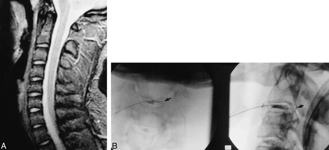
- fig 1.
Patient with 5/10 concordant, bilateral, occipital head, CVJ, and upper neck pain.
A, Sagittal MPGR image (3-mm thick, 1-mm gap, 20° flip angle, TE = 10) shows a completely normal-appearing C2–C3 disk.
B, C2–C3 diskogram in anteroposterior (left) and lateral (right) views shows uncovertebral fissure (arrow on left) along with full-thickness posterior tear and contrast leakage (arrow on right).
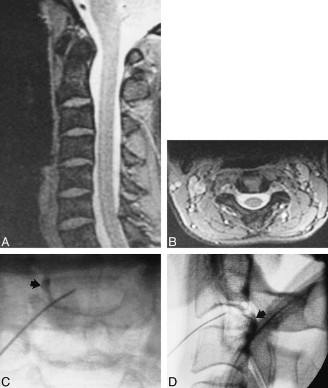
- fig 2.
Patient with 6.5/10 concordant, but mostly right-sided, occipital and mastoid pain with uncovertebral fissure.
A and B, Sagittal (A) and axial (B) MPGR images (3-mm thick, TE = 1015) show slight forward displacement of C2 on C3 (A). Axial image (B) appears normal. Because of the slight forward displacement of C2 on C3 (A), C2–C3 disk is judged to be abnormal.
C and D, Anteroposterior (C) and lateral (D) diskograms reveal an uncovertebral fissure and contrast leakage (arrows).
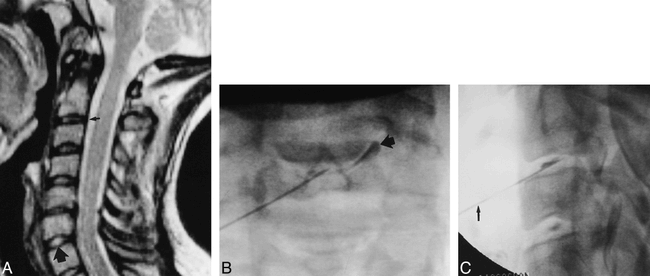
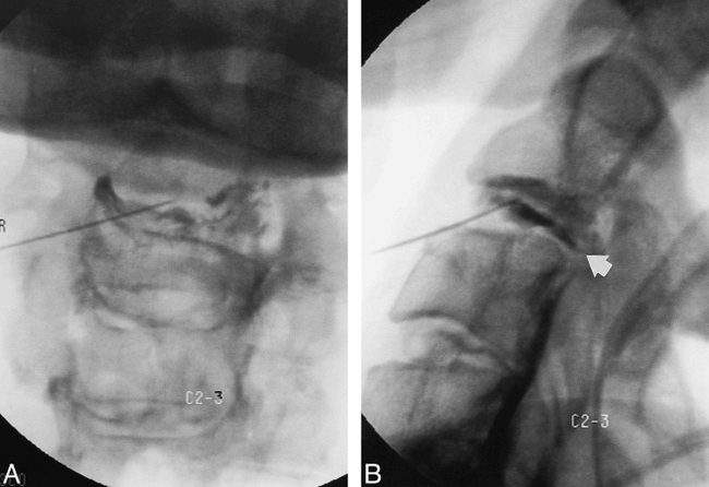
- fig 3.
Patient with 9/10 concordant unilateral mastoid and occipital head pain associated with uncovertebral tear.
A, Sagittal, 3-mm-thick MPGR midline image reveals relative dehydration of the C2–C3 disk (small arrow) as compared with the normally hydrated C6–C7 disk (large arrow).
B and C, Anteroposterior (B) and lateral (C) diskograms show an uncovertebral fissure (arrow in B). Note how the 25-gauge needle (arrow in C) enters the disk slightly from below.
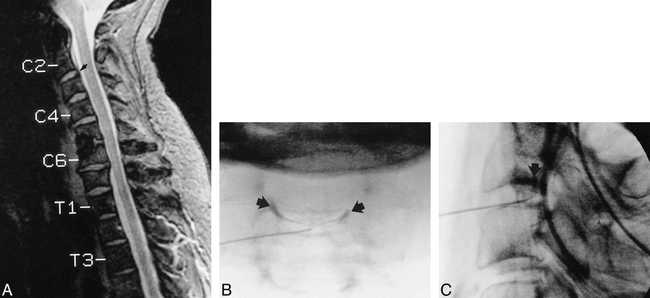
- fig 4.
Patient with 9/10 concordant bilateral neck, CVJ, and occipital head pain with slightly abnormal C2–C3 disk contour at MR imaging.
A, Sagittal midline, 3-mm-thick MPGR image shows slight dorsal bulging of the C2–C3 disk annulus (arrow). Note previous interbody fusion at C4–C5.
B and C, Anteroposterior (B) and lateral (C) diskograms reveal bilateral uncovertebral fissures (arrows in B). The fissures are superimposed on lateral view (arrow in C).
- fig 5.
Patient with 8.5/10 concordant, diffuse, bilateral CVJ, occipital, parietal, and temporal head pain associated with bilateral and posterior annular tears of the C2–C3 disk.
A and B, A large, broadly based defect is seen on both projections (arrow in B), extending into C2–C3 foramina (A).
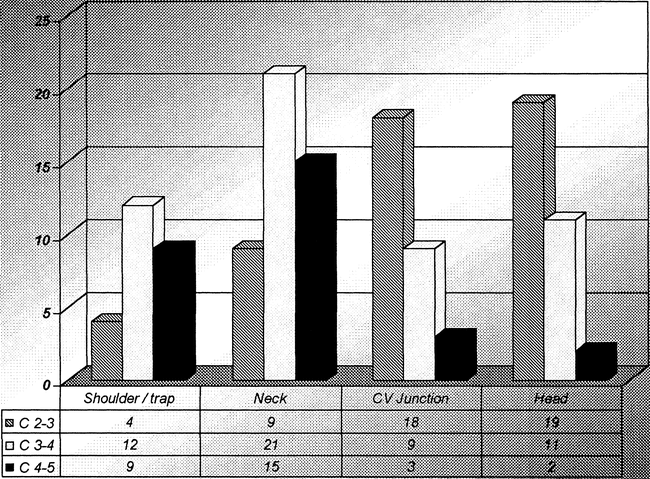
- fig 6.
Analysis of diskographically provoked responses at C2–C3, C3–C4, and C4–C5. Forty disks were studied at each level. Only intensely painful (≥ 7/10) and concordant disks were counted
- fig 7.
Specific locations of head pain perceptions from intensely painful, concordant disks


Tables

Breakdown of intensely painful, concordant head pain responses comparing C2–C3 alone, C2–C3 and some other disk(s), and disk(s) other than C2–C3


