
Article Figures & Data
Figures
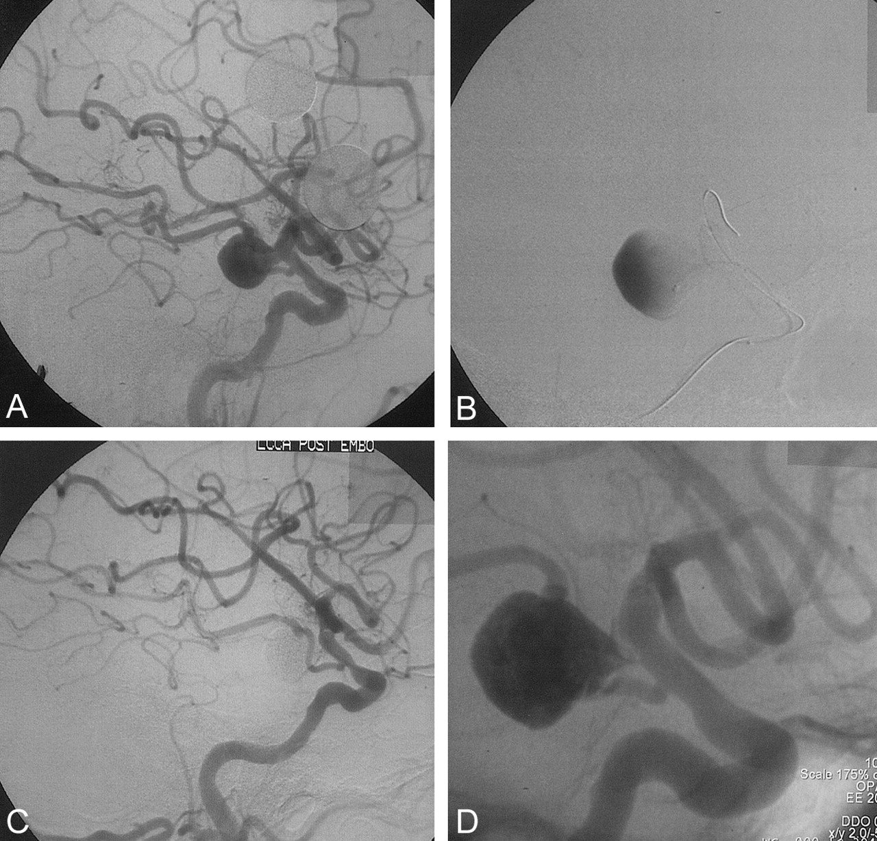
- Fig 1.
60-year-old male patient who presented with headache and in whom a large unruptured internal carotid aneurysm was found. Pretreatment, treatment, and follow-up images are presented.
A, Large internal carotid aneurysm before treatment.
B, Seal test before treatment of the aneurysm with Onyx. Microcatheter and balloon are in place with gentle contrast material injection into the aneurysm
C, Angiographic result immediately after Onyx treatment.
D, Follow-up angiogram 6 months after treatment, showing complete occlusion and intervening soft thickened tissue between vessel lumen and Onyx cast in the aneurysm
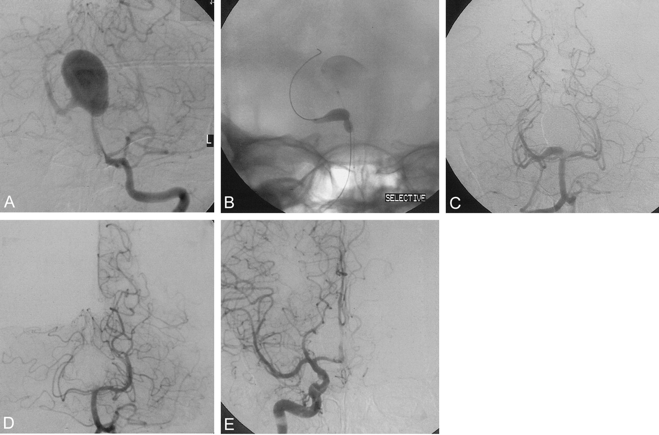
- Fig 2.
28-year-old hemophiliac patient with severe headaches but no evidence of subarachnoid hemorrhage has a wide-neck basilar bifurcation aneurysm involving the proximal right postcerebral artery.
A, Large basilar bifurcation and proximal posterior cerebral aneurysms.
B, Seal test, showing highly compliant Equinox balloon inflated in the basilar tip and in the right posterior cerebral artery with the balloon protecting the aneurysm neck, which takes up most of the proximal P1-segment vessel.
C, Immediate posttreatment image, showing reconstruction of the posterior cerebral artery segment.
D, Three-month follow-up angiogram, showing occlusion of the proximal posterior cerebral artery and the aneurysm.
E, Three-month right carotid angiogram, showing posterior cerebral perfusion via the posterior communicating artery. (Images courtesy of Mike Nelson, Leeds.)
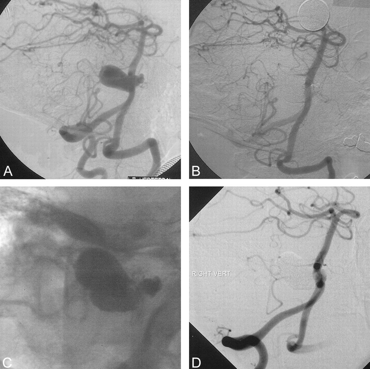
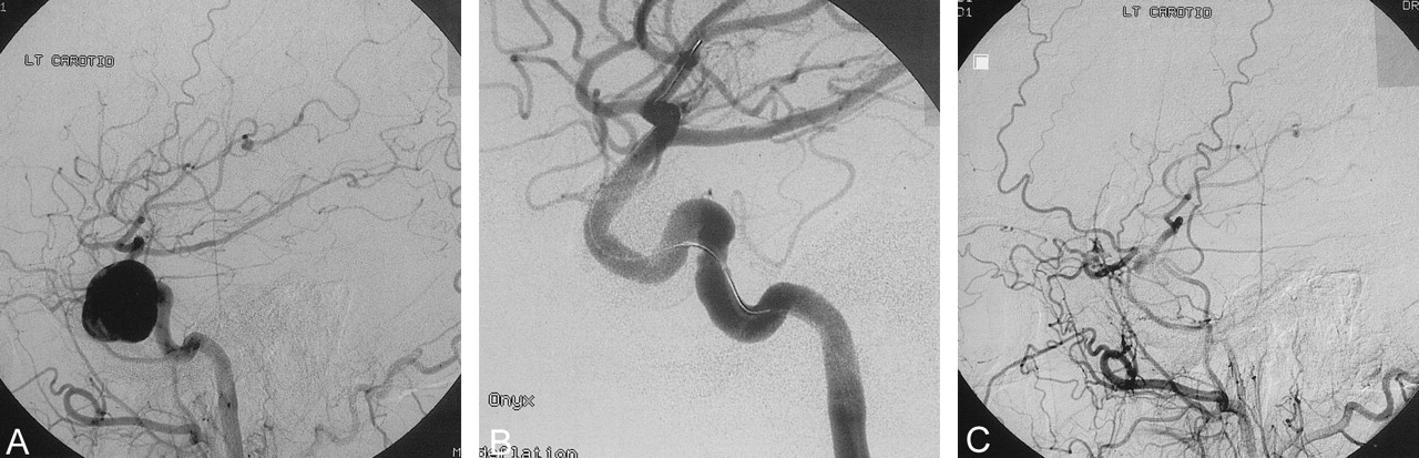
- Fig 3.
50-year-old male patient with leg weakness and a possible history of subarachnoid hemorrhage. This patient was found to have a vertebral basilar junction aneurysm compressing the medulla.
A, Pretreatment angiogram showing a large vertebrobasilar junction aneurysm treated on two occasions (treated with an INX stent at the second procedure after recurrence). Complete occlusion was noted at 6-month and 1-year follow-up after second procedure.
B, Angiogram obtained after second treatment at 3 months after early recurrence and after placement of an INX stent and re-treatment with Onyx.
C, Unsubtracted image obtained after second procedure, showing INX stent and second cast of Onyx in inferior recurrent aneurysm pocket.
D, Follow-up angiogram obtained 6 months after second treatment.
- Fig 4.
45-year-old patient presenting with a partial cranial nerve III palsy and headache. No anterior cerebral artery was present on the left side and the carotid only supplied the left middle cerebral territory.
A, Lateral arterial phase carotid angiogram obtained immediately before treatment.
B, Final angiography obtained after Onyx treatment balloon catheter still present in vessel.
C, Follow-up angiography 3 months after procedure. Patient developed a complete ophthalomoplegia immediately after the procedure, which had resolved entirely by 3 months when she was asymptomatic. Note extensive orbital collaterals filling middle cerebral territory. (Images courtesy of Peter Flynn and Steven Mckinstry, Royal Victoria Hospital, Belfast.)


Tables
Sex 79% Female Age (y) Median = 46 y, Mean = 45 y (Range: 6–76 y) Presentation SAH: Recent (<1 mo) 10 Late (>1 mo) 7 Incidental 29 Mass Effect (including CN palsies) 39 Headache alone 7 Seizures 2 Other: 6 Nasal Bleeding 3 F/U of traumatic CCF 2 Previous SAH 1 y ago 1 Previously Treated Coil 15 Clip 0 Wrap 2 Stent 3 Note.—SAH indicates subarachnoid hemorrhage; CN, cranial nerve; F/U, follow-up; and CCF, carotid cavernous fistula.
Location Carotid n = 93 Petrous 6 Cavernous 25 Petrous/Cavernous 2 Ophthalmic 47 Other intracranial sites 13 Posterior circulation n = 7 Vertebral 1 Vertebrobasilar 1 Superior cerebellar 1 Posterior cerebral 2 Basilar tip 2 Average aneurysm size (mm)* Median Range Dome height 15 4–40 Dome width 14 3–44 Neck width 7 1–25 Total Neck <4 mm Neck ≥4 mm Neck ≥4 mm or D:N <2 Aneurysm sac (mm) Small (<10 mm) 21 6 15 18 Large (10–25 mm) 60 3 57 57 Giant (>25 mm) 19 0 19 19 n = 100† 100 9 91 94 * 1 aneurysm nearly fusiform mc
† Includes 1 fusiform neck.
Small <10 mm Large 10–24 mm Giant >25 mm Postembolization occlusion rates n = 20 (%) n = 60 (%) n = 19 (%) Complete (100%) 15 (75) 33 (55) 19 (47) Subtotal (90–99%) 5 (25) 26 (43) 19 (47) Incomplete (<90%) — 1 (2) 1 (5) 3–6 month follow-up occlusion rates n = 15 (%) n = 51 (%) n = 15 (%) Complete (100%) 13 (86.7) 31 (61) 8 (53) Subtotal (90–99%) 1 (6.7) 12 (23) 4 (27) Incomplete (<90%) 1 (6.7) 7 (14) 2 (13) Not determinable 1 (2) 1 (7) 12-month follow-up occlusion rates After re-treatment, when applicable n = 14 (%) n = 39 (%) n = 14 (%) Complete (100%) 13 (93) 30 (77) 8 (57) Subtotal (90–99%) 0 5 (13) 2 (14) Incomplete (<90%) 1 (7) 2 (5) 2 (14) Not determinable 2 (5) 2 (14) Number of Procedures 108 Procedure time (Start time = time at which delivery catheter is filled with DSMO; Stop time = delivery catheter detachment from Oynx mass) Mean 95 min Range 15 min–360 min First 54 procedures Mean 101 min Range 15–300 min Second 54 procedures Mean 90 min Range 20–360 min Onyx volume Mean 1.875 cc Range 0.07–10.25 cc Treatment with Onyx + Stent Initial treatment 17 Re-treatment 5 Unruptured and Other Recent SAH At baseline n = 87 n = 10 Baseline Rankin ≥2 81 — Hunt & Hess grade 1 or 2 — 7 4 3 At discharge* with including re-treatment as applicable n = 86† n = 10 Rankin ≤2 76 (88%) 5 (95% CI 79–94) Rankin Unchanged/improved 75 — Rankin Worsened 8 (9%) — Death at discharge 3 1 At 3–6 months including re-treatment as applicable n = 82 n = 7 Rankin ≤2 74 (90%) 6 (95% CI 82–96) Rankin Unchanged/improved 74 7 Rankin Worsened 5 0 Death at discharge to 3 months 0 0 1 lost to f/u 1 no data at 3 mths At 12-Months or latest f/u, with re-treatment as applicable n = 83† n = 7 Rankin ≤2 76 (91.5%) 4 (95% CI 83–96) Rankin Unchanged/improved 75 6 Rankin Worsened 3 0 Death at 3 months to 12 months 2 1 * Discharge data missing in 1.
† Includes four patients with no 3–6 month follow-up Rankin score.
Patient Age (y)/Sex Size and Location of Aneurysm Description of Event and Cause of Event Cause Baseline Rankin or H& H Outcome of Event and mRS at Last Follow-Up Permanent morbidity 39/M Large carotid ophthalmic Ipsilateral visual loss due to leak in ophthalmic artery Device 0 mRS = 1 42/F Giant carotid cavernous Worsened CN III, IV, V, and VI palsy, resolution of CN v aplsy Procedure 1 mRS = 1 76/F Small posterior communicating Worsening of neurologic status (Grade, 2 H&H) SAH 2 Lost to follow-up 75/F Giant carotid cavernous Mild hemiparesis Device (Onyx) 1 mRS = 1 44/M Small carotid ophthalmic Numbness in L leg caused by senosis of M3 MCA Device (Onyx) 0 mRS = 1 56/M Giant carotid ophthalmic Onyx extravasation into subarachnoid space, SAH, severe hemiplegia Device (Onyx) 1 Ongoing at 12 mnths, mRS = 4 45/F Large carotid ophthalmic Blindness in R eye post procedure with retinal infarct Device (Onyx) 0 mRS = 1 55/F Large carotid ophthalmic Devloped delayed L MCA infarct 3 days afer retreatment with stent and Onyx caused aphasia and R hemiplegia Device (Stent) 2 Ongoing at 3 months, mRS = 4 Partial resolution by 1 year Transient neurologic morbidity 52/M Large carotid ophthalmic Developed symptomatic vasospasm with L hemiparesis; recent SAH Disease Grade 2 H&H mRS = 0 Resolved 47/F Large superior Developed transient hemiparesis Device (also had stent placement) 0 mRS = 0 Resolved 50/M Large vertebro-basilar junction Developed small SAH after catheter withdrawal; no neurologic deterioration; developed infected hematoma after 10 days retreatment procedure; required surgery Procedure 37/F Small carotid cavernous Developed confusion and ipsilateral hemiparesis headaches and nausea 1 week after treatment Unknown 1 mRS = 1 Resolved by 6 months 57/F Large carotid cavernous Procedural thrombus in left ICA; hemiparesis and aphasia; resolution of morbidity Procedure 0 mRS = 1 Resolved by 2 days 31/F Large carotid ophthalmic Transient hemiparesis Procedure 0 mRS = 0 Resolved 45/F Large carotid ophthalmic Developed worsened visual symptoms that subsequently improged Device 0 mRS = 1 Complete resolution and visual improvement 68/F Recurrent basilar tip Developed proximal brain stem stroke post procedure, probably thrombo-embolic, needed prolonged hospitalisation Procedure 1 mRS = 2 Neurological symptoms fully resolved by 1 year Optic nerve compression Baseline At Discharge 12-Month or Latest Follow-up No. of patients 14 Improved/resolved 2 6 Unchanged 11 7 Worsened 1 1 Additional patients 1 0* *1 no follow up to date Occulo-motor deficits Baseline At Discharge 12-Month or Latest Follow-up No. of patients 16 Improved/resolved 6 14 Unchanged 8 1 Worsened 1 1 1 = died prior discharge Hemiparesis or hemiplegia Baseline At Discharge 12-Month or Latest Follow-up No. of patients 8 Improved/resolved 1 5 Unchanged 5 1 Worsened 0 0 2* = died Headache (all), n = 57 at baseline Baseline At Discharge 12-Month or Latest Follow-up Mild/moderate No. of patients 48 Improved/resolved 43 42 Unchanged 2 1 Worsened 0 2 3 = died At Discharge 12-Month or Latest Follow-up Severe No. of patients 9 Improved/resolved 8 8 Unchanged 1 0 Worsened 0 0 1 = lost to follow up
In this issue


Jump to section
Related Articles
Cited By...
- An injectable shear-thinning biomaterial for endovascular embolization
- Treatment of experimental aneurysms with a new liquid embolic agent and a retrievable stent: proof of concept and feasibility study
- Influence of observers, threshold values, and measurement methods on volumetric analysis of cerebral aneurysms with three-dimensional rotational angiography
- Technology developments in endovascular treatment of intracranial aneurysms
- Guidelines for the Management of Patients With Unruptured Intracranial Aneurysms: A Guideline for Healthcare Professionals From the American Heart Association/American Stroke Association
- Preliminary in vivo evaluation of a novel intrasaccular cerebral aneurysm occlusion device
- The Maze-Making and Solving Technique for Coil Embolization of Large and Giant Aneurysms
- Predicting parent vessel patency and treatment durability: a proposed grading scheme for the immediate angiographic results following Onyx HD-500 embolization of intracranial aneurysms
- Optic pathway infarct after Onyx HD 500 aneurysm embolization: visual pathway ischemia from superior hypophyseal artery occlusion
- Computational Hemodynamics Analysis of Intracranial Aneurysms Treated with Flow Diverters: Correlation with Clinical Outcomes
- Optic pathway infarct after Onyx HD 500 aneurysm embolization: visual pathway ischemia from superior hypophyseal artery occlusion
- Onyx embolization for the endovascular treatment of infectious and traumatic aneurysms involving the cranial and cerebral vasculature
- The sea anchor technique: a novel method to aid in stent-assisted embolization of giant cerebral aneurysms
- Endovascular Treatment of Intracranial Aneurysms: Current Status
- Resolution of Mass Effect and Compression Symptoms following Endoluminal Flow Diversion for the Treatment of Intracranial Aneurysms
- Treatment of Intracranial Aneurysms Using the Pipeline Flow-Diverter Embolization Device: A Single-Center Experience with Long-Term Follow-Up Results
- Biomechanical attributes of microcatheters used in liquid embolization of intracranial aneurysms
- 1-Hexyl n-cyanoacrylate compound (Neucrylate™ AN), a new treatment for berry aneurysm. III: Initial clinical results
- 1-Hexyl n-cyanoacrylate compound (Neucrylate™ AN), a new berry aneurysm treatment. II. Rabbit implant studies: technique and histology
- Endovascular Treatment of Large and Giant Aneurysms
- Interventional neuroradiology
- Stroke Review: Advances in Interventional Neuroradiology 2004