
Article Figures & Data
Figures
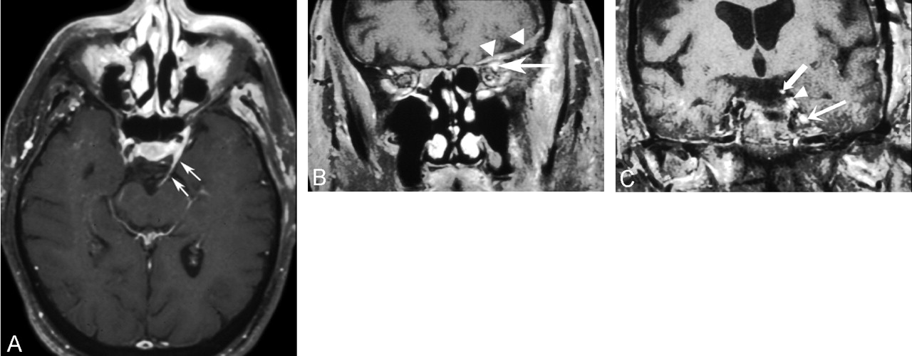
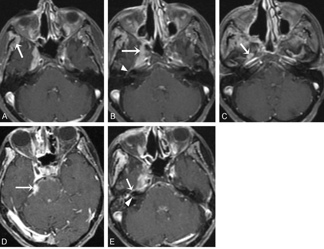
- Fig 1.
Patient 3, a 78-year-old man status post prior resection of a desmoplastic melanoma of the left forehead presents with numbness over left forehead and diplopia.
A, Contrast-enhanced axial T1-weighted image (600/11/2 [TR/TE/NEX]) with fat saturation demonstrates abnormal enhancement in the left CN III (arrows) and cavernous sinus. There is also enhancement in the left temporalis muscle secondary to denervation change.
B, Contrast-enhanced coronal T1-weighted image (600/11/2) with fat saturation demonstrates abnormal soft tissue prominence and enhancement at the level of the left frontal nerve (arrow). Increased signal intensity of the left temporalis muscle is consistent with denervation change. In addition, a small left subdural hematoma (arrowheads) is incidentally noted in this elderly patient. This demonstrated intrinsic T1 shortening consistent with blood products and resolved on a follow-up scan (not shown).
C, A more posterior contrast-enhanced coronal T1-weighted image (600/11/2) with fat saturation demonstrates enhancing soft tissue in Meckel’s cave on the left (lower arrow), consistent with involvement of the gasserian ganglion, as well as abnormal enhancement and enlargement of the left CN III (upper arrow) and abnormal enhancement and enlargement of the left CN VI (arrowhead) as well.
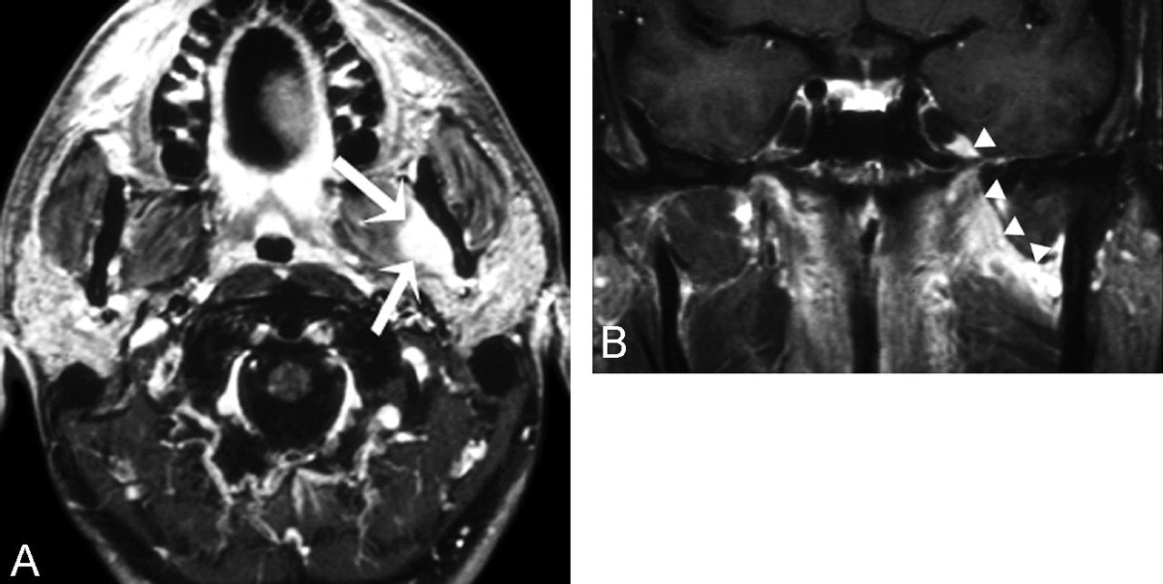
- Fig 2.
Patient 2, a 29-year-old man status post prior resection of a desmoplastic melanoma of the lower lip presents with decreased sensation over his left lower jaw.
A, Contrast-enhanced axial T1-weighted image (600/11/2) with fat saturation demonstrates a mass (arrows) just medial to the mandibular foramen, as well as subtle decrease in bulk of the left-sided muscles of mastication.
B, Contrast-enhanced coronal T1-weighted image (600/11/2) with fat saturation demonstrates enlargement and abnormal enhancement of the left CN V3 (arrowheads) from the level of the mandibular foramen up to the left Meckel’s cave, consistent with perineural spread of tumor.
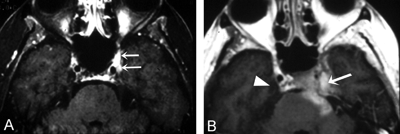
- Fig 3.
Patient 7, a 70-year-old woman status post prior resection of a left supraorbital lesion presents with a palsy of her left CN V1.
A, Contrast-enhanced axial T1-weighted MR image (600/11/2) with fat saturation demonstrates subtly asymmetrical enhancing soft tissue (anterior arrow) in the left superior orbital fissure/cavernous sinus, as well as the anterosuperior aspect of Meckel’s cave (posterior arrow).
B, Six months later, repeat contrast-enhanced axial T1-weighted image (600/11/2) demonstrates interval disease progression along the cisternal segment of the trigeminal nerve to involve the pons and cerebellar peduncle. Enhancing soft tissue completely fills Meckel’s cave on the left (arrow) as compared with the normal right side (arrowhead).
- Fig 4.
Patient 5, a 70-year-old woman with a history of desmoplastic melanoma involving the right nose, presents with right-sided facial pain.
A, Contrast-enhanced axial T1-weighted image (600/11/2) with fat saturation demonstrates a soft tissue mass in the right Meckel’s cave and cavernous sinus. There is enhancement of the right temporalis muscle secondary to denervation change (arrow).
B, A more inferior contrast-enhanced axial T1-weighted image (600/11/2) with fat saturation demonstrates a soft tissue mass enlarging the right foramen rotundum (arrow). In addition, there is questionable spread along the GSPN (arrowhead).
C, A more inferior contrast-enhanced axial T1-weighted image (600/11/2) with fat saturation demonstrates involvement of the foramen ovale, which is enlarged and shows abnormal enhancement (arrow). The patient received gamma knife radiosurgery to the right skull base and adjacent soft tissues and improved symptomatically. One year after treatment, however, the patient had interval progression of disease to involve CNs III–XII on the right, and a follow-up MR was performed.
D, Contrast-enhanced axial T1-weighted image (600/11/2) with fat saturation demonstrates an enhancing soft tissue mass (arrow) at the level of the root entry zone of right CN V.
E, A more inferior contrast-enhanced axial T1-weighted image (600/11/2) with fat saturation shows progression in the right cavernous sinus/Meckel’s cave region, as well as clear-cut enhancement of the right GSPN (arrow), with enhancement now extending into the distal right internal auditory canal (arrowhead).
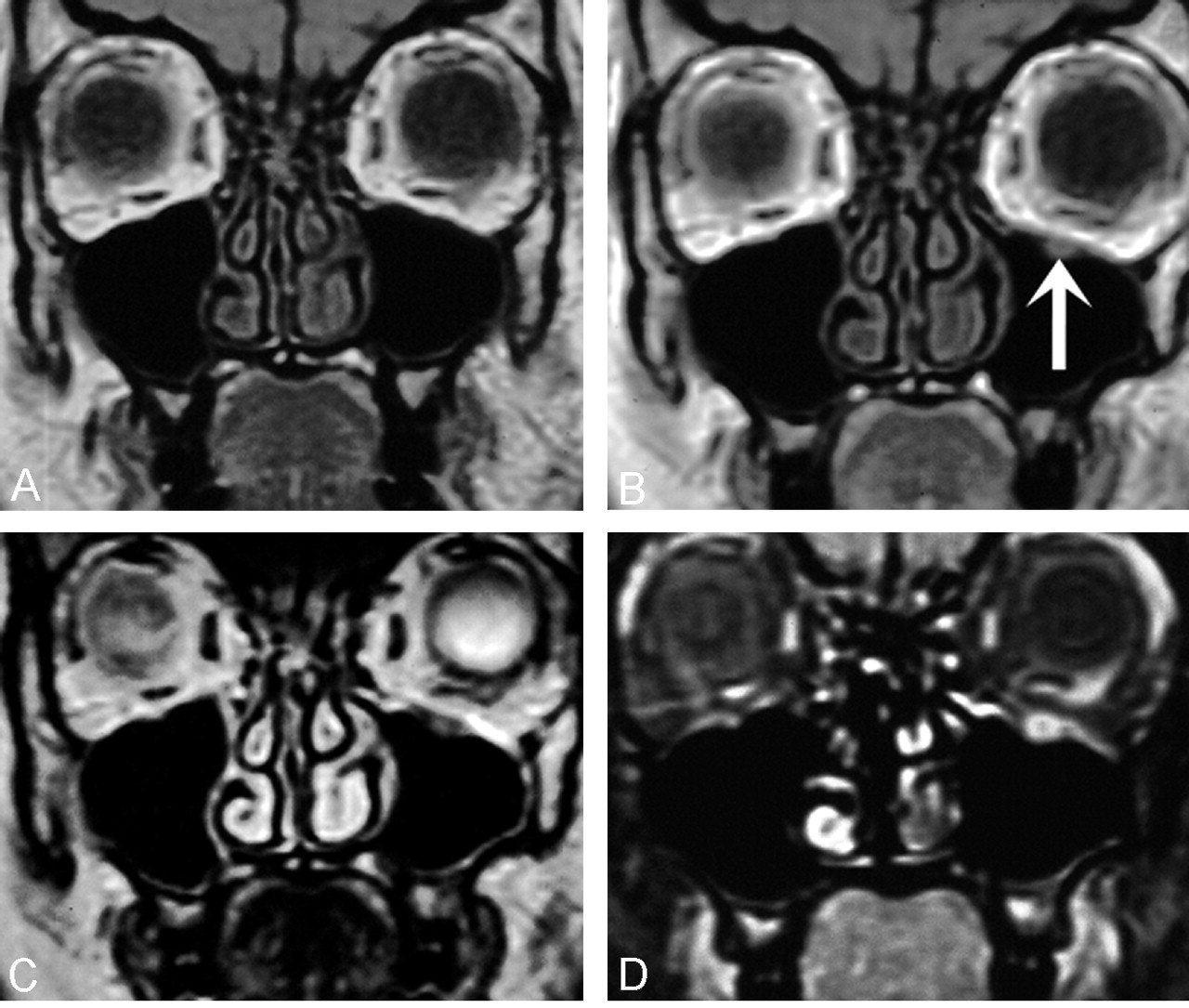
- Fig 5.
Patient 6, a 49-year-old woman presenting with a mass on her left upper lip. Biopsy confirmed melanoma, and she was treated with surgery and radiation. A follow-up scan was performed in April 2002, 5 months after her initial surgery, and further follow-up was performed in October 2002.
A, Coronal T1-weighted image (600/11/2) demonstrates no abnormality of the infraorbital nerve.
B, In follow-up 6 months later, the patient noted left cheek numbness and tingling, and a repeat MR was performed. Coronal T1-weighted image (600/11/2) was initially interpreted as negative, but further review demonstrates enlargement of the left infraorbital nerve (arrow).
C, Coronal T2-weighted image (4000/102/2) demonstrating that the enlarged left infraorbital nerve is low in signal intensity and not consistent with benign sinus contents adjacent to the infraorbital canal.
D, Contrast-enhanced coronal T1-weighted image (600/11/2) with fat saturation demonstrating clear cut enlargement and enhancement of the left infraorbital nerve. The patient was taken to repeat resection, and perineural spread of malignant melanoma was confirmed.


Tables
- TABLE 1:
Clinical characteristics of patients with perineural spread of malignant melanoma
Patient no. Age (years)/Sex Risk Factors Original Presenting Lesion Time to Develop Clinical Neuropathy Pathology Delay in Diagnosis of Perineural Disease Cranial Neuropathies Biopsy Years of Follow-Up Outcome 1 59/M Lentigo maligna Mass on lower lip 11 years Mucosal 13 months V2, V3, VII Mandibular nerve 4 AWD 2 29/M None known Mass on lower lip 3 years Desmoplastic 1 month V3 Inferior alveolar nerve 5 AWD 3 78/M Lentigo maligna Pigmented lesion over left forehead 1.5 years Desmoplastic 10 months III, IV, V1, VI, VII Supraorbital nerve, CT-FNA 2 DOD 4 41/F None Right nasopharyngeal mass 3 years Mucosal N/A V1, V2, VI, VII Infraorbital nerve 1 month Unknown 5 70/F Family history of melanoma Mass over right nose 2.5 years Desmoplastic 6 months III, IV, V, VI, VII, VIII, IX, X, XI, XII Infraorbital nerve, CT-FNA 5 DOD 6 49/F None Mass on upper lip 3 years Desmoplastic 1 month V2 Infraorbital nerve 1 AWD 7 70/F Lentigo maligna Left supraorbital skin mass 12 years Spindle-cell 11 months III, V1, VI Supraorbital nerve 2 DOD 8 64/M None Left infraorbital skin mass 3 years Desmoplastic 8 months V1, V2 Infraorbital nerve 2 DOD Note.—AWD, alive with disease; DOD, dead of disease; N/A, not applicable.
Patient No. Abnormal Cranial Nerves Nerve Enlargement Nerve Enhancement CS Mass MC Mass Denervation Change in Muscles of Mastication 1 V2, V3, VII Y Y Y Y Y 2 V3 Y Y N Y Y 3 III, IV, V1, V3, VI, VII Y Y Y Y Y 4 V1, V2, VI, VII Y Y Y Y N 5 III, IV, V3, VI, VII, VIII, IX, X, XI, XII Y Y Y Y Y 6 V2 Y Y N N N 7 III, V1, V3, VI Y Y Y Y Y 8 V1, V2 Y Y Y Y N Note.—CS, cavernous sinus; MC, Meckel’s cave; N, no; Y, yes.
In this issue


Jump to section
Related Articles
Cited By...
- An Imagers Guide to Perineural Tumor Spread in Head and Neck Cancers: Radiologic Footprints on 18F-FDG PET, with CT and MRI Correlates
- Visualization of the Peripheral Branches of the Mandibular Division of the Trigeminal Nerve on 3D Double-Echo Steady-State with Water Excitation Sequence
- Imaging Findings of Head and Neck Dermatofibrosarcoma Protuberans
- Imaging Lesions of the Cavernous Sinus