
Article Figures & Data
Figures
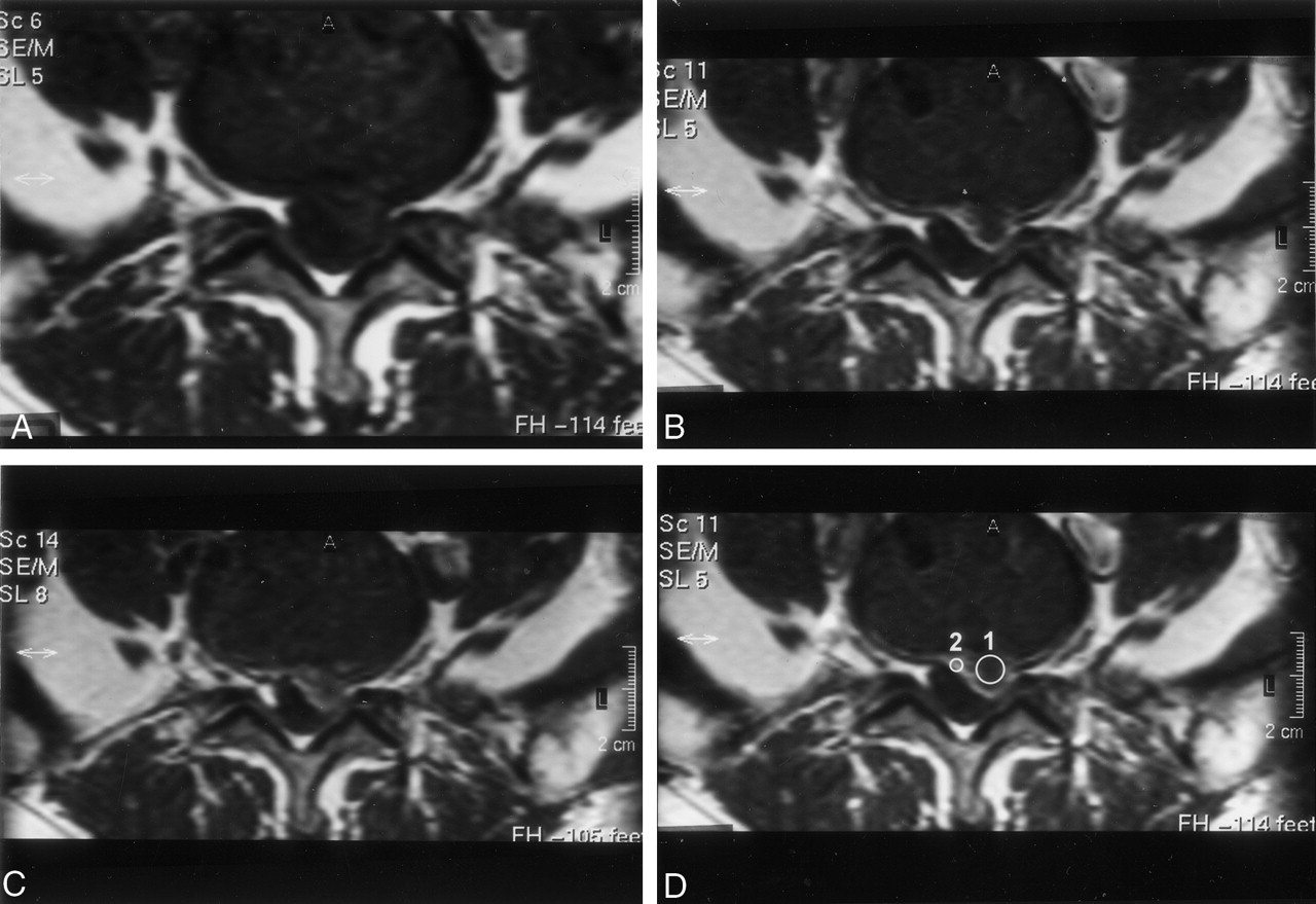
- Fig 1.
Axial view images of a patient with surgically confirmed recurrent herniated disk.
A, Obtained before the administration of gadopentetate dimeglumine (0.1 mmol/kg).
B, Obtained 5 min after the administration of gadopentetate dimeglumine (0.1 mmol/kg).
C, Obtained 50 min after the administration of gadopentetate dimeglumine (0.1 mmol/kg).
D, Obtained 5 min after the administration of contrast medium. Placement of cursor to measure recurrent disk (1) and scar (2) enhancement. Contrast ratio between scar and recurrent disk fragment is greater at 5 min than at 50 min.
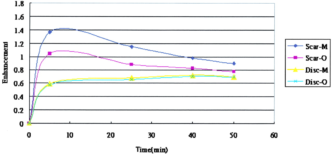
- Fig 2.
Graph of the average enhancement in scar and in recurrent disk fragment as a function of time after the injection of 0.1 mmol/kg gadopentetate dimeglumine (M) or gadodiamide (O). The scar tended to decrease in enhancement and the recurrent disk fragment tended to increase in enhancement with time.
- Fig 3.
MR images of a patient with recurrent herniated disk
A, Sagittal view unenhanced T2-weighted MR image.
B, Axial view image obtained before the IV administration of ionic contrast medium.
C, Axial view image obtained 5 min after the IV administration of ionic contrast medium.
D, Axial view image obtained 50 min after the IV administration of ionic contrast medium. Contrast ratio is greater after administration of ionic rather than nonionic contrast media at 50 min (see panel G).
E, Axial view image obtained before the IV administration of nonionic contrast medium.
F, Axial view image obtained 5 min after the IV administration of nonionic contrast medium.
G, Axial view image obtained 50 min after the IV administration of nonionic contrast medium. Contrast ratio is greater after administration of ionic rather than nonionic contrast media at 50 min.
- Fig 4.
Graph of the contrast ratio between scar and recurrent disk fragment as a function of time after injection of 0.1 mmol/kg ionic (M) and nonionic (O) contrast medium. The contrast radio tended to decrease with time (arithmetic mean ± SD, n = 20).


Tables
- TABLE 1:
Contrast enhancement in recurrent disk fragment and scar after IV injection of ionic and nonionic contrast media
Location Type of Contrast Medium Enhancement at Time after Injection (min) 5 25 40 50 Ionic 0.60 ± 0.10 0.68 ± 0.07 0.72 ± 0.07 0.70 ± 0.07 Disk Nonionic 0.58 ± 0.09 0.66 ± 0.08 0.71 ± 0.08 0.70 ± 0.08 Ionic 1.37 ± 0.26 1.15 ± 0.18 0.98 ± 0.17 0.90 ± 0.15 Scar Nonionic 1.04 ± 0.21 0.88 ± 0.15 0.83 ± 0.12 0.78 ± 0.11 * Enhancement is expressed as arithmetic mean ± SD (n = 20).
- TABLE 2:
Contrast between the scar and recurrent disk fragment after IV injection of ionic and nonionic contrast media
Type of Contrast Medium Contrast (SI scar/SI disk) at Time after Injection (min) 0 5 25 40 50 Ionic 1.32 ± 0.42 1.95 ± 0.80 1.66 ± 0.67 1.40 ± 0.42 1.32 ± 0.41 Nonionic 1.37 ± 0.61 1.93 ± 0.78 1.54 ± 0.44 1.30 ± 0.44 1.20 ± 0.56 Note.—SI indicates signal intensity.
* Contrast between scar and recurrent disc fragment is expressed as arithmetic mean ± SD (n = 20).
In this issue


Jump to section
Related Articles
Cited By...
- No citing articles found.