Advertisement

Main menu

  • Home
  • Content
    • Current Issue
    • Accepted Manuscripts
    • Article Preview
    • Past Issue Archive
    • AJNR Case Collection
    • Case of the Week Archive
    • Classic Case Archive
    • Case of the Month Archive
  • Special Collections
    • Spinal CSF Leak Articles (Jan 2020-June 2024)
    • 2024 AJNR Journal Awards
    • Most Impactful AJNR Articles
  • Multimedia
    • AJNR Podcast
    • AJNR Scantastics
    • Video Articles
  • For Authors
    • Submit a Manuscript
    • Author Policies
    • Fast publishing of Accepted Manuscripts
    • Graphical Abstract Preparation
    • Manuscript Submission Guidelines
    • Imaging Protocol Submission
    • Submit a Case for the Case Collection
  • About Us
    • About AJNR
    • Editorial Board
  • More
    • Become a Reviewer/Academy of Reviewers
    • Subscribers
    • Permissions
    • Alerts
    • Feedback
    • Advertisers
    • ASNR Home
  • Other Publications
    • ajnr

User menu

  • Alerts
  • Log in

Search

  • Advanced search
American Journal of Neuroradiology

American Journal of Neuroradiology

ASHNR American Society of Functional Neuroradiology ASHNR American Society of Pediatric Neuroradiology ASSR
  • Alerts
  • Log in

Advanced Search

  • Home
  • Content
    • Current Issue
    • Accepted Manuscripts
    • Article Preview
    • Past Issue Archive
    • AJNR Case Collection
    • Case of the Week Archive
    • Classic Case Archive
    • Case of the Month Archive
  • Special Collections
    • Spinal CSF Leak Articles (Jan 2020-June 2024)
    • 2024 AJNR Journal Awards
    • Most Impactful AJNR Articles
  • Multimedia
    • AJNR Podcast
    • AJNR Scantastics
    • Video Articles
  • For Authors
    • Submit a Manuscript
    • Author Policies
    • Fast publishing of Accepted Manuscripts
    • Graphical Abstract Preparation
    • Manuscript Submission Guidelines
    • Imaging Protocol Submission
    • Submit a Case for the Case Collection
  • About Us
    • About AJNR
    • Editorial Board
  • More
    • Become a Reviewer/Academy of Reviewers
    • Subscribers
    • Permissions
    • Alerts
    • Feedback
    • Advertisers
    • ASNR Home
  • Follow AJNR on Twitter
  • Visit AJNR on Facebook
  • Follow AJNR on Instagram
  • Join AJNR on LinkedIn
  • RSS Feeds

Welcome to the new AJNR, Updated Hall of Fame, and more. Read the full announcements.


AJNR is seeking candidates for the position of Associate Section Editor, AJNR Case Collection. Read the full announcement.


Getting new auth cookie, if you see this message a lot, tell someone!
Research ArticleBRAIN

Is Volume Transfer Coefficient (Ktrans) Related to Histologic Grade in Human Gliomas?

Tufail F. Patankar, Hamied A. Haroon, Samantha J. Mills, Danielle Balériaux, David L. Buckley, Geoff J.M. Parker and Alan Jackson
American Journal of Neuroradiology November 2005, 26 (10) 2455-2465;
Tufail F. Patankar
  • Find this author on Google Scholar
  • Find this author on PubMed
  • Search for this author on this site
Hamied A. Haroon
  • Find this author on Google Scholar
  • Find this author on PubMed
  • Search for this author on this site
Samantha J. Mills
  • Find this author on Google Scholar
  • Find this author on PubMed
  • Search for this author on this site
Danielle Balériaux
  • Find this author on Google Scholar
  • Find this author on PubMed
  • Search for this author on this site
David L. Buckley
  • Find this author on Google Scholar
  • Find this author on PubMed
  • Search for this author on this site
Geoff J.M. Parker
  • Find this author on Google Scholar
  • Find this author on PubMed
  • Search for this author on this site
Alan Jackson
  • Find this author on Google Scholar
  • Find this author on PubMed
  • Search for this author on this site
  • Article
  • Figures & Data
  • Info & Metrics
  • Responses
  • References
  • PDF
Loading

Article Figures & Data

Figures

  • Tables
  • Fig 1.
    • Download figure
    • Open in new tab
    • Download powerpoint
    Fig 1.

    Representative Ktrans maps from (A) a grade II fibrillary astrocytoma, (B) a grade III anaplastic astrocytoma, and (C) a grade IV glioblastoma multiforme. The white boxes enclose the tumor area in each image. Note that vasculature does not appear in these maps, and Ktrans values in normal brain are insignificant and consistent with noise. The Ktrans values in the grade II tumor (A) are insignificant corresponding to the lack of enhancement with contrast. The high-grade-defining necrotic core is clearly evident in the middle of the tumor in panel C. The heterogeneity of Ktrans is clearly evident in the enhancing tumor portion in panels B and C.
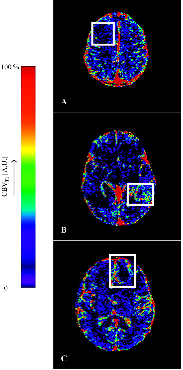
  • Fig 2.
    • Download figure
    • Open in new tab
    • Download powerpoint
    Fig 2.

    Representative CBV maps from (A) a grade II fibrillary astrocytoma, (B) a grade III anaplastic astrocytoma, and (C) a grade IV glioblastoma multiforme. The white boxes enclose the tumor area in each image. The normal cerebral vasculature is clearly seen on these maps, particularly the superior sagittal sinus and other major vessels. The grade II tumor in panel A homogeneously shows very low blood volume, which is below the measurement accuracy of the technique. The necrotic core is clearly evident in the middle of the tumor in panel C. The heterogeneity of CBV is clearly evident in the enhancing tumor portion in panels B and C and shows very different distributions to those in Fig 1 (A and B).
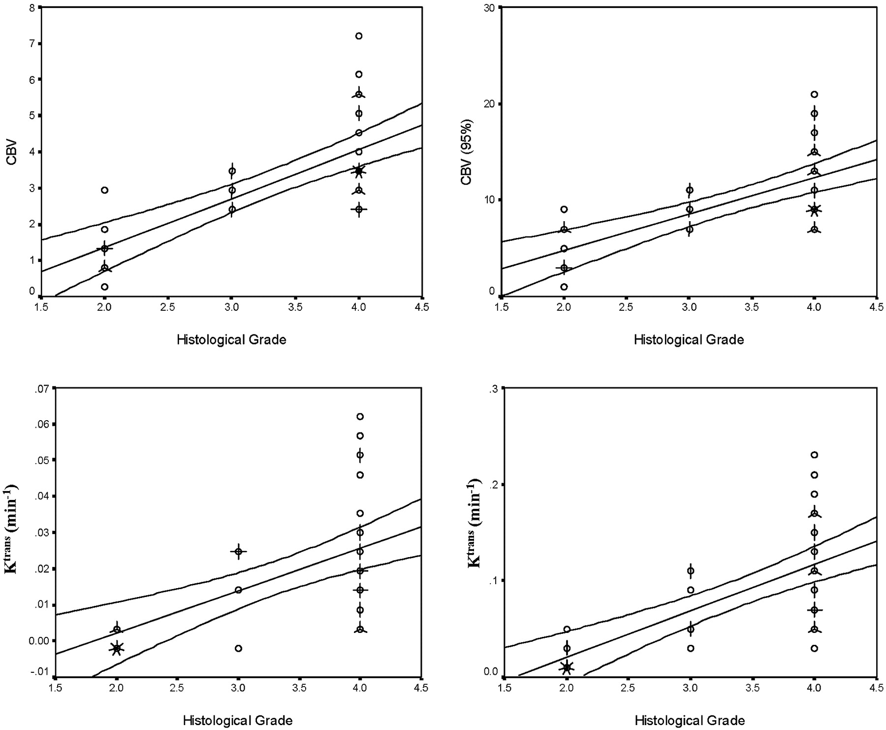
  • Fig 3.
    • Download figure
    • Open in new tab
    • Download powerpoint
    Fig 3.

    Scattergrams showing the relationships between histologic grade and median values of Ktrans, Ktrans (95%), CBV, and CBV (95%). Individual cases are indicated by circles, multiple cases are represented by the addition of “petals” to the glyph with the number of petals representing the number of cases. Lines indicate the optimal linear regression fit for the data and the 95th percentile confidence limits for the regression fit for the entire dataset. The correlation between grade and the median values of each of the parametric variables is significant (Ktrans, Ktrans [95%], CBV, and CBV [95%]; P < .01).

  • Fig 4.
    • Download figure
    • Open in new tab
    • Download powerpoint
    Fig 4.

    Scattergram showing the relationship between values of median CBV and Ktrans (95%) for all individual cases. The grade II tumors show lower values of both CBV and Ktrans (95%). Higher values are seen in grade III and IV tumors, but there is a considerable overlap in these distributions.
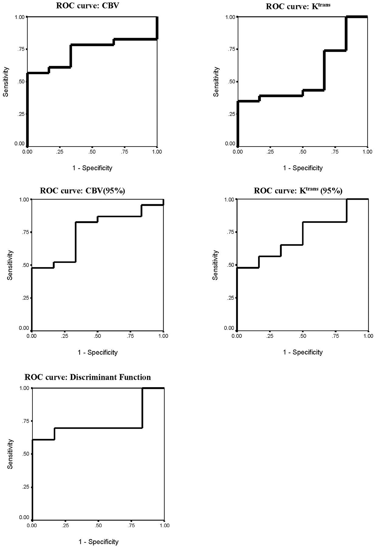
  • Fig 5.
    • Download figure
    • Open in new tab
    • Download powerpoint
    Fig 5.

    ROC analysis showing effect of using each individual variable or the discriminant function (C1) in differentiating between high- and low-grade tumors. The area under the ROC curve for high- versus low-grade is greatest for the discriminant function (0.993). Within the independent parametric variables the area was highest for Ktrans (95%) (0.986), though similarly high values were seen for Ktrans and CBV (0.979 and 0.966, respectively). (Areas under the ROC curves are shown in Table 7.)

  • Fig 6.
    • Download figure
    • Open in new tab
    • Download powerpoint
    Fig 6.

    ROC analysis for separation of grade III and IV tumors. Areas under the ROC curves are shown in Table 7.

Tables

  • Figures
    • View popup
    TABLE 1:

    Patient demographics

    Patient No./Sex/Age (y)Histological Grade
    1/M/52IV
    2/M/58IV
    3/F/31II
    4/F/46III
    5/M/75III
    6/M/55IV
    7/M/52IV
    8/M/53III
    9/M/53IV
    10/M/45IV
    11/M/54IV
    12/F/71IV
    13/M/64IV
    14/M/69IV
    15/M/69IV
    16/M/50IV
    17/M/55IV
    18/M/65IV
    19/F/56IV
    20/M/47II*
    21/F/46II*
    22/M/44II*
    23/F/34II*
    24/M/45II
    25/F/42II
    26/M/38III
    27/M/46III
    28/M/53IV
    29/M/58IV
    30/M/48IV
    31/F/40II*
    32/F/60IV
    33/M/55IV
    34/M/72II
    35/M/61IV
    36/M/50IV
    37/M/77IV
    38/M/42III
    39/F/33II
    • Note.—Grading is according to World Health Organisation criteria for histopathologic findings of tissue biopsy samples taken after imaging. In general, glioma grade II indicates fibrillary astrocytoma; III, anaplastic astrocytoma; IV, glioblastoma multiforme.

    • * Oligodendroglioma, but has been included.

    • View popup
    TABLE 2:

    Means and standard deviations of volume transfer coefficient (Ktrans) and cerebral blood volume (CBV) measurements in gliomas grouped by World Health Organization grade

    WHO Histological GradeNo. of PatientsKtrans (min−1)CBV (%)
    MeanSDMeanSD
    Median values*
        II100.0007120.0007121.2840.75
        III60.01850.01022.9950.44
        IV230.0250.01774.0221.352
    95% values
        II100.0180.01264.632.47
        III60.0730.0338.831.64
        IV230.1120.05512.244.37
    • Note.—Assumes that values are normally distributed.

    • View popup
    TABLE 3:

    Post hoc pairwise comparisons between grades (Tamahane test)

    Grade IIIGrade IV
    Grade II
        CBV<.001<.001
        KtransNS<.001
        CBV (95%)<.01<.001
        Ktrans (95%)<.05<.001
    Grade III
        CBV<.05
        KtransNS
        CBV (95%)<.05
        Ktrans (95%)NS
    • Note.—CBV indicates cerebral blood volume; Ktrans, volume transfer coefficient. Grading is according to World Health Organization criteria.

    • View popup
    TABLE 4:

    Correlation values (Spearman rho) and significance of the correlation between each of the four parameteric variables and grade

    Spearman rhoSignificance
    CBV0.719<.001
    Ktrans0.656<.001
    CBV (95%)0.718<.01
    Ktrans (95%)0.740<.001
    • Note.—CBV indicates cerebral blood volume; Ktrans, volume transfer coefficient.

    • View popup
    TABLE 5:

    Classification results using the canonical discriminate variable

    Predicted/ObservedGrade IIGrade IIIGrade IV
    Grade II9 (90)1 (10)0
    Grade III06 (100)0
    Grade IV09 (39.1)14 (60.9)
    • Note.—Numbers in parentheses represent percentages of correct classification. The total number of cases correctly classified is 74.4%

    • C1=0.695 (CBV) + 0.577 (Ktrans (95%)), where CBV indicate cerebral blood volume and Ktrans is the volume transfer coefficient.

    • View popup
    TABLE 6:

    Classification results using canonical discriminate functions derived using a leave-one-out (jackknife) cross-validation analysis

    Predicted/ObservedGrade IIGrade IIIGrade IV
    Grade II9 (90)1 (10)0
    Grade III1 (16.7)4 (66.7)1 (16.7)
    Grade IV09 (39.1)14 (60.9)
    • Note.—This approach can be expected to represent classifications more accurately that are expected in a larger population. Numbers in parentheses represent percentages. The number of cases correctly classified is 69.2%.

    • View popup
    TABLE 7:

    Results of the receiver operator curve analysis for each variable and for the discriminate function C1 in differentiating between low-grade tumors and grades III IV high-grade tumors

    High Versus Low GradeGrade III Versus Grade IV
    CBV0.966 (<.001)0.754 (NS)
    Ktrans0.979 (<.05)0.551 (NS)
    CBV (95%)0.955 (<.001)0.754 (NS)
    Ktrans (95%)0.986 (<.001)0.725 (NS)
    CI0.993 (<.001)0.732 (NS)
    • Note.—CBV indicates cerebral blood volume; Ktrans, volume transfer coefficient. Values are areas under ROC followed by significance (P values) in parentheses.

PreviousNext
Back to top

In this issue

American Journal of Neuroradiology: 26 (10)
American Journal of Neuroradiology
Vol. 26, Issue 10
1 Nov 2005
  • Table of Contents
  • Index by author
Advertisement
Print
Download PDF
Email Article

Thank you for your interest in spreading the word on American Journal of Neuroradiology.

NOTE: We only request your email address so that the person you are recommending the page to knows that you wanted them to see it, and that it is not junk mail. We do not capture any email address.

Enter multiple addresses on separate lines or separate them with commas.
Is Volume Transfer Coefficient (Ktrans) Related to Histologic Grade in Human Gliomas?
(Your Name) has sent you a message from American Journal of Neuroradiology
(Your Name) thought you would like to see the American Journal of Neuroradiology web site.
CAPTCHA
This question is for testing whether or not you are a human visitor and to prevent automated spam submissions.
Cite this article
Tufail F. Patankar, Hamied A. Haroon, Samantha J. Mills, Danielle Balériaux, David L. Buckley, Geoff J.M. Parker, Alan Jackson
Is Volume Transfer Coefficient (Ktrans) Related to Histologic Grade in Human Gliomas?
American Journal of Neuroradiology Nov 2005, 26 (10) 2455-2465;

Citation Manager Formats

  • BibTeX
  • Bookends
  • EasyBib
  • EndNote (tagged)
  • EndNote 8 (xml)
  • Medlars
  • Mendeley
  • Papers
  • RefWorks Tagged
  • Ref Manager
  • RIS
  • Zotero
0 Responses
Respond to this article
Share
Bookmark this article
Is Volume Transfer Coefficient (Ktrans) Related to Histologic Grade in Human Gliomas?
Tufail F. Patankar, Hamied A. Haroon, Samantha J. Mills, Danielle Balériaux, David L. Buckley, Geoff J.M. Parker, Alan Jackson
American Journal of Neuroradiology Nov 2005, 26 (10) 2455-2465;
del.icio.us logo Twitter logo Facebook logo Mendeley logo
  • Tweet Widget
  • Facebook Like
  • Google Plus One

Jump to section

  • Article
    • Abstract
    • Materials and Methods
    • Results
    • Discussion
    • Footnotes
    • References
  • Figures & Data
  • Info & Metrics
  • Responses
  • References
  • PDF

Related Articles

  • No related articles found.
  • PubMed
  • Google Scholar

Cited By...

  • Automated Processing of Dynamic Contrast-Enhanced MRI: Correlation of Advanced Pharmacokinetic Metrics with Tumor Grade in Pediatric Brain Tumors
  • Mitotic Activity in Glioblastoma Correlates with Estimated Extravascular Extracellular Space Derived from Dynamic Contrast-Enhanced MR Imaging
  • Dynamic Contrast-Enhanced MR Imaging in Head and Neck Cancer: Techniques and Clinical Applications
  • Comparison of the Diagnostic Accuracy of DSC- and Dynamic Contrast-Enhanced MRI in the Preoperative Grading of Astrocytomas
  • Pixel-by-Pixel Comparison of Volume Transfer Constant and Estimates of Cerebral Blood Volume from Dynamic Contrast-Enhanced and Dynamic Susceptibility Contrast-Enhanced MR Imaging in High-Grade Gliomas
  • Glioma: Application of Histogram Analysis of Pharmacokinetic Parameters from T1-Weighted Dynamic Contrast-Enhanced MR Imaging to Tumor Grading
  • Diagnostic Accuracy of Dynamic Contrast-Enhanced MR Imaging Using a Phase-Derived Vascular Input Function in the Preoperative Grading of Gliomas
  • Imaging biomarkers of angiogenesis and the microvascular environment in cerebral tumours
  • Biology, genetics and imaging of glial cell tumours
  • Multimodality Assessment of Brain Tumors and Tumor Recurrence
  • Enhancing Fraction in Glioma and Its Relationship to the Tumoral Vascular Microenvironment: A Dynamic Contrast-Enhanced MR Imaging Study
  • Enhancing Fraction Predicts Clinical Outcome following First-Line Chemotherapy in Patients with Epithelial Ovarian Carcinoma
  • Imaging Tumor Vascular Heterogeneity and Angiogenesis using Dynamic Contrast-Enhanced Magnetic Resonance Imaging
  • Crossref
  • Google Scholar

This article has not yet been cited by articles in journals that are participating in Crossref Cited-by Linking.

More in this TOC Section

  • SWI or T2*: Which MRI Sequence to Use in the Detection of Cerebral Microbleeds? The Karolinska Imaging Dementia Study
  • Progression of Microstructural Damage in Spinocerebellar Ataxia Type 2: A Longitudinal DTI Study
  • Usefulness of Quantitative Susceptibility Mapping for the Diagnosis of Parkinson Disease
Show more Brain

Similar Articles

Advertisement

Indexed Content

  • Current Issue
  • Accepted Manuscripts
  • Article Preview
  • Past Issues
  • Editorials
  • Editors Choice
  • Fellow Journal Club
  • Letters to the Editor

Cases

  • Case Collection
  • Archive - Case of the Week
  • Archive - Case of the Month
  • Archive - Classic Case

Special Collections

  • Special Collections

Resources

  • News and Updates
  • Turn around Times
  • Submit a Manuscript
  • Author Policies
  • Manuscript Submission Guidelines
  • Evidence-Based Medicine Level Guide
  • Publishing Checklists
  • Graphical Abstract Preparation
  • Imaging Protocol Submission
  • Submit a Case
  • Become a Reviewer/Academy of Reviewers
  • Get Peer Review Credit from Publons

Multimedia

  • AJNR Podcast
  • AJNR SCANtastic
  • Video Articles

About Us

  • About AJNR
  • Editorial Board
  • Not an AJNR Subscriber? Join Now
  • Alerts
  • Feedback
  • Advertise with us
  • Librarian Resources
  • Permissions
  • Terms and Conditions

American Society of Neuroradiology

  • Not an ASNR Member? Join Now

© 2025 by the American Society of Neuroradiology All rights, including for text and data mining, AI training, and similar technologies, are reserved.
Print ISSN: 0195-6108 Online ISSN: 1936-959X

Powered by HighWire