
Article Figures & Data
Figures
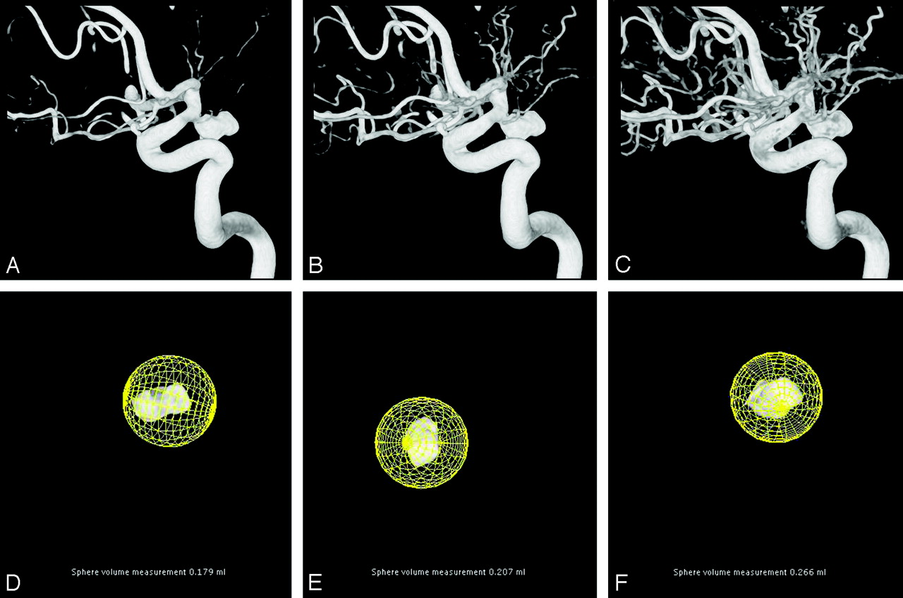
- Fig 1.
3D angiographic image of a posterior communicating artery aneurysm in 3 different manual threshold settings. All 3 images are visually acceptable to delineate vessel and aneurysm anatomy.
A, Low-threshold setting: not all small vessels are visible. After segmentation of the aneurysm, the calculated volume is 179 mm3 (D).
B, Intermediate-threshold setting: better visualization of small vessels. After segmentation of the aneurysm, the calculated volume is 207 mm3 (E).
C, High-threshold setting: exaggerated visibility of small vessels. After segmentation of the aneurysm, the calculated volume is 266 mm3 (F).
- Fig 2.
Two-dimensional illustration in a cut plane of the 3D dataset. The blue lines represent the contours in the cut plane of a blood vessel (ellipsoid) and an aneurysm (circle).
A, -B, Manual thresholding: different threshold levels change the contours of the vessel and the aneurysm.
Gradient-edge detection: voxels with high gradient values are white and voxels with low gradient values are black. In homogeneous parts of the volume, some gradient differences are present, caused by image noise (C). After the small gradient differences are filtered, the contours are unchanged (D).
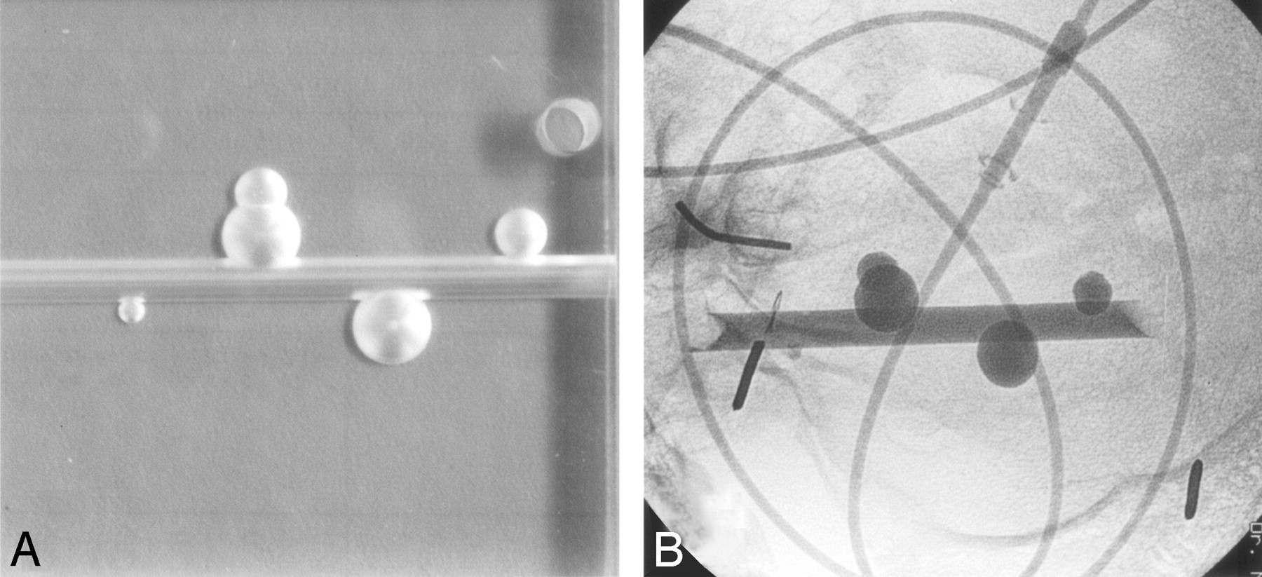
- Fig 3.
A, Four aneurysm phantoms on a parent artery in different shapes and sizes in Perspex.
B, Radiograph of aneurysm phantoms filled with contrast material in a human skull placed in a water-filled bucket. Injection lines filled with contrast material simulate blood vessels.
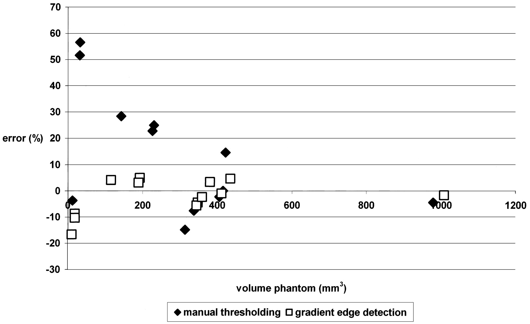
- Fig 4.
Graphic display of errors in volume measurement of 13 aneurysm phantoms with the manual threshold method and the gradient edge detection method.
- Fig 5.
Mountain plot of percentile for each ranked difference from the phantom volumes for the gradient edge detection method and the manual threshold method. Dotted line indicates manual threshold method; solid line, the gradient edge detection method.


In this issue


Jump to section
Related Articles
Cited By...
- 4D-DSA: Development and Current Neurovascular Applications
- Impact of image reconstruction parameters when using 3D DSA reconstructions to measure intracranial aneurysms
- Influence of observers, threshold values, and measurement methods on volumetric analysis of cerebral aneurysms with three-dimensional rotational angiography
- Endovascular treatment of intracranial aneurysms with detachable coils: correlation between aneurysm volume, packing, and angiographic recurrence
- Widening and High Inclination of the Middle Cerebral Artery Bifurcation Are Associated With Presence of Aneurysms
- AngioSuite: an accurate method to calculate aneurysm volumes and packing densities
- Stent-Assisted Coiling of Intracranial Bifurcation Aneurysms Leads to Immediate and Delayed Intracranial Vascular Angle Remodeling
- Durability of Treatment of Intracranial Aneurysms With Hydrocoils Is Not Different From Standard Platinum Coils