Advertisement

Main menu

  • Home
  • Content
    • Current Issue
    • Accepted Manuscripts
    • Article Preview
    • Past Issue Archive
    • AJNR Case Collection
    • Case of the Week Archive
    • Classic Case Archive
    • Case of the Month Archive
  • Special Collections
    • Spinal CSF Leak Articles (Jan 2020-June 2024)
    • 2024 AJNR Journal Awards
    • Most Impactful AJNR Articles
  • Multimedia
    • AJNR Podcast
    • AJNR Scantastics
    • Video Articles
  • For Authors
    • Submit a Manuscript
    • Author Policies
    • Fast publishing of Accepted Manuscripts
    • Graphical Abstract Preparation
    • Manuscript Submission Guidelines
    • Imaging Protocol Submission
    • Submit a Case for the Case Collection
  • About Us
    • About AJNR
    • Editorial Board
  • More
    • Become a Reviewer/Academy of Reviewers
    • Subscribers
    • Permissions
    • Alerts
    • Feedback
    • Advertisers
    • ASNR Home
  • Other Publications
    • ajnr

User menu

  • Alerts
  • Log in

Search

  • Advanced search
American Journal of Neuroradiology

American Journal of Neuroradiology

ASHNR American Society of Functional Neuroradiology ASHNR American Society of Pediatric Neuroradiology ASSR
  • Alerts
  • Log in

Advanced Search

  • Home
  • Content
    • Current Issue
    • Accepted Manuscripts
    • Article Preview
    • Past Issue Archive
    • AJNR Case Collection
    • Case of the Week Archive
    • Classic Case Archive
    • Case of the Month Archive
  • Special Collections
    • Spinal CSF Leak Articles (Jan 2020-June 2024)
    • 2024 AJNR Journal Awards
    • Most Impactful AJNR Articles
  • Multimedia
    • AJNR Podcast
    • AJNR Scantastics
    • Video Articles
  • For Authors
    • Submit a Manuscript
    • Author Policies
    • Fast publishing of Accepted Manuscripts
    • Graphical Abstract Preparation
    • Manuscript Submission Guidelines
    • Imaging Protocol Submission
    • Submit a Case for the Case Collection
  • About Us
    • About AJNR
    • Editorial Board
  • More
    • Become a Reviewer/Academy of Reviewers
    • Subscribers
    • Permissions
    • Alerts
    • Feedback
    • Advertisers
    • ASNR Home
  • Follow AJNR on Twitter
  • Visit AJNR on Facebook
  • Follow AJNR on Instagram
  • Join AJNR on LinkedIn
  • RSS Feeds

Welcome to the new AJNR, Updated Hall of Fame, and more. Read the full announcements.


AJNR is seeking candidates for the position of Associate Section Editor, AJNR Case Collection. Read the full announcement.


OtherHEAD AND NECK

Agenesis of Bilateral Internal Carotid Arteries and Posterior Fossa Abnormality in a Patient with Facial Capillary Hemangioma: Presumed Incomplete Phenotypic Expression of PHACE Syndrome

Young-Cheol Weon, Jin-Il Chung, Hyung-Jin Kim and Hong sik Byun
American Journal of Neuroradiology November 2005, 26 (10) 2635-2639;
Young-Cheol Weon
  • Find this author on Google Scholar
  • Find this author on PubMed
  • Search for this author on this site
Jin-Il Chung
  • Find this author on Google Scholar
  • Find this author on PubMed
  • Search for this author on this site
Hyung-Jin Kim
  • Find this author on Google Scholar
  • Find this author on PubMed
  • Search for this author on this site
Hong sik Byun
  • Find this author on Google Scholar
  • Find this author on PubMed
  • Search for this author on this site
  • Article
  • Figures & Data
  • Info & Metrics
  • Responses
  • References
  • PDF
Loading

Article Figures & Data

Figures

  • Fig 1.
    • Download figure
    • Open in new tab
    • Download powerpoint
    Fig 1.

    On axial (A) and coronal (B) T2-weighted images, hypoplasia of the right cerebellar hemisphere and absence of the inferior cerebellar vermis are present. Note that the abnormally dilated basilar artery with numerous small signal intensity void structures corresponds with the collateral networks at the prepontine cistern. On MR angiography (C, -D), both internal carotid arteries are not seen, and the distal portion of internal carotid arteries is reconstituted from the external carotid arteries.
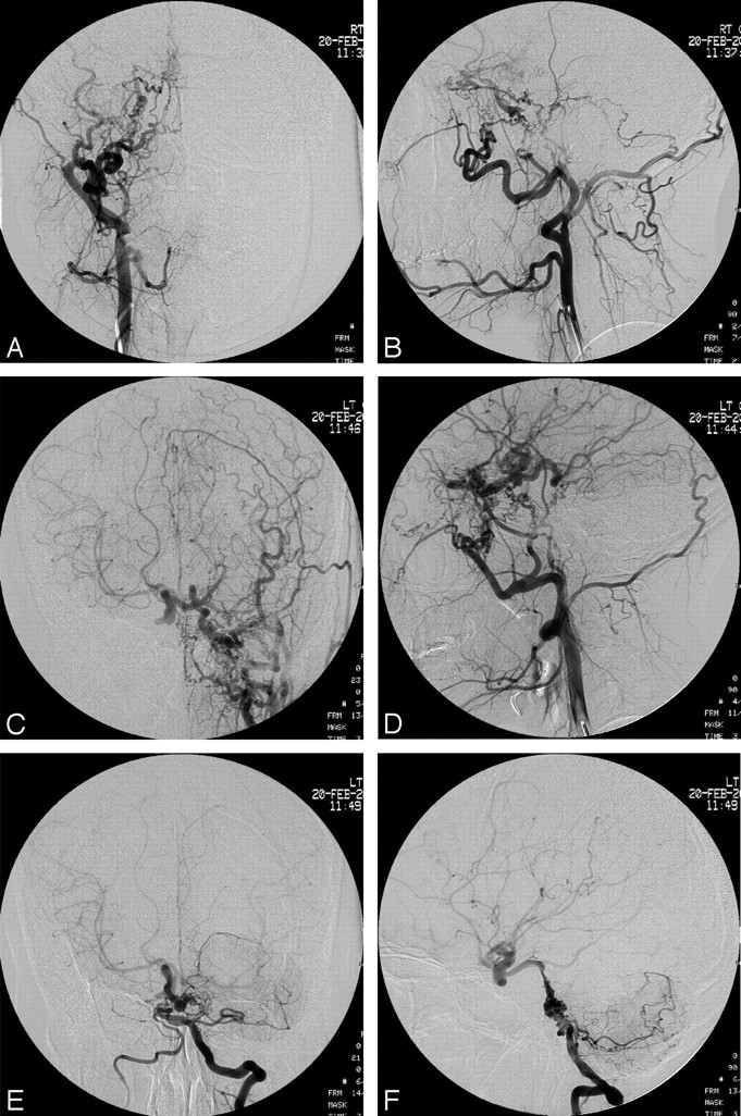
  • Fig 2.
    • Download figure
    • Open in new tab
    • Download powerpoint
    Fig 2.

    Injection of right external carotid artery (A and B) and left external carotid artery (C and D) demonstrates the reconstitution of the distal internal carotid artery by branches of the internal maxillary arteries, middle meningeal arteries, deep temporal arteries, artery of the foramen rotundum, ascending pharyngeal arteries, and collaterals of the occipital artery. Injection of the left vertebral artery (E and F) shows the discontinuation between the vertebral arteries and the abnormally dilated proximal basilar artery with collateral branches mainly from the left vertebral artery.
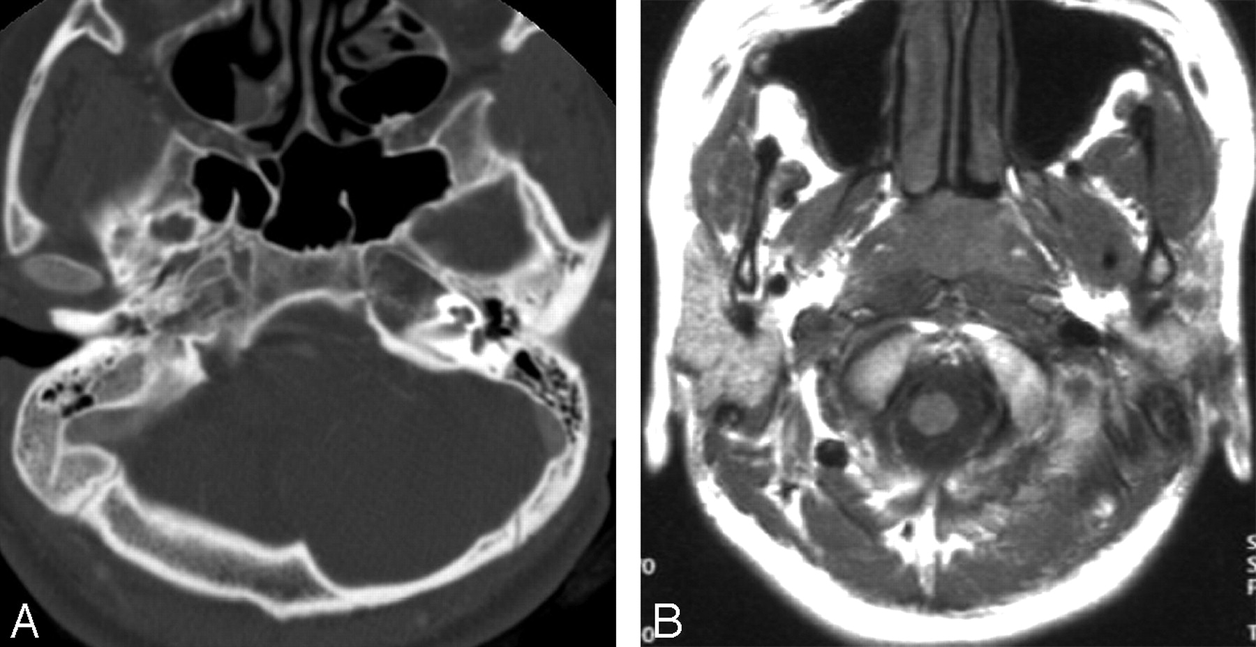
  • Fig 3.
    • Download figure
    • Open in new tab
    • Download powerpoint
    Fig 3.

    Normal internal carotid arteries are not seen in the carotid spaces, and no carotid canals are noted on the bone window setting of the brain CT (A) and T1-weighted brain MRI (B).
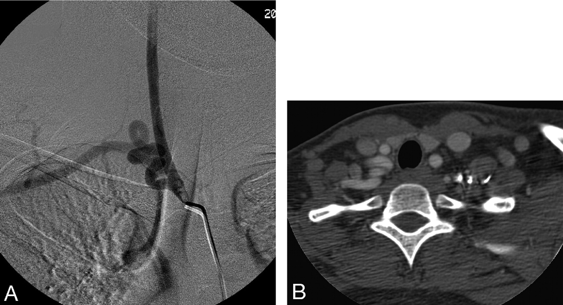
  • Fig 4.
    • Download figure
    • Open in new tab
    • Download powerpoint
    Fig 4.

    Injection of the innominate artery (A) and CT scan obtained at the level of thoracic inlet (B) demonstrate the unusual subclavian artery turning like a screw.

PreviousNext
Back to top

In this issue

American Journal of Neuroradiology: 26 (10)
American Journal of Neuroradiology
Vol. 26, Issue 10
1 Nov 2005
  • Table of Contents
  • Index by author
Advertisement
Print
Download PDF
Email Article

Thank you for your interest in spreading the word on American Journal of Neuroradiology.

NOTE: We only request your email address so that the person you are recommending the page to knows that you wanted them to see it, and that it is not junk mail. We do not capture any email address.

Enter multiple addresses on separate lines or separate them with commas.
Agenesis of Bilateral Internal Carotid Arteries and Posterior Fossa Abnormality in a Patient with Facial Capillary Hemangioma: Presumed Incomplete Phenotypic Expression of PHACE Syndrome
(Your Name) has sent you a message from American Journal of Neuroradiology
(Your Name) thought you would like to see the American Journal of Neuroradiology web site.
CAPTCHA
This question is for testing whether or not you are a human visitor and to prevent automated spam submissions.
Cite this article
Young-Cheol Weon, Jin-Il Chung, Hyung-Jin Kim, Hong sik Byun
Agenesis of Bilateral Internal Carotid Arteries and Posterior Fossa Abnormality in a Patient with Facial Capillary Hemangioma: Presumed Incomplete Phenotypic Expression of PHACE Syndrome
American Journal of Neuroradiology Nov 2005, 26 (10) 2635-2639;

Citation Manager Formats

  • BibTeX
  • Bookends
  • EasyBib
  • EndNote (tagged)
  • EndNote 8 (xml)
  • Medlars
  • Mendeley
  • Papers
  • RefWorks Tagged
  • Ref Manager
  • RIS
  • Zotero
0 Responses
Respond to this article
Share
Bookmark this article
Agenesis of Bilateral Internal Carotid Arteries and Posterior Fossa Abnormality in a Patient with Facial Capillary Hemangioma: Presumed Incomplete Phenotypic Expression of PHACE Syndrome
Young-Cheol Weon, Jin-Il Chung, Hyung-Jin Kim, Hong sik Byun
American Journal of Neuroradiology Nov 2005, 26 (10) 2635-2639;
del.icio.us logo Twitter logo Facebook logo Mendeley logo
  • Tweet Widget
  • Facebook Like
  • Google Plus One

Jump to section

  • Article
    • Abstract
    • Case Report
    • Discussion
    • Acknowledgments
    • References
  • Figures & Data
  • Info & Metrics
  • Responses
  • References
  • PDF

Related Articles

  • No related articles found.
  • PubMed
  • Google Scholar

Cited By...

  • Concomitant carotid aplasia and basilar artery occlusion in a child with PHACES syndrome
  • The Cerebral Vasculopathy of PHACES Syndrome
  • Crossref
  • Google Scholar

This article has not yet been cited by articles in journals that are participating in Crossref Cited-by Linking.

More in this TOC Section

  • Correlation of Apparent Diffusion Coefficient at 3T with Prognostic Parameters of Retinoblastoma
  • Parathyroid Lesions: Characterization with Dual-Phase Arterial and Venous Enhanced CT of the Neck
  • MR Diagnosis of Facial Neuritis: Diagnostic Performance of Contrast-Enhanced 3D-FLAIR Technique Compared with Contrast-Enhanced 3D-T1-Fast-Field Echo with Fat Suppression
Show more Head and Neck

Similar Articles

Advertisement

Indexed Content

  • Current Issue
  • Accepted Manuscripts
  • Article Preview
  • Past Issues
  • Editorials
  • Editors Choice
  • Fellow Journal Club
  • Letters to the Editor

Cases

  • Case Collection
  • Archive - Case of the Week
  • Archive - Case of the Month
  • Archive - Classic Case

Special Collections

  • Special Collections

Resources

  • News and Updates
  • Turn around Times
  • Submit a Manuscript
  • Author Policies
  • Manuscript Submission Guidelines
  • Evidence-Based Medicine Level Guide
  • Publishing Checklists
  • Graphical Abstract Preparation
  • Imaging Protocol Submission
  • Submit a Case
  • Become a Reviewer/Academy of Reviewers
  • Get Peer Review Credit from Publons

Multimedia

  • AJNR Podcast
  • AJNR SCANtastic
  • Video Articles

About Us

  • About AJNR
  • Editorial Board
  • Not an AJNR Subscriber? Join Now
  • Alerts
  • Feedback
  • Advertise with us
  • Librarian Resources
  • Permissions
  • Terms and Conditions

American Society of Neuroradiology

  • Not an ASNR Member? Join Now

© 2025 by the American Society of Neuroradiology All rights, including for text and data mining, AI training, and similar technologies, are reserved.
Print ISSN: 0195-6108 Online ISSN: 1936-959X

Powered by HighWire