
Article Figures & Data
Figures
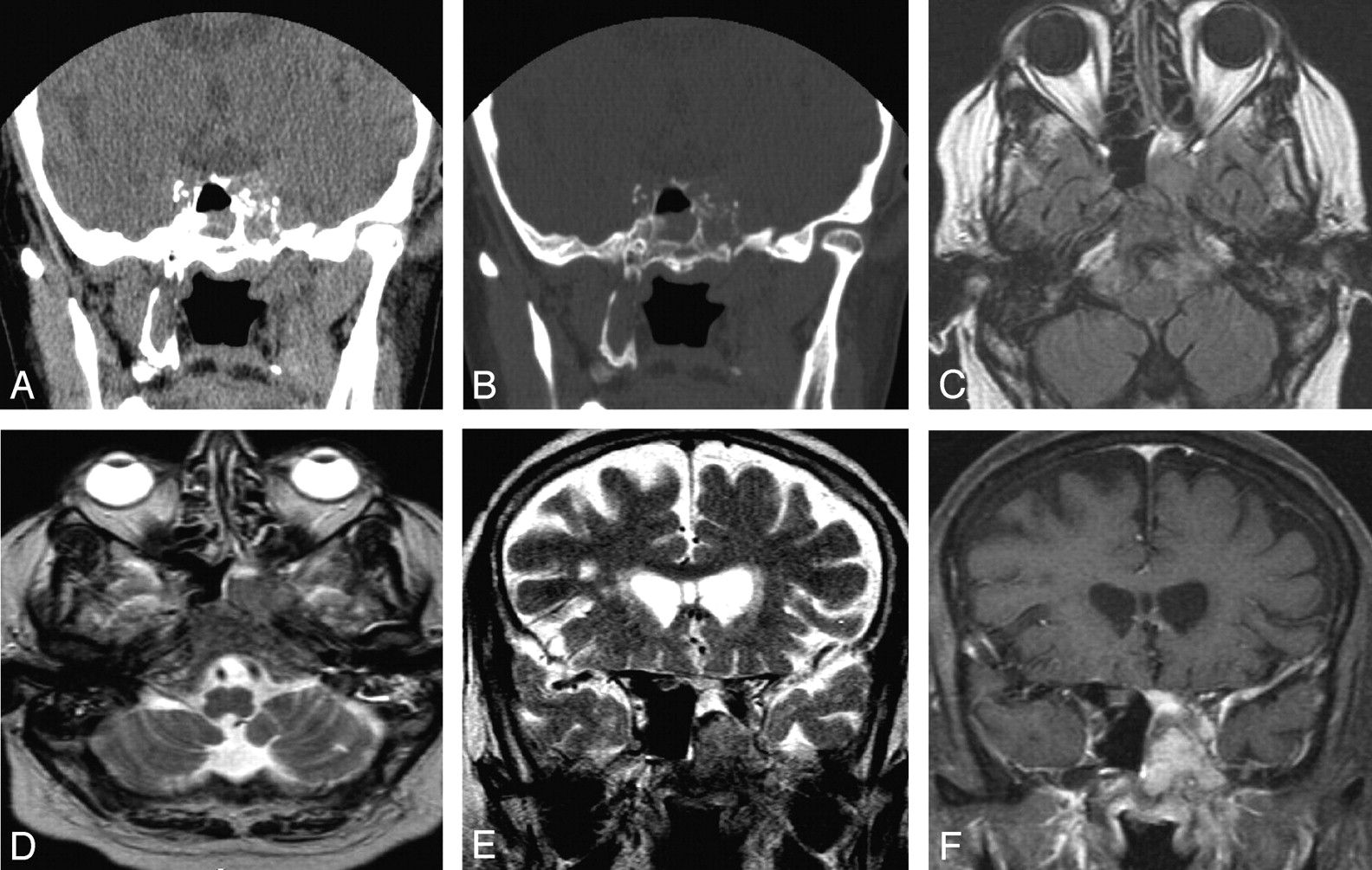
- Fig 1.
Coronal NCCT of PNS at the level of sphenoid sinus (A and B), demonstrating a hyperattenuated mass in the left side of sphenoid sinus with destruction of the lateral sinus wall. Mucosal thickening from inflammatory disease was also seen in the right side of sphenoid sinus. Follow-up MR imaging with contrast redemonstrates the mass, appearing iso-hyperintense on axial T1-weighted image with extension in the left side of clivus (C). The mass appears hypointense on axial and coronal T2-weighted images (D and E) with diffuse homogenous enhancement in the mass and adjoining dura in left temporal fossa on postgadolinium T1-weighted images. Also seen is left parasellar extension into the cavernous sinus and the base of skull, in the region of foramen ovale.
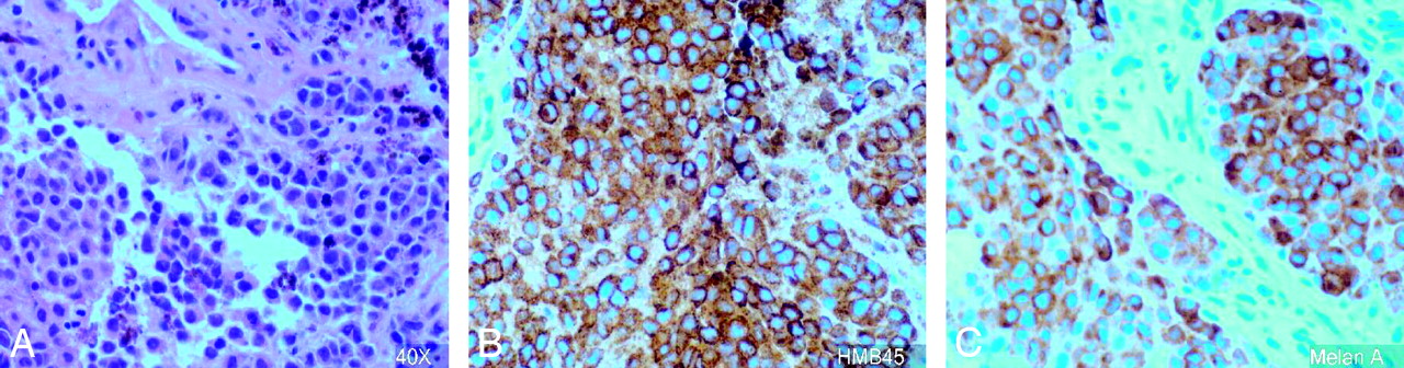
- Fig 2.
Histopathology slide (magnification ×40) demonstrates melanoma cells (A), positive for HMB45 (B) and melan A (C).
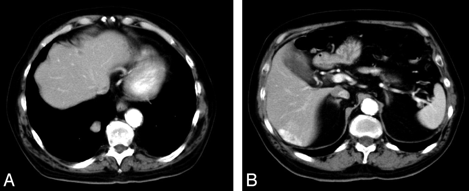
- Fig 3.
Hypervascular lesions in the liver and right lobe lung of lung suggest metastasis.


