
Article Figures & Data
Figures
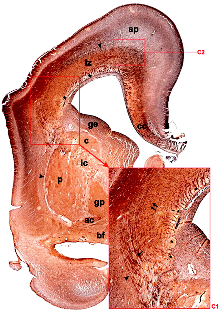
- Fig 1.
The coronal section through the telencephalon of 18-week-old human fetus, Gallyas silver staining. Note that fiber bundles and their crossings are located in the fetal “white matter”—ie, in periventricular region (C1, C2, and asterisks)—and the intermediate zone (iz), whereas the subplate zone (sp) is characterized by a loose (“isotropic”) argirophilic network of fibers. The fetal “white matter” consists of tangentially stratified fiber bundles such as external capsule (arrowheads), corpus callosum radiation (cc), thalamocortical projection fibers (arrow), and the deep system (double arrows; see insert C1). In addition, a prominent periventricular system of unstained and transversely or obliquely cut fiber bundles (row of asterisks) is situated in the subventricular zone. Abbreviations for this and subsequent figures: a = amygdala; ac = anterior commissure; bf = basal forebrain; c = caudate nucleus; C1 –C6 = crossroad areas; cc = corpus callosum; ge = ganglionic eminence; gp = globus pallidus; ic = internal capsule; iz = intermediate zone; p = putamen; sp = subplate zone; th = thalamus.
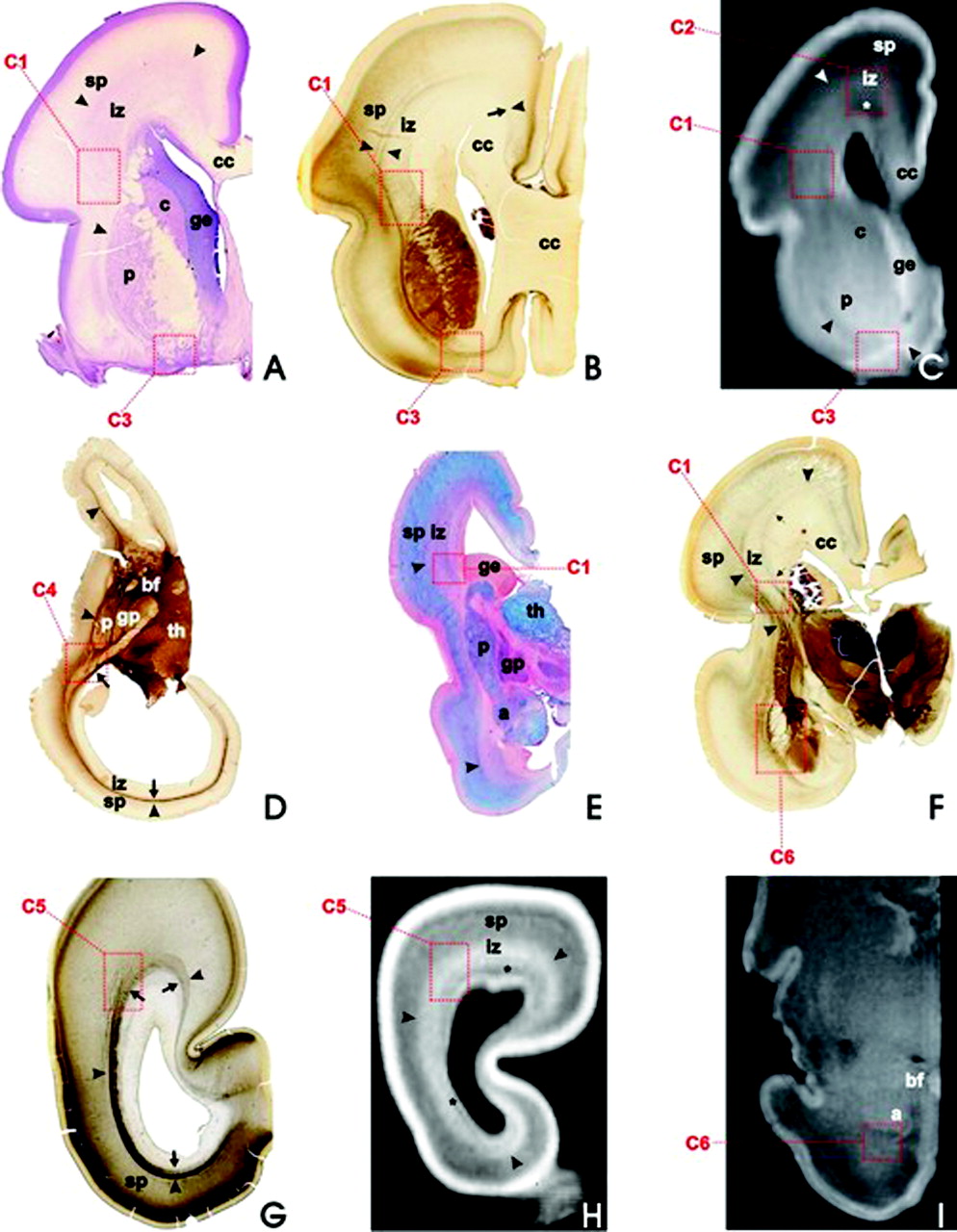
- Fig 2.
During the fetal lamination stage (16–24 weeks of gestation), the basic fiber-architectonic pattern is characterized by predominantly tangential organization (axon strata) and prominent crossroads (C1–C6, dashed squares) of projection, association, and commissural fibers, as revealed on coronal (A–C and E–I) and horizontal (D) sections; Nissl-stained (A), AChE-stained (B, D, F, G), PAS-AB-stained (E), and T1-weighted MR imaging sections (C, H, I); in fetuses aged 16 weeks (D), 18 weeks (G, H), 19 weeks (A, E), 21 weeks (B, C), and 24 weeks (F). After 28 weeks of gestation, MR imaging signal intensity changes with consequent blurring of laminar pattern, as revealed by T1-weighted MR coronal section through the cerebrum of 30-week-old fetus (I). Asterisks in C and H mark the periventricular-subventricular fiber system.
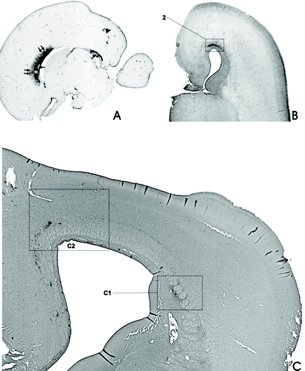
- Fig 3.
In vivo acquired fast T2-weighted MR images (HASTE sequence) in 14 POW (A and B) and 26 POW (C) fetuses reveal the same typical fetal lamination pattern of the cerebral wall as T1-weighted MR images of postmortem specimens of equivalent developmental ages. In panel A (and its inlet), the vertical bar denotes the thickness (approximately 5 mm) of the entire telencephalic wall. Inhomogeneous, poorly delineated, spotlike area (dotted rectangle marked as C1 on panels B and C) of higher MR imaging signal intensity corresponds to the main crossroad area. Larger, rectangulary-marked spotlike area above corresponds to C2 crossroad area. In panel C, asterisk marks the fibrillar periventricular-subventricular zone, while the subplate zone is situated between 2 arrows. Other abbreviations as in Fig 1. For further details, see text.
- Fig 4.
During the stage of typical fetal lamination, various immunocytochemical markers selectively label discrete contingents of fibers within the stratified periventricular fetal WM (iz): MAP-1b (A, 18-week-old fetus) and SMI-312 (C, 20-week-old fetus) label the cytoskeletal components of many growing axons, whereas the prominent periventricular system of fiber bundles is stained by SNAP-25 antibody (B, 18-week-old fetus, arrows). These markers are also visualizing periventricular fiber crossroad areas (C1, C2, C3, C6). Note that CS–56 antibody (D, 18-week-old fetus) labels the extracellular matrix, but is not expressed within the periventricular fiber system (asterisks). Arrowheads denote the capsula externa system, which marks the border between the fetal WM (iz) and the subplate zone (sp), which is part of the neocortical anlage.
- Fig 5.
SEMA3A immunoreactivity (A, 20-week-old fetus) is restricted to median and paramedian parts of the callosal radiation, where it is located in both cell bodies and the extracellular matrix in the ventral part of the corpus callosum (asterisk), and in the fornix system (arrow). The SEMA3A immunoreactivity is also located in the roof of the anterior horn of the lateral ventricle, corresponding to the frontal crossroad area C2 (dashed square in B, 20-week-old fetus). In addition, the expression of CD68 marker for microglia (C, 18-week-old fetus) is most prominent in the frontal crossroad area C2 (dark dots).
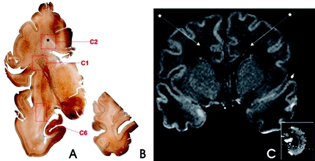
- Fig 6.
The outlines of the corona radiata appear after 28 weeks of gestation. Gallyas staining (A and B, 33-week-old preterm neonate) and MR image (C, 36-week-old neonate). The peduncular part of the corona radiata corresponds to the major frontal crossroad area (C1 in A); however, the centrum semiovale (asterisk in A) still displays signs of immaturity and corresponds to the crossroad area C2. This is manifested as pale (ie, poorly myelinated) area in Gallyas-stained sections (C2 in A) and as patches of decreased MR imaging signal intensity in T1-weighted MR images (C, asterisks). Note that tangential stratification of sagittally oriented fibers is still well preserved in the occipital white matter (B) and that temporal crossroad area (C6 in A) is still poorly myelinated in the preterm neonate.
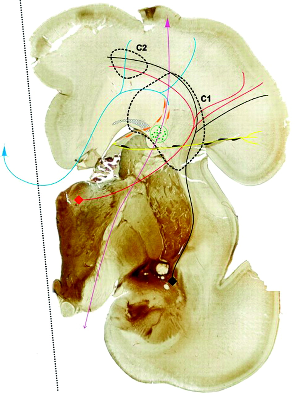
- Fig 7.
Summary diagram, superposed on AChE-stained coronal section through a telencephalon of 28-week-old human fetus constructed on the basis of our data and evidence from current literature. Thick black dashed lines delineate the first (C1) and second (C2) frontal crossroad area. The honeycomb pattern area denotes the deep periventricular system of fiber bundles; and circle with green dots developing fronto-occipital system, both containing SNAP-25 immunoreactive fibers. Colored lines denote systems of projection, association and commissural fibers passing through the crossroads (with triangles or quadrangles depicting cell bodies of origin), as follows: black = basal forebrain afferents; red = thalamocortical afferents; blue = callosal fibers; violet = corticofugal efferents. Note that both radially migrating neurons (black profiles along the yellow radial glial fiber) and tangentially migrating neurons (orange profiles) pass through the major crossroad (C1) area, which is located at the main predilection site of hypoxic-ischemic lesion in preterm infants.


In this issue


Jump to section
Related Articles
Cited By...
- White matter tract crossing and bottleneck regions in the fetal brain
- Neuronal Coupling Modes Show Differential Development in the Early Cortical Activity Networks of Human Newborns
- Neurodevelopmental Patterns of Early Postnatal White Matter Maturation Represent Distinct Underlying Microstructure and Histology
- Diagnostic Value of Brain Calcifications in Adult-Onset Leukoencephalopathy with Axonal Spheroids and Pigmented Glia
- High Signal Intensity on T2-Weighted MR Imaging at Term-Equivalent Age in Preterm Infants Does Not Predict 2-Year Neurodevelopmental Outcomes