
Article Figures & Data
Figures
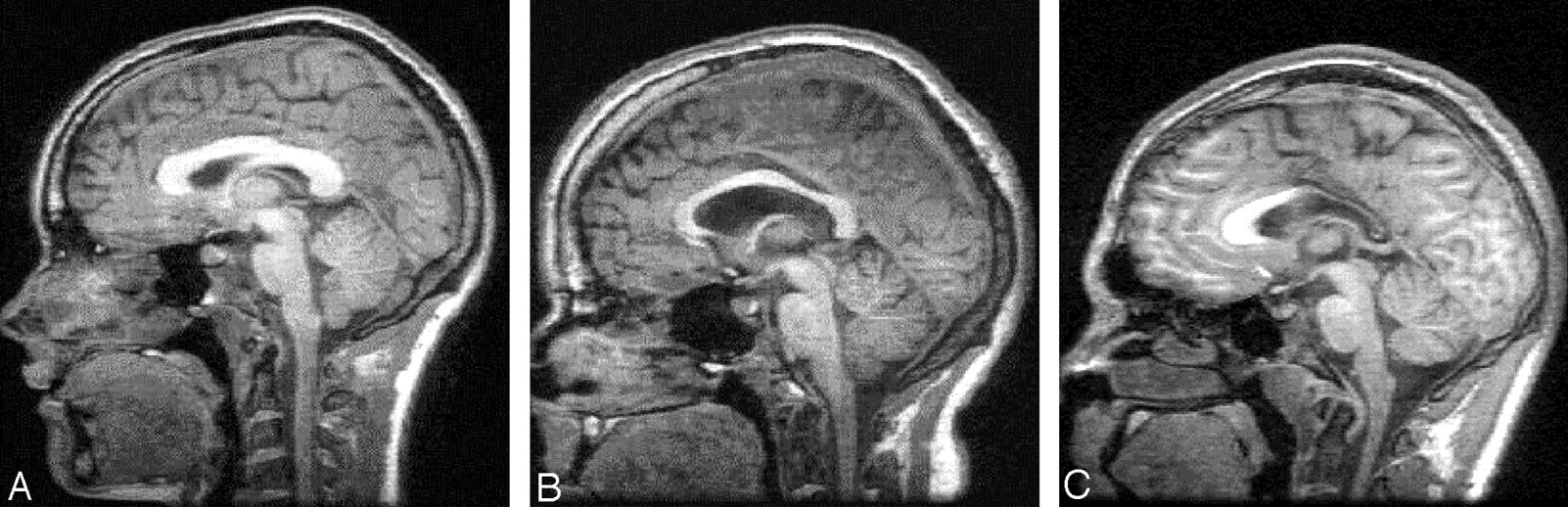
- Fig 1.
Examples of absent (A), moderate (B), and marked (C) thinning of the corpus callosum on midsaggital MR images.
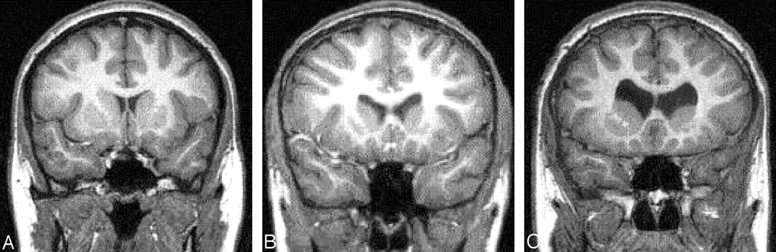
- Fig 2.
Examples of absent (A), moderate (B), and marked (C) enlargement of the frontal horns of the lateral ventricles. These images also show absent (A), moderate (B), and marked (C) blunting of the lateral angles of the frontal horns of the lateral ventricles.
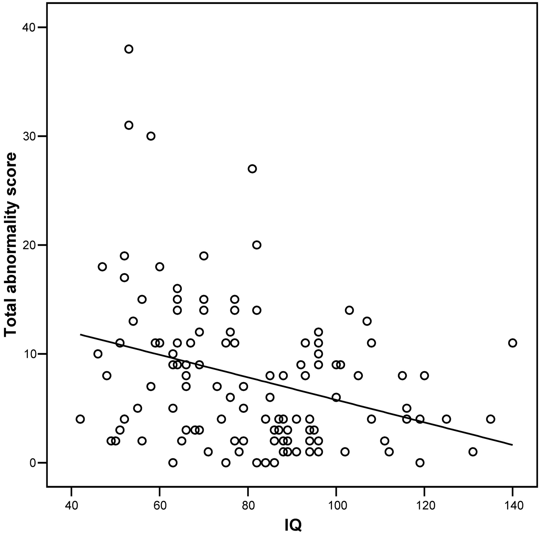
- Fig 3.
Scatterplot of total abnormality score against IQ for all 120 subjects. There is a significant negative correlation for total abnormality score versus IQ (r = −0.320; P < .001).


Tables
- TABLE 1:
Structural anomalies assessed on MR imaging and their observed frequencies among subjects and controls
Observed Frequencies Moderate Anomaly Marked Anomaly Subjects (n = 80) Controls (n = 40) Subjects (n = 80) Controls (n = 40) Lateral ventricle Blunting of lateral angles of midpoint of body of lateral ventricles 30 12 13 1 Blunting of lateral angles of frontal horns of lateral ventricles 31 12 13 1 Enlargement of frontal horns of lateral ventricles 37 14 7 0 Enlargement of body of lateral ventricles 40 12 7 0 Enlargement of occipital horns of lateral ventricles 28 12 10 0 Asymmetry of lateral ventricle 28 12 3 0 Abnormal shape of lateral ventricle 32 10 6 0 Third ventricle Widening of anterior portion of third ventricle 22 6 3 0 Widening throughout third ventricle 21 6 3 0 Cortex Enlargement of cortical sulci 26 9 3 0 Enlargement of subarachnoid spaces 22 9 3 0 Enlargement of interhemispheric fissure 7 1 1 0 Enlargement of subarachnoid cisterns 23 10 2 0 Temporal lobe Abnormal shape of hippocampus 1 0 4 0 Abnormal gyral pattern of hippocampus 4 0 1 0 Reduced size of hippocampus 2 0 3 0 Enlargement of temporal horns 12 10 5 0 White matter abnormalities Corpus callosal dysgenesis 0 0 0 0 Corpus callosal thinning 17 2 6 0 Corpus callosal notching 17 9 2 0 Focal high-intensity white matter lesions 4 0 3 0 White matter high intensity of myelination delay 1 0 0 0 White matter volume loss 4 1 5 0 Virchow-Robin spaces 38 24 0 0 Other abnormalities Cavum septi pellucidi or cavum vergae 16 10 3 0 Aqueduct stenosis 0 0 0 0 Arachnoid cysts 2 0 1 0 Porencephalic cysts 0 0 1 0 Cerebellar tonsil through occipital foramen* 14 10 2 2 Pachygyria 0 0 0 0 Polymicrogyria 0 0 0 0 Gray matter heterotopia 1 1 0 0 Schizencephaly 0 0 0 0 Enlarged cisterna magna 20 6 2 0 Brain stem anomalies 1 0 0 0 Skull shape anomalies 1 0 0 0 * As a guide for this item, “moderate” represents a displacement of 0–5 mm below the basio-opisthion line, and “marked” represents a displacement >5 mm.
- TABLE 2:
Item-by-item intraclass correlation (ICC) values for interobserver and intraobserver agreement
Feature Interobserver Agreement Intraobserver Agreement Lateral ventricle Blunting of lateral angles of midpoint of body of lateral ventricles 0.95 0.90 Blunting of lateral angles of frontal horns of lateral ventricles 0.91 0.85 Enlargement of frontal horns of lateral ventricles 0.79 0.62 Enlargement of body of lateral ventricles 0.80 0.62 Enlargement of occipital horns of lateral ventricles 0.57 0.82 Asymmetry of lateral ventricle 0.75 0.91 Abnormal shape of lateral ventricle 0.47 0.80 Third ventricle Widening of anterior portion of third ventricle 0.74 0.52 Widening throughout third ventricle 0.77 0.52 Cortex Enlargement of cortical sulci 0.58 0.75 Enlargement of subarachnoid spaces 0.64 0.83 Enlargement of interhemispheric fissure 0.51 1 Enlargement of subarachnoid cisterns 0.54 0.73 Temporal lobe Abnormal shape of hippocampus 0.82 1 Abnormal gyral pattern of hippocampus 0.72 1 Reduced size of hippocampus 0.65 1 Enlargement of temporal horns 0.61 0.90 White matter abnormalities Corpus callosal thinning 0.75 0.90 Corpus callosal notching 0.72 0.89 Focal high-intensity white matter lesions 0.51 0.94 White matter volume loss 0.68 1 Virchow-Robin spaces 0.55 0.73 Other abnormalities Cavum septi pellucidi or cavum vergae 0.70 —* Arachnoid cysts 0.94 1 Cerebellar tonsil through occipital foramen 0.87 0.85 Enlarged cisterna magna 0.69 0.58 Total abnormality score 0.93 0.97 Note.—This table excludes 10 items for which the observed frequency was <2% in 120 scans.
*ICC could not be calculated due to 0 variance within one set of observations.


