
Abstract
BACKGROUND AND PURPOSE: Meningoencephalitis can severely damage the developing brain. Preterms are more prone for nosocomial infections with pathogens other than Group B streptococci and Escherichia coli. In this report we focus on the deleterious clinical course and imaging characteristics of proven Bacillus cereus meningoencephalitis.
METHODS: We collected 3 cases of proven Bacillus cereus meningoencephalitis. In the medical records we focused on prenatal, perinatal, and postnatal risk factors. Imaging data of several brain ultrasounds, MR images, and diffusion-weighted images were reevaluated.
RESULTS: The ultrasound and MR images show a typical pattern of mainly hemorrhagic and early cavitating, selective white matter destruction.
CONCLUSION: Knowledge of this paradigm of acquired brain injury may help to better understand the natural course of these severe neonatal infections.
Infections of the neonatal central nervous system (CNS) may have major clinical consequences (1). Sheth (2) found an incidence of CNS infection of 1.3% in a group of 4164 neonates in a neonatal intensive care unit (2) Recently Stoll et al described an incidence of 1.4% of late-onset meningitis among very-low-birth-weight infants (3).
Group B beta-hemolytic streptococci and E coli are the most common causes of meningoencephalitis in the neonatal period (4, 5). Bacteria that typically account for meningitis in older age groups (Haemophilus influenzae type B, Neisseria meningitidis, and Streptococcus pneumoniae) are infrequent in the neonatal population.
Serratia marcescens, or Citrobacter, may be considered as a causal micro-organism in cases of hemorrhagic meningoencephalitis (6, 7). If the culture of blood or CSF shows gram-positive rods, Listeria monocytogenes is the most likely causative agent. It is important to note that Bacillus cereus, though rare, can also cause hemorrhagic meningoencephalitis. B cereus is a common gram-positive food bacteria, is a member of the nonanthrax Bacillus species, and can induce food poisoning (8). Local infections of this species have been described, especially a destructive eye infection that can lead to orbital abscess and endophthalmitis (9). Systemic infections affecting the central nervous system are less well recognized. This may be because microbiologists often regard B cereus as a contaminant.
During the past several years, we have treated 3 preterm infants with severe hemorrhagic meningoencephalitis in the course of a B cereus infection. The infants had a stereotyped disease course, and their imaging templates showed similarities. To facilitate recognition of this entity, we describe the clinical course and evolution of brain imaging findings.
Patients and Methods
We studied 3 preterm infants with severe hemorrhagic meningoencephalitis due to a B cereus infection. The microbiologic confirmation of the diagnosis was established in all infants in blood cultures and in 2 cases in CSF. All 3 had the same clinical course: an uncomplicated delivery, no perinatal hypoxia, and uneventful first days. In this period, routine sonography of the brain was performed. The first day of disease signs ranged from 5 to 13. All infants had signs and symptoms of meningitis (CSF showed leukocytosis, low glucose, and increased protein). From the first day of symptoms, a deleterious clinical course was seen, with thrombocytopenia, hypotension, elevated serum C-reactive protein and seizures. Despite appropriate therapy, all died within 5 days (Table 1). In this period, almost-daily sonography of the brain was performed to evaluate the progress of meningoencephalitis.
Summary of three cases with a destructive hemorrhagic meningoencephalitis
MR imaging was performed at 1.0T and 1.5T during the acute illness, 1–2 days after the onset of symptoms. Our routine MR examination include axial T1-weighted and T2-weighted images and axial or coronal fluid-attenuated inversion recovery (FLAIR) images. Contrast-enhanced T1-weighted images in the axial plane were added. Axial diffusion-weighted imaging (DWI) was performed by using a single-shot echoplanar spin-echo technique on the 1.5T machine and multiple-shot echoplanar spin-echo technique on the 1.0T machine. Diffusion gradients were applied in x, y, and z directions with 4 b values ranging between 0 and 1000 seconds/mm2 at 1.5 T and 0 and 756 seconds/mm2 at 1.0 T. With these data, apparent diffusion coefficient (ADC) maps were calculated.
Results
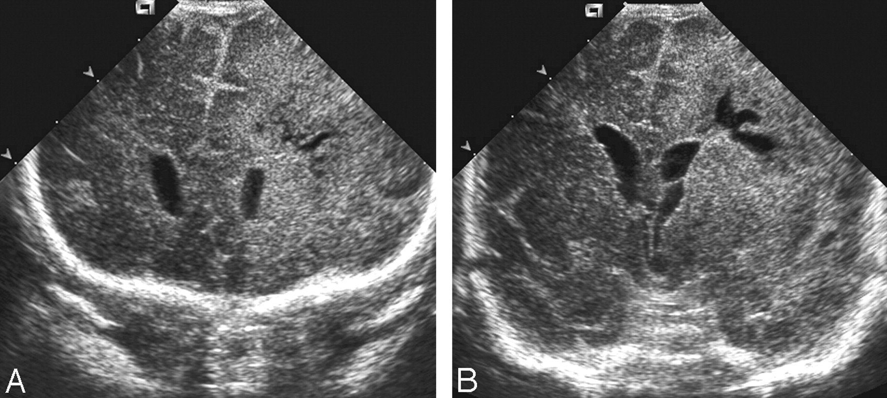
We saw no abnormalities on brain sonography in the uneventful period in all 3 infants. In the first 2 days of clinical symptoms, brain sonograms delineated widespread abnormalities of white matter, preferring periventricular, as well as subcortical, white matter. In time this white matter area showed signs of central necrosis and a rim of hyperechogenicity around it (Fig 1A and -B). The white matter destruction was asymmetrical in all cases. Sparing of adjacent cortex and deep gray matter was conspicuous. In only one case the basal ganglia showed hyperechogenicity. The cortical sparing can be seen on both sonographic and conventional MR images. Superior sagittal, straight, and transverse sinuses were patent on sonography. In one case, brain sonography showed intraventricular septa and thickened ependyma due to ventriculitis (case 3).

Case 3. A, Coronal sonography (8.5 MHz) on day 15 with hyperechogenicity of subcortical white matter with a shift of the midline and the beginning of necrosis of the left hemisphere. B, Coronal sonography on day 23 showing thickening of ependyma and septae due to ventriculitis.
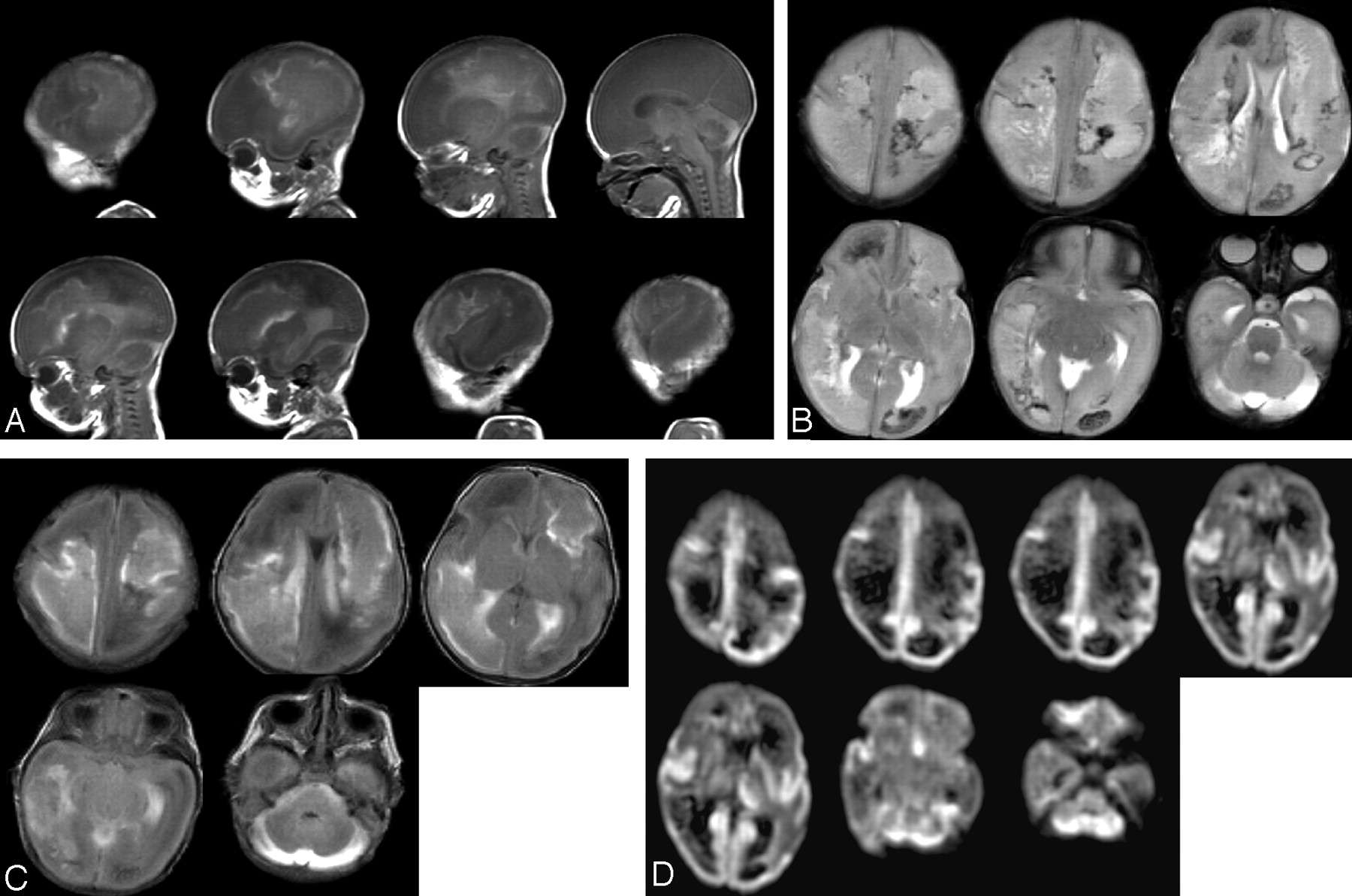
The MR images performed in the acute period had characteristics of hemorrhage in white matter on the T1-weighted, T2-weighted, and FLAIR images (Fig 2A–C). In the first infant (case 1), diffusion was restricted in the basal ganglia and in perisylvian and calcarine cortex (Fig 2D). These injury sites were not conspicuous on conventional T1-weighted and T2-weighted images. In the second infant (case 2), T2-weighted MR images showed (hemorrhagic) areas of low signal intensity in white matter, especially frontoparietal on the left side and parietooccipital on the right side (Fig 3A and -B). DWI confirmed limited water diffusion in these areas, but additional lesions were detected. Low ADCs were also seen in the left parietal cortex, insular area, calcarine cortex, and thalamus at the left side and at the level of the mesencephalon (Fig 3C).

Case 1. MR on day 7 has characteristics of hemorrhage in the white matter on (A) sagittal T1-weighted (TR 780 ms; TE 14 ms) and (B) axial T2-weighted (TR 3000 ms; TE 120 ms) images. C, Axial FLAIR (TR 9000 ms; TE 105 ms) image shows the extensive hemorrhagic meningoencephalitis. D, DWI (EPI sequence TE 128 ms; b = 1000 mm2/s) also showed restricted diffusion at the thalamus and cortex at the perisylvian and calcarine area and cerebellum.

Case 2. T1-weighted (A) (TR 780 ms; TE 14 ms) with hyperintensities right parietal and left frontal and T2-weighted (B) (TR 3000 ms; TE 120 ms) images, showing diffuse hypointensities at the level of the centrum semiovale, right frontal and left occipital and some hyperintensity right periventricular occipitally. On DWI (C) images (EPI sequence TE 128 ms; b = 1000 mm2/s), hyperintensity is seen on the left parietal, insular area, calcarine cortex, and thalamus at the left side and at the level of the mesencephalon.
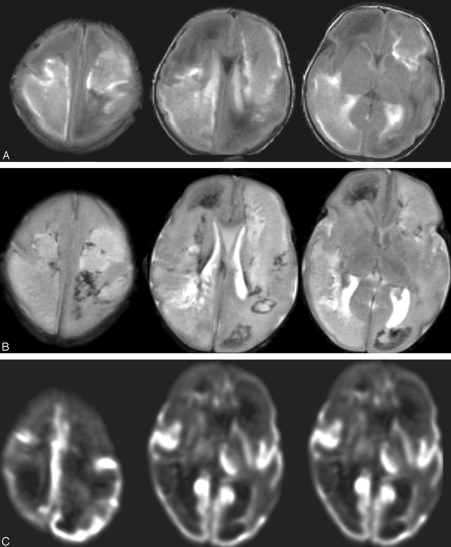
The T2-weighted MR images in the third infant (case 3) showed confluent hypointense areas in white matter indicative of massive hemorrhagic destruction in both hemispheres, in the areas seen with sonography (Fig 4A–C). These areas of destruction were not compatible with patterns seen in arterial as well as venous infarction. Superior sagittal, straight, and transverse sinuses were patent. Cortical sparing can be seen on MR images, though the right occipital lobe is subtlely abnormal on the T2-weighted images in Fig 4B. A rim enhancement around the ventricles was seen on the T1-weighted SE with gadolinium, which is indicative of ventriculitis. DWI showed an additional ischemic area at the calcarine cortex at the right side (Fig 4D).

Case 3. On day 15 axial T1-weighted SE (A), axial T2 TSE (B), and coronal FLAIR (C) images show hemorrhagic liquefactive necrosis of the left hemisphere (case 3). D, Diffusion-weighted image (TR, 1440 ms; TE, 146 ms; b = 756 mm2/s) shows restrictive diffusion in the calcarine area.
Discussion
Neonatal meningoencephalitis caused by B cereus is rare. In most cases, the infection is fatal because of extensive damage and necrosis of infected tissue caused by the toxins produced by B cereus. The organism itself swarms out from veins into tissue. Senesi et al suggest that swarm-cell differentiation is coupled with virulence in this organism (10). The chemotactic activity in B cereus depends on the capability for swarming. The enterotoxin, phospholipases, proteases, and hemolysins induce a widespread liquefactive necrosis. Recognition of this pattern of damage might also be important because the B cereus infection is in some cases a nosocomial infection with serious consequences for the neonatal intensive care unit. Van der Zwet et al have described an outbreak of B cereus infection on a neonatal ward due to colonization of the balloon masks (11).
Hendrickx et al (1981) described the destruction by B cereus especially in the cortex and basal ganglia (12). In 2 of our cases, and in the one described by Chu et al (2001), the destruction is more extensive in subcortical and periventricular white matter, sparing the cortex (13). This preference could be explained by spread of infection along white matter tracks (14).
In the recent literature, few reports concern imaging in patients with neonatal (mainly gram-negative: Proteus, Serratia, Citrobacter) bacterial meningoencephalitis (15). Serratia marcescens is the most common cause of hemorrhagic meningoencephalitis in infants (6). There are some reports of B cereus as a causative agent of a meningoencephalitis (Table 2). In most reports, imaging was restricted to sonography and/or CT (13).
Reports in the literature of infection of the central nervous system with Bacillus cereus
The cases presented here demonstrate the value of serial sonography to detect brain destruction in a neonate with signs of sepsis and/or convulsions and normal early prior scans.
The supratentorial periventricular, as well as subcortical white matter, seems to be primarily affected. Later, the destructive infection will also involve the cortex. Our 3 cases with DWI demonstrated not only the abnormal diffusion in the brain parenchyma, but also lesions that were not detected on conventional MR images. In Fig 2D, low ADCs were especially in the frontal white matter, more periventricular than subcortical. The cortex seemed to be relatively spared. The cortex of the occipital lobes seemed to be most affected. The low ADC was most likely due to cytotoxic edema (16–18). In cases of hemorrhagic meningoencephalitis, however, a careful interpretation of DWI is needed because of possible paramagnetic effects of blood deposits disturbing the images (19). Nevertheless, analysis of routine MR images in our 3 cases makes it clear that changes on DWI are in most linked to restricted diffusion rather than hemorrhage. In our cases, the additional abnormalities picked up by DWI did not change treatment.
Bacterial white matter destruction has to be differentiated from periventricular leukomalacia (PVL) in preterm infants. This is an ongoing process that in many cases starts with symmetrical flaring in the periventricular white matter within the first days of life sometimes evolving into cystic destruction of the white matter (20). The condition is bilateral and almost symmetrical, preferentially damaging posterior frontal and parietal regions. In our cases, brain ultrasounds in the first days of life were normal, which makes the diagnosis of PVL of prenatal origin unlikely.
Thrombosis of the deep cerebral veins can also cause destruction of the white matter, the deep gray matter, basal ganglia, and thalamus (21). In our cases, these structures are initially spared and the destruction was primarily in the white matter and in one hemisphere. Furthermore, no signs of sagittal sinus or deep venous thrombosis were observed on sonography.
Birth asphyxia (22) will show transition from abnormal hyperechogenicity in major arterial areas to slow (in the course of a few weeks) destruction. This is in contrast to very rapid destruction of an initially normal brain in bacterial encephalitis; however, none of our preterm infants met the criteria of birth asphyxia (Table 1). The abnormal hyperechogenicity seen in the basal ganglia in case 1 is probably due to direct extension of the necrotic infection in the surrounding white matter as depicted with MR.
Viral encephalitis is another important differential diagnosis. Herpes simplex, especially, can cause massive destruction of the white matter in the neonatal period. The cortex and deep gray nuclei may also be affected, because of infarction caused by vasculitis and obstruction of small vessels, but also neuronal apoptosis. In none of our cases was H simplex or another virus detected with PCR or culture. Another cause of white matter destruction in the neonatal period could be a mitochondrial disease. In almost all cases, this is symmetrical and not hemorrhagic (23). The deep gray matter is often involved, or the brain stem. Both were not initially affected in our cases.
In the literature, all infections with B cereus in preterm infants followed a devastating course: one had cerebral palsy as a late sequel, and all the others died. There may be more risk factors for brain destruction besides the factors described in Table 1 and the toxins of B cereus itself. These other predisposing factors are vasculitis, vasospasm, hydrocephalus, and diminished cerebrovascular autoregulation, which is often seen in premature infants. The occurrence of many of those risk factors in our cases is probably the reason for the bad outcome of this hemorrhagic meningoencephalitis caused by B cereus (Table 1).
In conclusion, B cereus can cause a severe late-onset hemorrhagic meningoencephalitis in preterm infants. We emphasize the importance of early and serial brain sonography in neonates, especially preterm infants with signs of sepsis and convulsions. Serial sonography showed that the hemorrhagic meningoencephalitis affects first the white matter and later the basal ganglia and cortex.
If sonography shows clear abnormalities, MR imaging should be performed for better delineation of the extent of brain damage, which can be of help for decision making. DWI might have additional value, although in hemorrhagic meningoencephalitis diffusion values may be flawed by paramagnetic artifacts of the blood residue.
References
- Received November 9, 2004.
- Accepted after revision April 1, 2005.
- Copyright © American Society of Neuroradiology


