Advertisement

Main menu

  • Home
  • Content
    • Current Issue
    • Accepted Manuscripts
    • Article Preview
    • Past Issue Archive
    • AJNR Case Collection
    • Case of the Week Archive
    • Classic Case Archive
    • Case of the Month Archive
  • Special Collections
    • Spinal CSF Leak Articles (Jan 2020-June 2024)
    • 2024 AJNR Journal Awards
    • Most Impactful AJNR Articles
  • Multimedia
    • AJNR Podcast
    • AJNR Scantastics
    • Video Articles
  • For Authors
    • Submit a Manuscript
    • Author Policies
    • Fast publishing of Accepted Manuscripts
    • Graphical Abstract Preparation
    • Manuscript Submission Guidelines
    • Imaging Protocol Submission
    • Submit a Case for the Case Collection
  • About Us
    • About AJNR
    • Editorial Board
  • More
    • Become a Reviewer/Academy of Reviewers
    • Subscribers
    • Permissions
    • Alerts
    • Feedback
    • Advertisers
    • ASNR Home
  • Other Publications
    • ajnr

User menu

  • Alerts
  • Log in

Search

  • Advanced search
American Journal of Neuroradiology

American Journal of Neuroradiology

ASHNR American Society of Functional Neuroradiology ASHNR American Society of Pediatric Neuroradiology ASSR
  • Alerts
  • Log in

Advanced Search

  • Home
  • Content
    • Current Issue
    • Accepted Manuscripts
    • Article Preview
    • Past Issue Archive
    • AJNR Case Collection
    • Case of the Week Archive
    • Classic Case Archive
    • Case of the Month Archive
  • Special Collections
    • Spinal CSF Leak Articles (Jan 2020-June 2024)
    • 2024 AJNR Journal Awards
    • Most Impactful AJNR Articles
  • Multimedia
    • AJNR Podcast
    • AJNR Scantastics
    • Video Articles
  • For Authors
    • Submit a Manuscript
    • Author Policies
    • Fast publishing of Accepted Manuscripts
    • Graphical Abstract Preparation
    • Manuscript Submission Guidelines
    • Imaging Protocol Submission
    • Submit a Case for the Case Collection
  • About Us
    • About AJNR
    • Editorial Board
  • More
    • Become a Reviewer/Academy of Reviewers
    • Subscribers
    • Permissions
    • Alerts
    • Feedback
    • Advertisers
    • ASNR Home
  • Follow AJNR on Twitter
  • Visit AJNR on Facebook
  • Follow AJNR on Instagram
  • Join AJNR on LinkedIn
  • RSS Feeds

Welcome to the new AJNR, Updated Hall of Fame, and more. Read the full announcements.


AJNR is seeking candidates for the position of Associate Section Editor, AJNR Case Collection. Read the full announcement.


Research ArticleBRAIN

Assessing Global Invasion of Newly Diagnosed Glial Tumors with Whole-Brain Proton MR Spectroscopy

Benjamin A. Cohen, Edmond A. Knopp, Henry Rusinek, James S. Babb, David Zagzag and Oded Gonen
American Journal of Neuroradiology October 2005, 26 (9) 2170-2177;
Benjamin A. Cohen
  • Find this author on Google Scholar
  • Find this author on PubMed
  • Search for this author on this site
Edmond A. Knopp
  • Find this author on Google Scholar
  • Find this author on PubMed
  • Search for this author on this site
Henry Rusinek
  • Find this author on Google Scholar
  • Find this author on PubMed
  • Search for this author on this site
James S. Babb
  • Find this author on Google Scholar
  • Find this author on PubMed
  • Search for this author on this site
David Zagzag
  • Find this author on Google Scholar
  • Find this author on PubMed
  • Search for this author on this site
Oded Gonen
  • Find this author on Google Scholar
  • Find this author on PubMed
  • Search for this author on this site
  • Article
  • Figures & Data
  • Info & Metrics
  • Responses
  • References
  • PDF
Loading

Article Figures & Data

Figures

  • Tables
  • Fig 1.
    • Download figure
    • Open in new tab
    • Download powerpoint
    Fig 1.

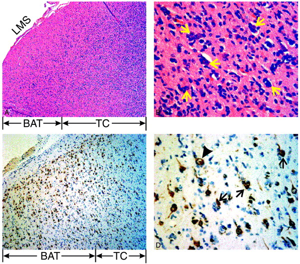
    Histopathology of the invasive edge of the glioblastoma multiforme from patient 12 in the Table.

    Top left, A, Low power, ×50, hematoxylin and eosin stain depicting tumor core (TC) and brain adjacent to tumor (BAT). Note the high cellularity of the core in the white matter, with invading cells spreading as far as the leptomeningeal space (LMS).

    Top right, B, High power, ×400, of the BAT emphasizing perineuronal satellitosis—glioma cells around neuronal cell bodies (arrows).

    Bottom left, C, Medium power, ×100, with a monoclonal antibody (NeuN) staining neurons.

    Bottom right, D, High power, ×400, view of C highlighting immunoreactive neurons (arrows) surrounded by infiltrating tumor cells. These vacuolated neurons appear unhealthy, and therefore, are likely to contain less NAA. A healthy neuron is indicated for comparison (arrowhead).

  • Fig 2.
    • Download figure
    • Open in new tab
    • Download powerpoint
    Fig 2.

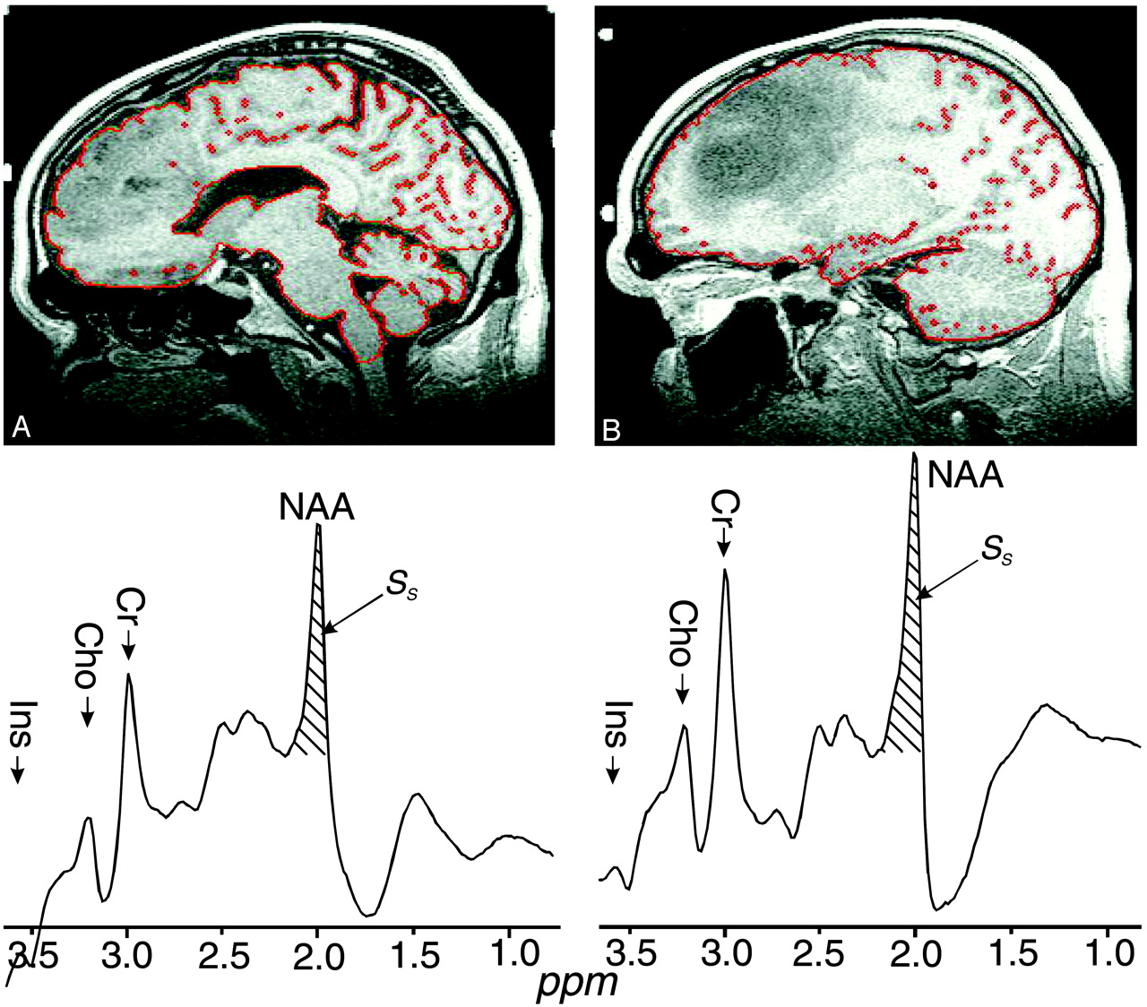
    Top, A and B, Segmented preoperative T1-weighted sagittal section for 2 patients, 15 and 5 in the Table, respectively. The red sulci and ventricle outline is the output of the tissue/CSF segmentation process. Patient A had a 41-cm3 grade III tumor, 3.0% of his brain volume, and 37% WBNAA deficit. Patient B had a 93-cm3 grade II tumor, 6.2% of his brain volume, but only half the WBNAA decline (∼20%).

    Bottom, A′ and B′, Corresponding whole-head 1H-MR spectroscopy. The hatched regions indicate the peak-areas used to obtain QNAA of Equation (1). Note the overall similarity, indicating cross-sectional reproducibility, and good lipid suppression. Also note that, because localization relies on knowledge that NAA is found only in neuronal cells, it cannot quantify the brain’s contribution to metabolites also present in other tissue types (eg, choline, creatine, and so forth).

  • Fig 3.
    • Download figure
    • Open in new tab
    • Download powerpoint
    Fig 3.

    First, 2nd (median), and 3rd quartiles (box) and ±95% (whiskers) of WBNAA in the 17 controls, patients day of surgery (n = 17) and 1 day postsurgery (n = 14). Note that pre- and postsurgical WBNAA are significantly different from the controls, but not each other.

  • Fig 4.
    • Download figure
    • Open in new tab
    • Download powerpoint
    Fig 4.

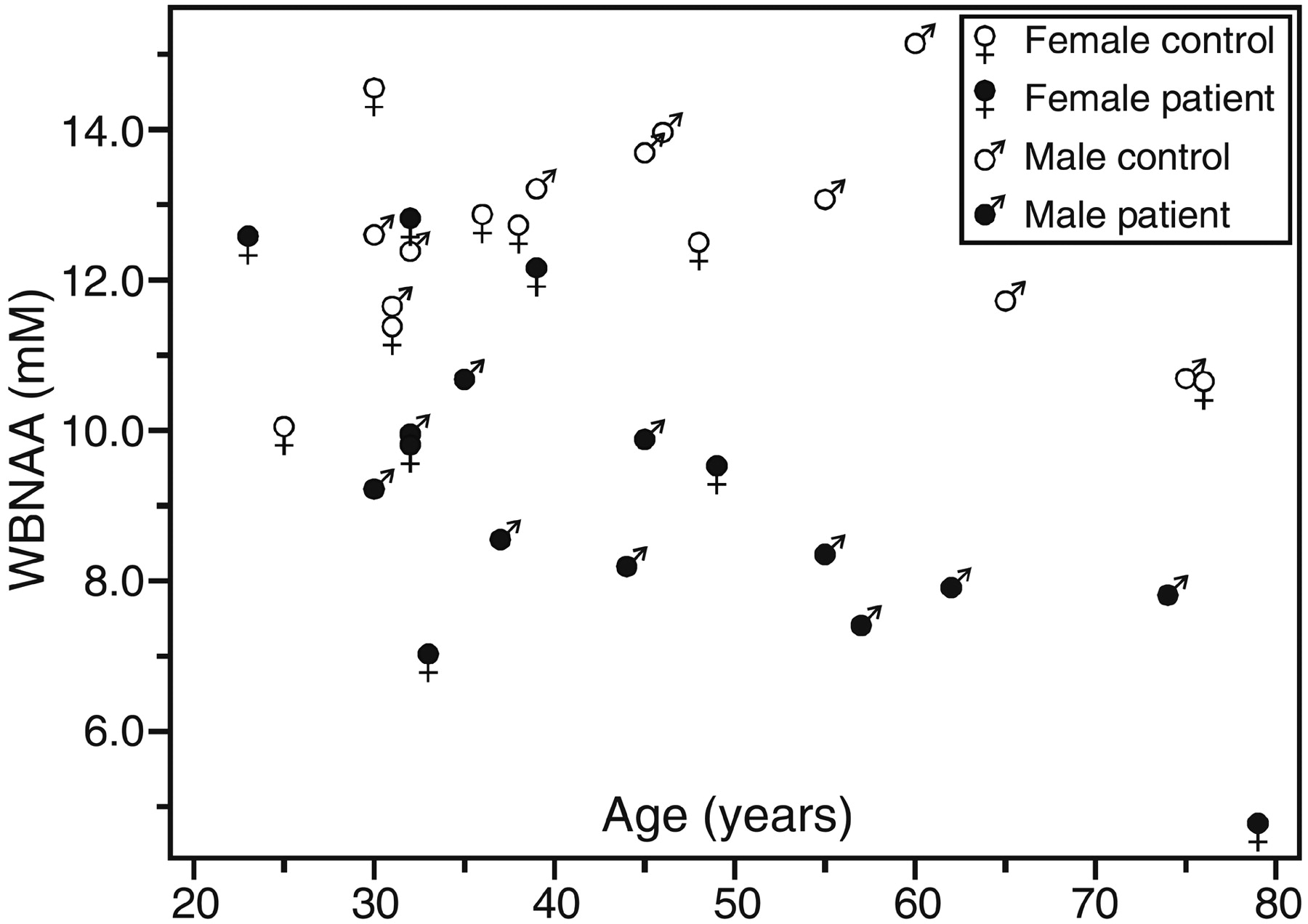
    Scatter plot of WBNAA versus age for the patients before surgery and their age- and sex-matched controls. Note that the patients’ WBNAA deficit is significant (P < .0005), even after taking into account age (P = .007) and sex effects (P > .05).

  • Fig 5.
    • Download figure
    • Open in new tab
    • Download powerpoint
    Fig 5.

    First, 2nd (median), and 3rd quartiles (box) and ±95% (whiskers) of preoperative WBNAA in the 9 patients with low-grade tumors compared with their matched controls (left) and 8 patients with high-grade tumors compared with their matched controls (right). Note preoperative WBNAA was lower in patients with high-grade (8.3 ± 1.8 mmol/L) than in those with low-grade (10.0 ± 2.1 mmol/L) tumors.

Tables

  • Figures
  • Details of study participants

    Patient No./Age (y)/SexWBNAA (mM)Histology (Grade)Control
    PreoperationPostoperationAge (y)/SexWBNAA (mM)
        1/23/F12.613.7GGNC-DNT (I)25/F10.1
        2/30/M9.28.7Mixed GN (II)30/M12.6
        3/32/F12.8N/AMixed GGNC (II)31/F11.4
        4/32/F9.99.2DFA (II)30/F14.6
        5/32/M10.09.8GGNC (II)31/M11.7
        6/33/F7.08.2Mixed GN (II)36/F12.9
        7/35/M10.811.2GBM (IV)32/M12.4
        8/37/M8.68.5Mixed GN (II)39/M13.2
        9/39/F12.210.9Mixed GN (II)38/F12.7
        10/44/M8.28.4Mixed GGNC (II)45/M13.7
        11/45/M9.98.5Anaplastic Mixed GN (III)46/M14.0
        12/49/F9.510.3GBM (IV)48/F12.5
        13/55/M8.510.6Anaplastic mixed GN (III)55/M13.1
        14/57/M7.46.7GBM (IV)60/M15.1
        15/62/M7.911.2AO (III)65/M11.7
        16/74/M7.8N/AGBM (IV)75/M10.7
        17/79/F4.8N/AAnaplastic GGNC (III)76/F10.7
    Avg (mean ± SD)9.2 ± 2.19.7 ± 1.812.5 ± 1.4
    • Note.—N/A indicates biopsy only or insufficient data due to patient movement; GGNC, ganglioglioneurocytoma; DNT, dysembryoplastic neuroepithelial tumor; GN, glioneurocytoma; DFA, diffuse fibrillary astrocytoma; GBM, glioblastoma multiforme; AO, anaplastic oligodendroglioma.

PreviousNext
Back to top

In this issue

American Journal of Neuroradiology: 26 (9)
American Journal of Neuroradiology
Vol. 26, Issue 9
1 Oct 2005
  • Table of Contents
  • Index by author
Advertisement
Print
Download PDF
Email Article

Thank you for your interest in spreading the word on American Journal of Neuroradiology.

NOTE: We only request your email address so that the person you are recommending the page to knows that you wanted them to see it, and that it is not junk mail. We do not capture any email address.

Enter multiple addresses on separate lines or separate them with commas.
Assessing Global Invasion of Newly Diagnosed Glial Tumors with Whole-Brain Proton MR Spectroscopy
(Your Name) has sent you a message from American Journal of Neuroradiology
(Your Name) thought you would like to see the American Journal of Neuroradiology web site.
CAPTCHA
This question is for testing whether or not you are a human visitor and to prevent automated spam submissions.
Cite this article
Benjamin A. Cohen, Edmond A. Knopp, Henry Rusinek, James S. Babb, David Zagzag, Oded Gonen
Assessing Global Invasion of Newly Diagnosed Glial Tumors with Whole-Brain Proton MR Spectroscopy
American Journal of Neuroradiology Oct 2005, 26 (9) 2170-2177;

Citation Manager Formats

  • BibTeX
  • Bookends
  • EasyBib
  • EndNote (tagged)
  • EndNote 8 (xml)
  • Medlars
  • Mendeley
  • Papers
  • RefWorks Tagged
  • Ref Manager
  • RIS
  • Zotero
0 Responses
Respond to this article
Share
Bookmark this article
Assessing Global Invasion of Newly Diagnosed Glial Tumors with Whole-Brain Proton MR Spectroscopy
Benjamin A. Cohen, Edmond A. Knopp, Henry Rusinek, James S. Babb, David Zagzag, Oded Gonen
American Journal of Neuroradiology Oct 2005, 26 (9) 2170-2177;
del.icio.us logo Twitter logo Facebook logo Mendeley logo
  • Tweet Widget
  • Facebook Like
  • Google Plus One

Jump to section

  • Article
    • Abstract
    • Methods
    • Results
    • Discussion
    • Conclusion
    • Footnotes
    • References
  • Figures & Data
  • Info & Metrics
  • Responses
  • References
  • PDF

Related Articles

  • No related articles found.
  • PubMed
  • Google Scholar

Cited By...

  • N-Acetylaspartate and 2-Hydroxyglutarate Assessed in Human Brain Tissue by Mass Spectrometry as Neuronal Markers of Oncogenesis
  • Crossref
  • Google Scholar

This article has not yet been cited by articles in journals that are participating in Crossref Cited-by Linking.

More in this TOC Section

  • Fast Contrast-Enhanced 4D MRA and 4D Flow MRI Using Constrained Reconstruction (HYPRFlow): Potential Applications for Brain Arteriovenous Malformations
  • Quiet PROPELLER MRI Techniques Match the Quality of Conventional PROPELLER Brain Imaging Techniques
  • Predictors of Reperfusion in Patients with Acute Ischemic Stroke
Show more Brain

Similar Articles

Advertisement

Indexed Content

  • Current Issue
  • Accepted Manuscripts
  • Article Preview
  • Past Issues
  • Editorials
  • Editors Choice
  • Fellow Journal Club
  • Letters to the Editor

Cases

  • Case Collection
  • Archive - Case of the Week
  • Archive - Case of the Month
  • Archive - Classic Case

Special Collections

  • Special Collections

Resources

  • News and Updates
  • Turn around Times
  • Submit a Manuscript
  • Author Policies
  • Manuscript Submission Guidelines
  • Evidence-Based Medicine Level Guide
  • Publishing Checklists
  • Graphical Abstract Preparation
  • Imaging Protocol Submission
  • Submit a Case
  • Become a Reviewer/Academy of Reviewers
  • Get Peer Review Credit from Publons

Multimedia

  • AJNR Podcast
  • AJNR SCANtastic
  • Video Articles

About Us

  • About AJNR
  • Editorial Board
  • Not an AJNR Subscriber? Join Now
  • Alerts
  • Feedback
  • Advertise with us
  • Librarian Resources
  • Permissions
  • Terms and Conditions

American Society of Neuroradiology

  • Not an ASNR Member? Join Now

© 2025 by the American Society of Neuroradiology All rights, including for text and data mining, AI training, and similar technologies, are reserved.
Print ISSN: 0195-6108 Online ISSN: 1936-959X

Powered by HighWire