
Article Figures & Data
Figures
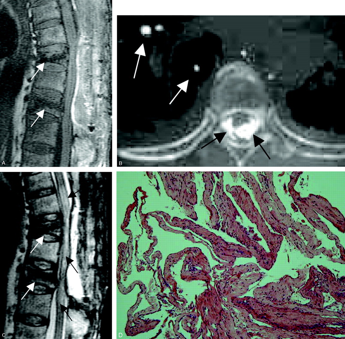
- Fig 1.
Case 1. A, Sagittal reconstructed CT scan performed same day as vertebroplasty shows postvertebroplasty appearance with hyperattenuated bone cement in T8, T10, and L1 vertebral bodies. Bone cement filled the anterior vertebral body, the posterior vertebral body, and the epidural space (arrows). The technical flaw in this case was allowing the bone cement to fill the posterior vertebral body and continue filling in the spinal canal.
B, Axial view at T7/T8 disk level performed same day as vertebroplasty shows bone cement in the epidural space (black arrows) and pulmonary arteries (white arrows).
C, T2-weighted MR imaging performed 50 days after vertebroplasty shows low-signal-intensity bone cement inside the anterior and posterior aspects of T8, T10, and L1 vertebral bodies (white arrows) and postlaminectomy appearance with bloody fluid collection (white arrowheads) causing posterior epidural compression to the spinal cord. There is high-signal- intensity change in the spinal cord on the T2-weighted images, because of compressive myelopathy or previous thermal injury. The posterior epidural compression and signal intensity change of the spinal cord are similar to the MR imaging performed 5 days after the second decompressive surgery. These changes were not present on MR imaging performed 2 days before vertebroplasty.
D, Fibrosis of arachnoid membrane (H&E stain).
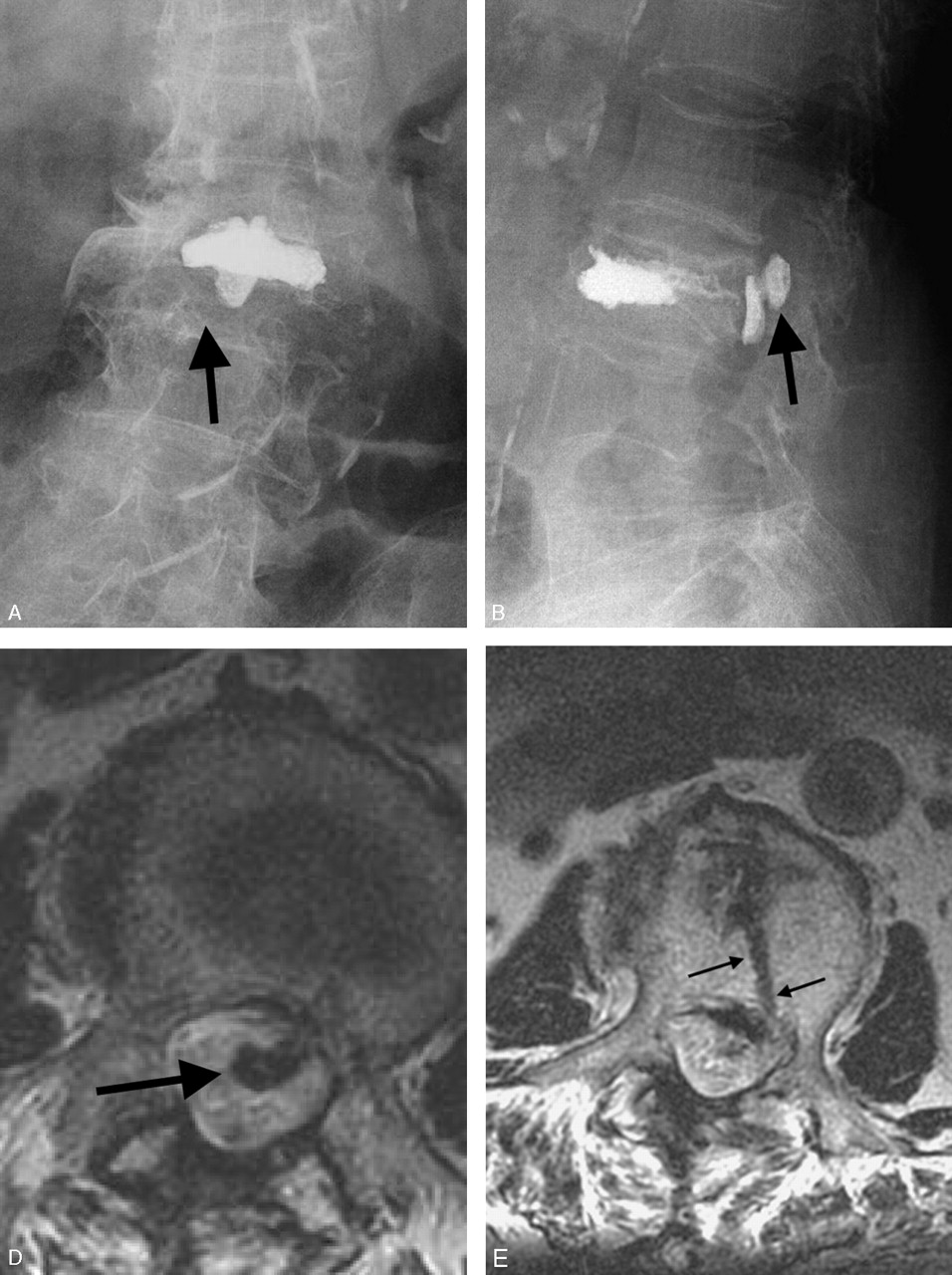
- Fig 2.
Case 2. Lumbar spine plain films and MR imaging were taken 8 months after vertebroplasty.
A, AP view conventional radiograph of lumbar spine.
B, Lateral view conventional radiograph of lumbar spine.
C, Sagittal MR imaging.
D and E, Axial MR imaging.
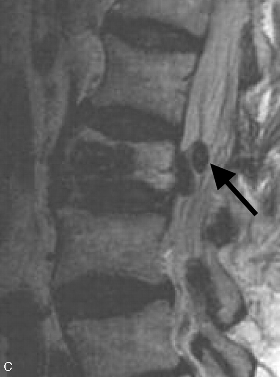
There is hyperattenuated bone cement in L2 vertebral body and in the spinal canal (arrows) on the anteroposterior and lateral conventional radiographs (A and B). On the proton-weighted image (C) and T2-weighted image (D and E), bone cement is low in signal intensity. Bone cement is found inside the dural sac in C and D (arrow). Needle tract can be identified as a low-signal-intensity channel (arrows) in the vertebral body extending posteriorly to the epidural space (E). The technical flaw in this case was puncturing the epidural space with the needle allowing cement to extend posteriorly to the epidural space along the needle tract.
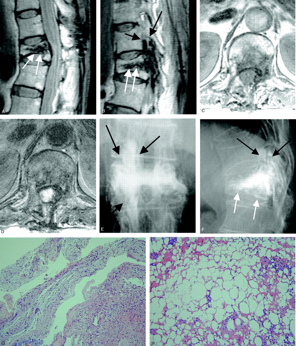
- Fig 3.
Case 3. MR imaging taken 73 days after vertebroplasty (A–D) and conventional radiographs of lumbar spine (E and F) taken 86 days after vertebroplasty. Dense bone cement is found in the anterior and posterior aspects of the vertebral body (white arrows), epidural space (short arrow), and intervertebral foramen (arrows). The technical flaw of this vertebroplasty was use of the wrong needle and possibly drilling past the epidural space.
G, Arachnoid membrane shows active fibrosis and thickening (H&E stain).
H, Epidural soft tissue shows fat necrosis and focal chronic inflammatory cell infiltration (H&E stain).


In this issue


Jump to section
Related Articles
Cited By...
- Large intraspinal cement leak during multilevel cement-augmented screw fixation
- Pulmonary embolism with coexistent incidental pulmonary cement embolism post vertebroplasty
- Sacral bone cyst treatment resulting in paraplegia
- Use and evaluation of a semi-permeable mesh implant in vertebral augmentation for the treatment of painful osteoporotic vertebral compression fractures