Advertisement

Main menu

  • Home
  • Content
    • Current Issue
    • Accepted Manuscripts
    • Article Preview
    • Past Issue Archive
    • AJNR Case Collection
    • Case of the Week Archive
    • Classic Case Archive
    • Case of the Month Archive
  • Special Collections
    • Spinal CSF Leak Articles (Jan 2020-June 2024)
    • 2024 AJNR Journal Awards
    • Most Impactful AJNR Articles
  • Multimedia
    • AJNR Podcast
    • AJNR Scantastics
    • Video Articles
  • For Authors
    • Submit a Manuscript
    • Author Policies
    • Fast publishing of Accepted Manuscripts
    • Graphical Abstract Preparation
    • Manuscript Submission Guidelines
    • Imaging Protocol Submission
    • Submit a Case for the Case Collection
  • About Us
    • About AJNR
    • Editorial Board
  • More
    • Become a Reviewer/Academy of Reviewers
    • Subscribers
    • Permissions
    • Alerts
    • Feedback
    • Advertisers
    • ASNR Home
  • Other Publications
    • ajnr

User menu

  • Alerts
  • Log in

Search

  • Advanced search
American Journal of Neuroradiology

American Journal of Neuroradiology

ASHNR American Society of Functional Neuroradiology ASHNR American Society of Pediatric Neuroradiology ASSR
  • Alerts
  • Log in

Advanced Search

  • Home
  • Content
    • Current Issue
    • Accepted Manuscripts
    • Article Preview
    • Past Issue Archive
    • AJNR Case Collection
    • Case of the Week Archive
    • Classic Case Archive
    • Case of the Month Archive
  • Special Collections
    • Spinal CSF Leak Articles (Jan 2020-June 2024)
    • 2024 AJNR Journal Awards
    • Most Impactful AJNR Articles
  • Multimedia
    • AJNR Podcast
    • AJNR Scantastics
    • Video Articles
  • For Authors
    • Submit a Manuscript
    • Author Policies
    • Fast publishing of Accepted Manuscripts
    • Graphical Abstract Preparation
    • Manuscript Submission Guidelines
    • Imaging Protocol Submission
    • Submit a Case for the Case Collection
  • About Us
    • About AJNR
    • Editorial Board
  • More
    • Become a Reviewer/Academy of Reviewers
    • Subscribers
    • Permissions
    • Alerts
    • Feedback
    • Advertisers
    • ASNR Home
  • Follow AJNR on Twitter
  • Visit AJNR on Facebook
  • Follow AJNR on Instagram
  • Join AJNR on LinkedIn
  • RSS Feeds

Welcome to the new AJNR, Updated Hall of Fame, and more. Read the full announcements.


AJNR is seeking candidates for the position of Associate Section Editor, AJNR Case Collection. Read the full announcement.


Research ArticleHEAD & NECK

Imaging and Clinical Characteristics of Temporal Bone Meningioma

B.E. Hamilton, K.L. Salzman, N. Patel, R.H. Wiggins, A.J. Macdonald, C. Shelton, R.C. Wallace, J. Cure and H.R. Harnsberger
American Journal of Neuroradiology November 2006, 27 (10) 2204-2209;
B.E. Hamilton
  • Find this author on Google Scholar
  • Find this author on PubMed
  • Search for this author on this site
K.L. Salzman
  • Find this author on Google Scholar
  • Find this author on PubMed
  • Search for this author on this site
N. Patel
  • Find this author on Google Scholar
  • Find this author on PubMed
  • Search for this author on this site
R.H. Wiggins III
  • Find this author on Google Scholar
  • Find this author on PubMed
  • Search for this author on this site
A.J. Macdonald
  • Find this author on Google Scholar
  • Find this author on PubMed
  • Search for this author on this site
C. Shelton
  • Find this author on Google Scholar
  • Find this author on PubMed
  • Search for this author on this site
R.C. Wallace
  • Find this author on Google Scholar
  • Find this author on PubMed
  • Search for this author on this site
J. Cure
  • Find this author on Google Scholar
  • Find this author on PubMed
  • Search for this author on this site
H.R. Harnsberger
  • Find this author on Google Scholar
  • Find this author on PubMed
  • Search for this author on this site
  • Article
  • Figures & Data
  • Info & Metrics
  • Responses
  • References
  • PDF
Loading

Article Figures & Data

Figures

  • Tables
  • Fig 1.
    • Download figure
    • Open in new tab
    • Download powerpoint
    Fig 1.

    Coronal graphic depicts characteristic findings in tegmen tympani meningioma. Tumor arises in the middle cranial fossa and spreads inferomedially through the tegmen tympani to the middle ear cavity. En plaque intracranial enhancement is typical (straight white arrows). Ossicles are encased by tumor (curved white arrow), without destruction (reprinted with permission from Amirsys14).

  • Fig 2.
    • Download figure
    • Open in new tab
    • Download powerpoint
    Fig 2.

    A, Coronal unenhanced CT scan reveals typical thickening and trabecular hyperostosis of the tegmen tympani (black star). Note the soft-tissue mass in the middle ear cavity (white star), which encases the ossicles but lacks erosive or destructive change.

    B, Axial unenhanced CT scan shows ossicular encasement without erosive or destructive changes (black arrow). The involved calvaria (white arrows) is characterized by preserved internal architecture (trabecular hyperostosis) and irregularity of the inner table.

    C, Coronal contrast-enhanced MR image shows typical en plaque enhancement along the floor of the middle cranial fossa (straight white arrows) in a tegmen tympani meningioma. Note thickened enhancing tegmen (white star) and enhancing tissue surrounding the ossicular chain (curved white arrow).

    D, Axial contrast-enhanced MR image of the tegmen tympani meningioma shows tumor enhancement in the middle ear cavity. Note the intact appearance of the ossicular chain (open white arrow), seen through the enhancement.

  • Fig 3.
    • Download figure
    • Open in new tab
    • Download powerpoint
    Fig 3.

    Coronal graphic depicts findings in JF meningioma: location primary to the JF (white star) with superolateral vector of spread of the soft-tissue mass into the middle ear cavity (black arrow) (reprinted with permission from Amirsys14).

  • Fig 4.
    • Download figure
    • Open in new tab
    • Download powerpoint
    Fig 4.

    A, Coronal unenhanced CT scan shows a mixed permeative/sclerotic appearance of the involved skull base (white star) that is typical for JF meningioma. Bony changes mildly narrow the mastoid segment of the facial nerve canal (open white arrow).

    B, Axial unenhanced CT scan again demonstrates the characteristic mixed permeative/sclerotic appearance and centrifugal involvement of the skull base (black star). Note intact-appearing cortical margins (black arrows). Although not a common finding, calcification of the soft-tissue mass (white arrow) is a fairly specific finding of meningioma when present.

    C, Coronal enhanced MR image demonstrates an enhancing mass primarily located in the jugular foramen (white arrow), with centrifugal involvement of the surrounding skull base and superolateral soft-tissue spread to the middle ear cavity (black star). Note intense intraosseous enhancement relative to the extracranial component and largely preserved intrinsic bony architecture (black arrows).

    D, Axial enhanced MR image demonstrates an intensely enhancing mass surrounding the intact-appearing ossicular chain (open white arrow). Note the intracranial enhancing dural tail (straight white arrow). Secondary extension to the internal auditory canal (curved white arrow) is also present in this case.

  • Fig 5.
    • Download figure
    • Open in new tab
    • Download powerpoint
    Fig 5.

    Coronal graphic depicts findings in IAC meningioma: tumor in the cochlea and vestibular apparatus (black arrows). Note the intracranial component in the IAC and cerebellopontine angle (black star) (reprinted with permission from Amirsys14).

  • Fig 6.
    • Download figure
    • Open in new tab
    • Download powerpoint
    Fig 6.

    A, Coronal enhanced MR image of an IAC meningioma demonstrates intense enhancement of the intralabyrinthine structures (curved white arrow). The mass extends from the IAC and cerebellopontine angle (open white arrow).

    B, Axial enhanced MR image of an IAC meningioma demonstrates intense enhancement of the cochlea (curved white arrow) and vestibular apparatus (open white arrow). The large IAC and cerebellopontine angle enhancing mass (straight white arrow) are clues to the diagnosis.
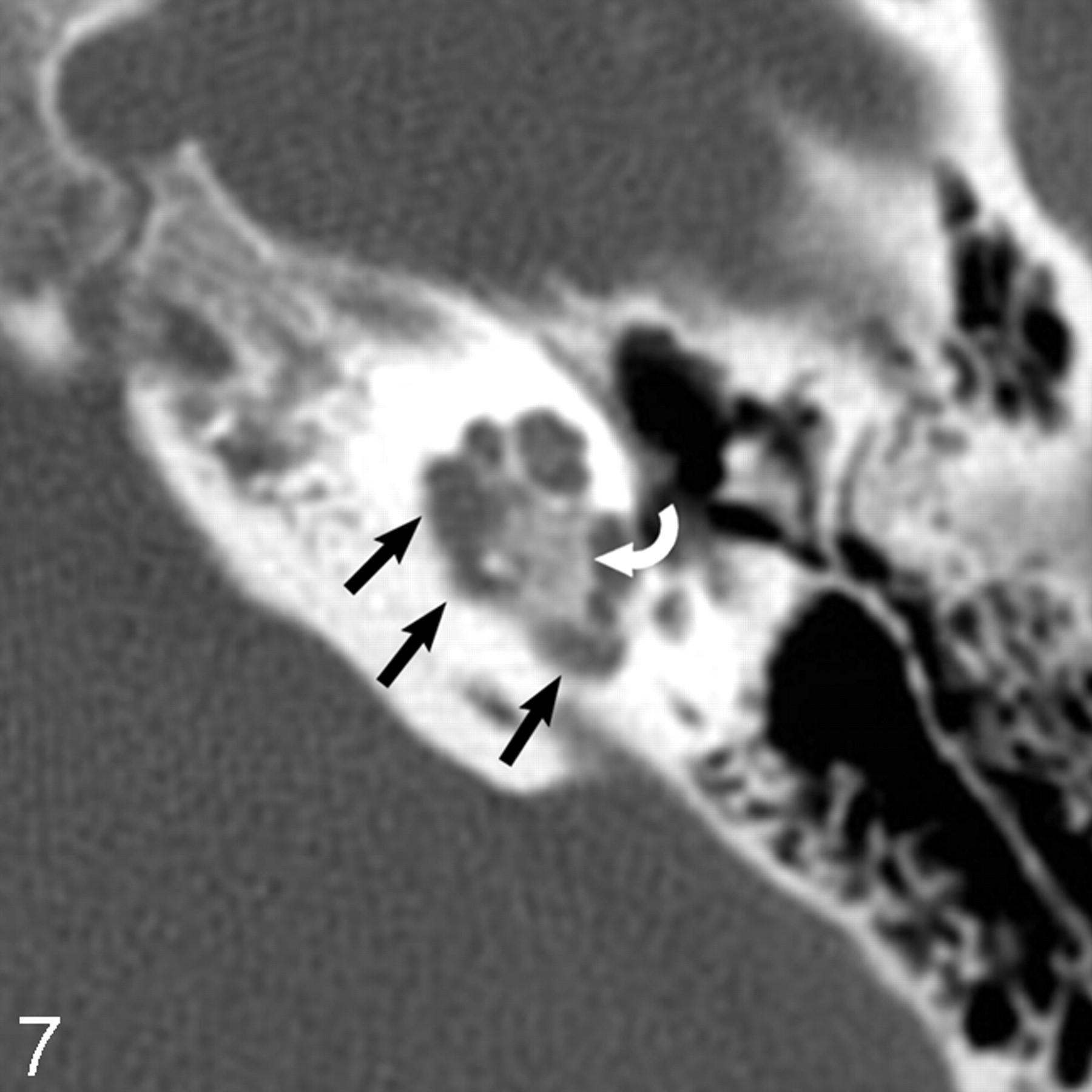
  • Fig 7.
    • Download figure
    • Open in new tab
    • Download powerpoint
    Fig 7.

    Axial unenhanced CT scan demonstrates pericochlear bone lucency (straight black arrows). Calcification of the mass (curved white matter) suggests the diagnosis of meningioma, as seen in other locations.

Tables

  • Figures
  • Clinical and imaging features of temporal bone meningioma

    Age/SexCHLSNHLOtoscopy FindingsFacial N SxVest SxMR: Dural TailMR: Assoc FeaturesCT: Bone ChangeCT: Facial CanalCT: Mass Ca++CT: Oss
    TT
        Left69/M++Gray RT mass−Dizzy for 2 years+−TH+−+
        Right50/F+−U−Dizzy+Vasogenic EdemaTH+−+
        Right65/M−+U−Dizzy, Vertigo, Ataxia+Larger Mass MCFMixed TH + lucency+−+
        Left46/F+−Vascular RT mass−−+−TH−−+
        Right57/F++Vascular RT mass+Nystag+−TH+−+
        Left60/MUUU−U+Larger Mass MCFTH−−U
    TT + JF
        Right56/F−+U+Dizzy+Spread to IACMixed TH-(TT) Perm–Scler (JF)+++
    JF
        Right31/M++Purple RT mass−Dizzy+Spread to CPAMixed Perm–Scler−+−
        Right65/F++Red RT mass−Dizzy+Spread to CPAMixed Perm–Scler−++
        Right53/F++Vascular RT mass−Dizzy+Spread to CPA + IACMixed Perm–Scler+++
        Left59/F++Red RT mass−Dizzy+Spread to CPAMixed Perm–Scler+−+
    IAC
        Right56/M++U−U+Larger CPA massU−U−
        Left18/F−+U−Tinn−−Min lucency−+−
    • Note:—Clinical & imaging features are displayed according to location. Clinical features are shown in bold; imaging findings are seen in the portion to the right. TT indicates tegmen tympani; JF, jugular foramen; IAC, internal auditory canal; CPA, cerebellopontine angle; MCF, middle cranial fossa; CHL, conductive hearing loss; SNHL, sensorineural hearing loss; TH, trabecular hyperostosis; RT, retrotympanic; Nystag, nystagmus; Tinn, tinnitus; Vest Sx, Vestibular symptoms; Perm –Scler, permeative –sclerotic; Min, minimal; U, unknown; CT Oss, presence of ossicular abutment or encasement by soft tissue; CT Facial Canal, presence of bony facial canal encroachment by hyperostosis; Ca++, calcification; M, male; F, female; Assoc, associated; N, nerve; +, positive features; –, absent findings.

PreviousNext
Back to top

In this issue

American Journal of Neuroradiology: 27 (10)
American Journal of Neuroradiology
Vol. 27, Issue 10
November 2006
  • Table of Contents
  • Index by author
Advertisement
Print
Download PDF
Email Article

Thank you for your interest in spreading the word on American Journal of Neuroradiology.

NOTE: We only request your email address so that the person you are recommending the page to knows that you wanted them to see it, and that it is not junk mail. We do not capture any email address.

Enter multiple addresses on separate lines or separate them with commas.
Imaging and Clinical Characteristics of Temporal Bone Meningioma
(Your Name) has sent you a message from American Journal of Neuroradiology
(Your Name) thought you would like to see the American Journal of Neuroradiology web site.
CAPTCHA
This question is for testing whether or not you are a human visitor and to prevent automated spam submissions.
Cite this article
B.E. Hamilton, K.L. Salzman, N. Patel, R.H. Wiggins, A.J. Macdonald, C. Shelton, R.C. Wallace, J. Cure, H.R. Harnsberger
Imaging and Clinical Characteristics of Temporal Bone Meningioma
American Journal of Neuroradiology Nov 2006, 27 (10) 2204-2209;

Citation Manager Formats

  • BibTeX
  • Bookends
  • EasyBib
  • EndNote (tagged)
  • EndNote 8 (xml)
  • Medlars
  • Mendeley
  • Papers
  • RefWorks Tagged
  • Ref Manager
  • RIS
  • Zotero
0 Responses
Respond to this article
Share
Bookmark this article
Imaging and Clinical Characteristics of Temporal Bone Meningioma
B.E. Hamilton, K.L. Salzman, N. Patel, R.H. Wiggins, A.J. Macdonald, C. Shelton, R.C. Wallace, J. Cure, H.R. Harnsberger
American Journal of Neuroradiology Nov 2006, 27 (10) 2204-2209;
del.icio.us logo Twitter logo Facebook logo Mendeley logo
  • Tweet Widget
  • Facebook Like
  • Google Plus One

Jump to section

  • Article
    • Abstract
    • Materials and Methods
    • Results
    • Discussion
    • Conclusion
    • References
  • Figures & Data
  • Info & Metrics
  • Responses
  • References
  • PDF

Related Articles

  • No related articles found.
  • PubMed
  • Google Scholar

Cited By...

  • Improved Detection of Transosseous Meningiomas Using 68Ga-DOTATATE PET/CT Compared with Contrast-Enhanced MRI
  • A Compartment-Based Approach for the Imaging Evaluation of Tinnitus
  • Crossref
  • Google Scholar

This article has not yet been cited by articles in journals that are participating in Crossref Cited-by Linking.

More in this TOC Section

  • WHO Classification Update: Nasal&Skull Base Tumors
  • Peritumoral Signal in Vestibular Schwannomas
  • Chondrosarcoma vs Synovial Chondromatosis: Imaging
Show more HEAD & NECK

Similar Articles

Advertisement

Indexed Content

  • Current Issue
  • Accepted Manuscripts
  • Article Preview
  • Past Issues
  • Editorials
  • Editors Choice
  • Fellow Journal Club
  • Letters to the Editor

Cases

  • Case Collection
  • Archive - Case of the Week
  • Archive - Case of the Month
  • Archive - Classic Case

Special Collections

  • Special Collections

Resources

  • News and Updates
  • Turn around Times
  • Submit a Manuscript
  • Author Policies
  • Manuscript Submission Guidelines
  • Evidence-Based Medicine Level Guide
  • Publishing Checklists
  • Graphical Abstract Preparation
  • Imaging Protocol Submission
  • Submit a Case
  • Become a Reviewer/Academy of Reviewers
  • Get Peer Review Credit from Publons

Multimedia

  • AJNR Podcast
  • AJNR SCANtastic
  • Video Articles

About Us

  • About AJNR
  • Editorial Board
  • Not an AJNR Subscriber? Join Now
  • Alerts
  • Feedback
  • Advertise with us
  • Librarian Resources
  • Permissions
  • Terms and Conditions

American Society of Neuroradiology

  • Not an ASNR Member? Join Now

© 2025 by the American Society of Neuroradiology All rights, including for text and data mining, AI training, and similar technologies, are reserved.
Print ISSN: 0195-6108 Online ISSN: 1936-959X

Powered by HighWire