Advertisement

Main menu

  • Home
  • Content
    • Current Issue
    • Accepted Manuscripts
    • Article Preview
    • Past Issue Archive
    • AJNR Case Collection
    • Case of the Week Archive
    • Classic Case Archive
    • Case of the Month Archive
  • Special Collections
    • Spinal CSF Leak Articles (Jan 2020-June 2024)
    • 2024 AJNR Journal Awards
    • Most Impactful AJNR Articles
  • Multimedia
    • AJNR Podcast
    • AJNR Scantastics
    • Video Articles
  • For Authors
    • Submit a Manuscript
    • Author Policies
    • Fast publishing of Accepted Manuscripts
    • Graphical Abstract Preparation
    • Manuscript Submission Guidelines
    • Imaging Protocol Submission
    • Submit a Case for the Case Collection
  • About Us
    • About AJNR
    • Editorial Board
  • More
    • Become a Reviewer/Academy of Reviewers
    • Subscribers
    • Permissions
    • Alerts
    • Feedback
    • Advertisers
    • ASNR Home
  • Other Publications
    • ajnr

User menu

  • Alerts
  • Log in

Search

  • Advanced search
American Journal of Neuroradiology

American Journal of Neuroradiology

ASHNR American Society of Functional Neuroradiology ASHNR American Society of Pediatric Neuroradiology ASSR
  • Alerts
  • Log in

Advanced Search

  • Home
  • Content
    • Current Issue
    • Accepted Manuscripts
    • Article Preview
    • Past Issue Archive
    • AJNR Case Collection
    • Case of the Week Archive
    • Classic Case Archive
    • Case of the Month Archive
  • Special Collections
    • Spinal CSF Leak Articles (Jan 2020-June 2024)
    • 2024 AJNR Journal Awards
    • Most Impactful AJNR Articles
  • Multimedia
    • AJNR Podcast
    • AJNR Scantastics
    • Video Articles
  • For Authors
    • Submit a Manuscript
    • Author Policies
    • Fast publishing of Accepted Manuscripts
    • Graphical Abstract Preparation
    • Manuscript Submission Guidelines
    • Imaging Protocol Submission
    • Submit a Case for the Case Collection
  • About Us
    • About AJNR
    • Editorial Board
  • More
    • Become a Reviewer/Academy of Reviewers
    • Subscribers
    • Permissions
    • Alerts
    • Feedback
    • Advertisers
    • ASNR Home
  • Follow AJNR on Twitter
  • Visit AJNR on Facebook
  • Follow AJNR on Instagram
  • Join AJNR on LinkedIn
  • RSS Feeds

Welcome to the new AJNR, Updated Hall of Fame, and more. Read the full announcements.


AJNR is seeking candidates for the position of Associate Section Editor, AJNR Case Collection. Read the full announcement.


Getting new auth cookie, if you see this message a lot, tell someone!
OtherHEAD & NECK

Unilateral Submandibular Gland Aplasia Associated with Ipsilateral Sublingual Gland Hypertrophy

A. Srinivasan, J.S. Moyer and S.K. Mukherji
American Journal of Neuroradiology November 2006, 27 (10) 2214-2216;
A. Srinivasan
  • Find this author on Google Scholar
  • Find this author on PubMed
  • Search for this author on this site
J.S. Moyer
  • Find this author on Google Scholar
  • Find this author on PubMed
  • Search for this author on this site
S.K. Mukherji
  • Find this author on Google Scholar
  • Find this author on PubMed
  • Search for this author on this site
  • Article
  • Figures & Data
  • Info & Metrics
  • Responses
  • References
  • PDF
Loading

Abstract

SUMMARY: Congenital absence of the major salivary glands is an infrequent disorder. Clinically, patients may be asymptomatic or may present with dryness of the mouth, difficulty in chewing and swallowing, and dental caries. The absence of the submandibular gland may be associated with hypertrophy of the contralateral submandibular gland. We report a case of ipsilateral sublingual hypertrophy associated with unilateral submandibular aplasia.

Congenital absence of the salivary glands is an infrequent disorder, which has been described to affect the parotid or submandibular glands. Congenital absence of a major salivary gland is associated with multiple other developmental anomalies, especially in the face.1,2 Clinically, patients may be asymptomatic or may present with dryness of the mouth, difficulty in chewing and swallowing, and dental caries.3,4 The most common pattern seen is absence of the submandibular gland, which may be associated with hypertrophy of the contralateral submandibular gland.1–4 We report a case of incidentally detected unilateral submandibular gland aplasia, with ipsilateral sublingual gland hypertrophy mimicking a mass, demonstrated on CT and MR imaging. To the best of our knowledge, this is the first report of ipsilateral sublingual hypertrophy associated with unilateral submandibular aplasia.

Case Report

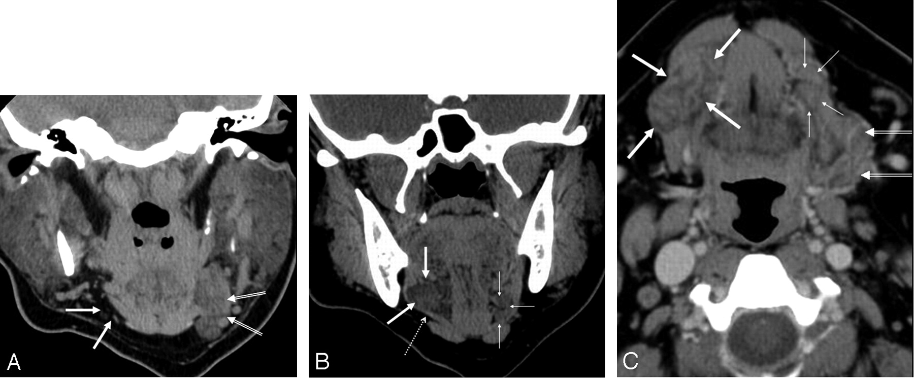
A 35-year-old woman who had facial pressure and rhinorrhea was referred by her internist for a CT scan of the paranasal sinuses to evaluate possible recurrent sinusitis. Apart from extensive mucosal disease, the CT also demonstrated incidental absence of the right submandibular gland, with a “mass” in the right sublingual space in the expected location of the right sublingual gland (Fig 1A, -B). There were no calculi or other lesions, and the mass was above the mylohyoid muscle. The possibility of hypertrophy of the ipsilateral sublingual gland was considered. Other differential diagnoses included an inflamed sublingual gland, postinflammatory enlargement, or a benign minor salivary gland tumor such as a benign mixed tumor. Squamous cell carcinoma of the floor of the mouth was also considered but was thought unlikely because of the low attenuation of the lesion.

Fig 1.
  • Download figure
  • Open in new tab
  • Download powerpoint
Fig 1.

A, Coronal noncontrast maxillofacial CT image reveals absence of the right submandibular gland (broad arrows). The contralateral submandibular gland is visualized in the left submandibular space (double arrows).

B, Coronal noncontrast maxillofacial CT image shows a well-defined low-attenuation mass (broad arrows) in the right sublingual space located lateral to the genioglossus-geniohyoid complex and bowing the mylohyoid inferiorly (dotted arrow). The normal left sublingual gland is visualized in its expected location (thin arrows).

C, Axial contrast-enhanced CT image demonstrates a low-attenuation mildly enhancing mass in the right sublingual region (broad arrows). The contralateral submandibular gland (double arrows) shows attenuation characteristics similar to those of the mass. The normal left sublingual space is also demonstrated (thin arrows).

The patient was referred to an otorhinolaryngologist for further assessment. Her history was unremarkable except for very occasional dysphagia. Clinical examination of the oral cavity and oropharynx did not reveal any palpable masses. There was no palpable neck adenopathy, and indirect laryngoscopy showed a normal larynx with mobile cords and a clear base of the tongue.

She underwent a contrast-enhanced neck CT, which revealed that the mass enhanced, with the degree of enhancement similar to that of the contralateral submandibular gland (Fig 1C). The margins of the mass were well defined, and there was no evidence of invasion of the adjacent soft tissue or inner cortex of the mandible.

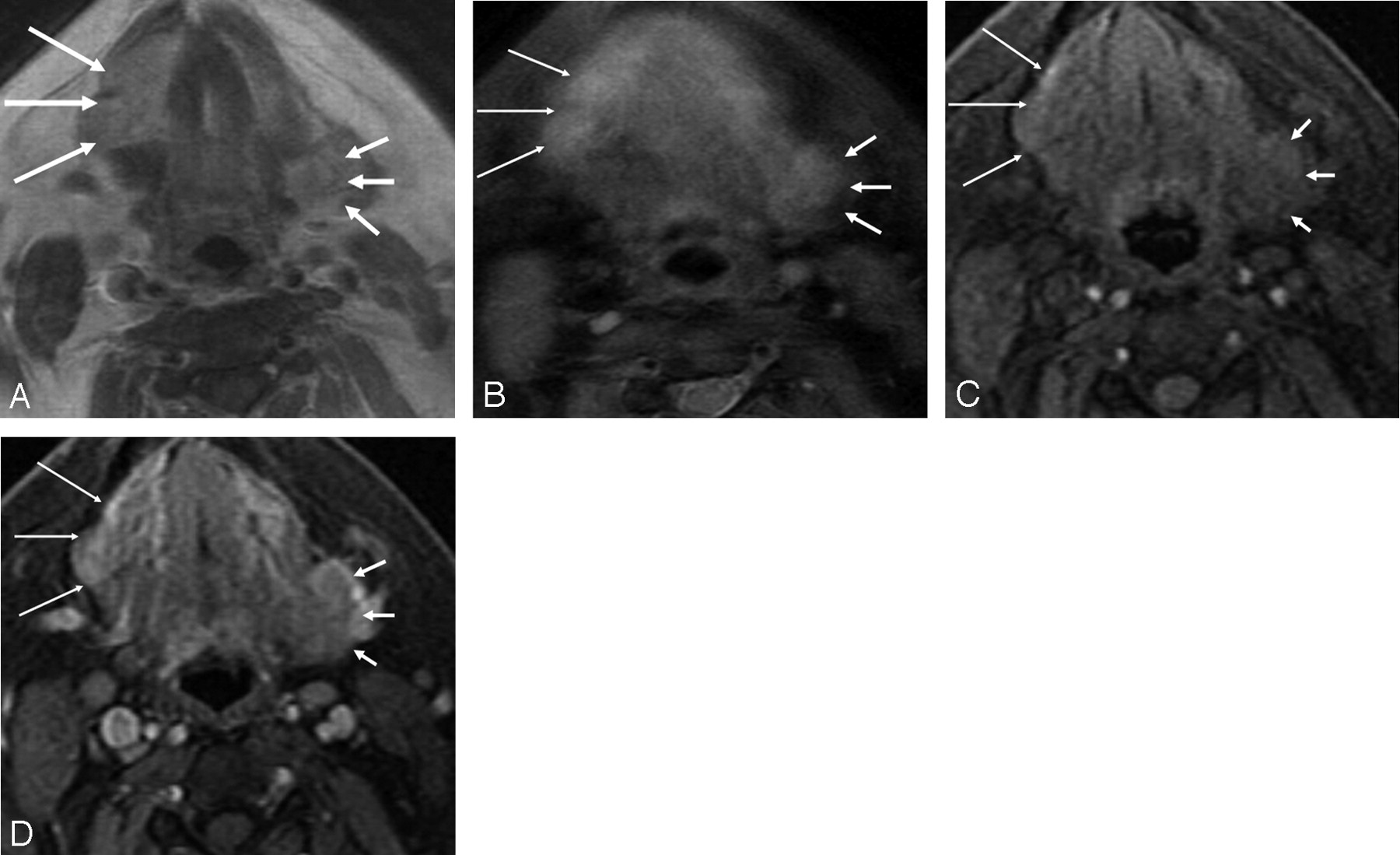
Contrast-enhanced MR imaging was performed for further characterization. The study demonstrated that the sublingual mass had T1-weighted (Fig 2A) and T2-weighted (Fig 2B) signal intensity characteristics similar to those of the contralateral submandibular gland. The dynamic contrast-enhanced MR images showed that the enhancement of the mass was similar to that of the contralateral sublingual and submandibular glands (Fig 2C, -D). On the basis of these findings, the imaging results were thought to be indicative of asymmetric sublingual gland hypertrophy.

Fig 2.
  • Download figure
  • Open in new tab
  • Download powerpoint
Fig 2.

The right sublingual mass (long arrows) reveals similar signal intensity similar to that of the contralateral submandibular gland (short arrows) on axial spin-echo nonenhanced T1-weighted (A) and fat-saturated T2-weighted (B) images. Dynamic contrast-enhanced fat-saturated gradient-echo images (C and D) reveal the enhancement pattern of the mass (long arrows) to be similar to that of the left submandibular gland (short arrows).

Discussion

The salivary glands arise as buds from the epithelial lining of the oral cavity. These primordial buds grow and extend into the underlying mesenchymal tissue as a solid core of cells. The developing tissue then undergoes extensive branching and eventually develops lumina. The glands are divided into lobules by the surrounding mesenchyme, which also envelopes them to form a capsule. The parotid glands appear during the fourth week of gestation in the angle between the maxillary process and the mandibular arch. The submandibular glands appear in the sixth week, and the sublingual glands arise during the ninth week between the tongue and the mandibular arch.5–7

Congenital absence of the salivary glands is infrequent and more often involves multiple major salivary glands.1,2 Isolated unilateral major salivary gland aplasia is a rare entity with only a few cases reported in the literature to date.4,8–10 The aplasia is likely due to arrest in organogenesis, but the exact etiology is unknown.9 The unique feature of our patient is the demonstrable ipsilateral sublingual gland hypertrophy associated with the submandibular gland aplasia, which, to the best of our knowledge, has not been reported previously.4 The enlargement of the sublingual gland is likely due to compensatory hypertrophy.

Salivary gland aplasia may be associated with first branchial arch defects in the Treacher-Collins syndrome (mandibulofacial dysostosis) or orbital abnormalities like lacrimal hypoplasia, canalicular atresia, and absence of the lacrimal puncta.1,2,11,12 Absence of salivary glands may also be seen as part of the lacrimoauriculodentodigital syndrome, characterized by hypoplasia, aplasia or atresia of the lacrimal system, deafness and ear malformations, and dental and digital anomalies.13 In our patient, there were no such associated anomalies.

The clinical picture of patients with salivary gland aplasia is variable. They may be asymptomatic or may complain of dry mouth, have difficulty in chewing and swallowing, and have an increased incidence of dental caries, which is thought to be due to reduction of the protective effect of saliva within the oral cavity.1,6,14 Our patient presented with symptoms of recurrent sinusitis, and a detailed history revealed only occasional dysphagia, with no other symptoms related to the gland aplasia. Her relative lack of salivary gland–related symptoms was presumably due to the unilateral nature of the aplasia, with the other major salivary glands being present, and compensatory hypertrophy of the ipsilateral sublingual gland.

Salivary gland aplasia can be diagnosed with a variety of imaging techniques, which include CT, MR imaging, sonography, sialography, or nuclear medicine (technetium T99m-pertechnetate) studies.1,2,4,6,10,15–18 In our patient, we were able to confirm the diagnosis of compensatory sublingual gland hypertrophy by using a combination of CT and MR imaging, including dynamic contrast-enhanced sequences. The imaging findings confirmed the presence of a hypertrophied sublingual gland in association with the absence of the ipsilateral submandibular gland and excluded a floor of the mouth tumor. Radiologists should be familiar with this entity and its imaging findings to prevent unnecessary biopsy of this benign condition.

References

  1. ↵
    McDonald FG, Mantas J, McEwen CG, et al. Salivary gland aplasia: an ectodermal disorder? J Oral Pathol 1986;15:115–17
    CrossRefPubMed
  2. ↵
    Higashino H, Horii T, Ohkusa Y, et al. Congenital absence of lacrimal puncta and of all major salivary glands: case report and literature review. Clin Pediatr (Phila) 1987;26:366–68
    Abstract/FREE Full Text
  3. ↵
    Whyte AM, Hayward MW. Agenesis of the salivary glands: a report of two cases. Br J Radiol 1989;62:1023–26
    Abstract/FREE Full Text
  4. ↵
    Yilmaz MD, Yucel A, Derekoy S, et al. Unilateral aplasia of the submandibular gland. Eur Arch Otorhinolaryngol 2002;259:554–56
    PubMed
  5. ↵
    Collins P. Embryology and development. In: Williams PL, ed. Gray’s Anatomy: The Anatomical Basis of Medicine and Surgery. 38th ed. New York: Churchill Livingstone;1995 :175
  6. ↵
    Wiesenfeld D, Ferguson MM, Allan CJ, et al. Bilateral parotid gland aplasia. Br J Oral Surg 1983;21:175–78
    CrossRefPubMed
  7. ↵
    Johns ME. The salivary glands: anatomy and embryology. Otolaryngol Clin North Am 1977;10:261–71
    PubMed
  8. ↵
    Abdel-Dayem HM. Congenital absence of submaxillary gland detected on 99m Tc-pertechnetate thyroid imaging. Clin Nucl Med 1978;3:442
    CrossRefPubMed
  9. ↵
    Kubo S, Abe K, Ureshino T, et al. Aplasia of the submandibular gland: a case report. J Craniomaxillofac Surg 1990;18:119–21
    PubMed
  10. ↵
    Garcia-Consuegra L, Gutierrez LJ, Castro JM, et al. Congenital unilateral absence of the submandibular gland. J Oral Maxillofac Surg 1999;57:344–46
    CrossRefPubMed
  11. ↵
    Caccamise WC, Townes PL. Congenital absence of the lacrimal puncta associated with alacrima and aptyalism. Am J Ophthalmol 1980;89:62–65
    PubMed
  12. ↵
    Cohen M. Mandibulofacial dysostosis. In: Bergsma D, ed. Birth Defect. Baltimore: Williams and Wilkins;1974 :465–74
  13. ↵
    Hollister DW, Klein SH, De Jager HJ, et al. The lacrimo-auriculo-dento-digital syndrome. J Pediatr 1973;83:438–44
    CrossRefPubMed
  14. ↵
    O’Malley AM, Macleod RI, Welbury RR. Congenital aplasia of major salivary glands in a 4-year-old child. Int J Paediatr Dent 1993;3:141–44
    PubMed
  15. ↵
    Smith NJ, Smith PB. Congenital absence of major salivary glands. Br Dent J 1977;142:259–60
    CrossRefPubMed
  16. Sucupira MS, Weinreb JW, Camargo EE, et al. Salivary gland imaging and radionuclide dacryocystography in agenesis of salivary glands. Arch Otolaryngol 1983;109:197–98
    CrossRefPubMed
  17. Myers MA, Youngberg RA, Bauman JM. Congenital absence of the major salivary glands and impaired lacrimal secretion in a child: case report. J Am Dent Assoc 1994;125:210–12
    Abstract
  18. ↵
    Yoshiura K, Yamada M, Yamada N. Demonstration of congenital absence of the major salivary glands by computed tomography. Dentomaxillofac Radiol 1990;2:77–78
    PubMed
  • Received October 12, 2005.
  • Accepted after revision November 9, 2005.
  • Copyright © American Society of Neuroradiology
View Abstract
PreviousNext
Back to top

In this issue

American Journal of Neuroradiology: 27 (10)
American Journal of Neuroradiology
Vol. 27, Issue 10
November 2006
  • Table of Contents
  • Index by author
Advertisement
Print
Download PDF
Email Article

Thank you for your interest in spreading the word on American Journal of Neuroradiology.

NOTE: We only request your email address so that the person you are recommending the page to knows that you wanted them to see it, and that it is not junk mail. We do not capture any email address.

Enter multiple addresses on separate lines or separate them with commas.
Unilateral Submandibular Gland Aplasia Associated with Ipsilateral Sublingual Gland Hypertrophy
(Your Name) has sent you a message from American Journal of Neuroradiology
(Your Name) thought you would like to see the American Journal of Neuroradiology web site.
CAPTCHA
This question is for testing whether or not you are a human visitor and to prevent automated spam submissions.
Cite this article
A. Srinivasan, J.S. Moyer, S.K. Mukherji
Unilateral Submandibular Gland Aplasia Associated with Ipsilateral Sublingual Gland Hypertrophy
American Journal of Neuroradiology Nov 2006, 27 (10) 2214-2216;

Citation Manager Formats

  • BibTeX
  • Bookends
  • EasyBib
  • EndNote (tagged)
  • EndNote 8 (xml)
  • Medlars
  • Mendeley
  • Papers
  • RefWorks Tagged
  • Ref Manager
  • RIS
  • Zotero
0 Responses
Respond to this article
Share
Bookmark this article
Unilateral Submandibular Gland Aplasia Associated with Ipsilateral Sublingual Gland Hypertrophy
A. Srinivasan, J.S. Moyer, S.K. Mukherji
American Journal of Neuroradiology Nov 2006, 27 (10) 2214-2216;
del.icio.us logo Twitter logo Facebook logo Mendeley logo
  • Tweet Widget
  • Facebook Like
  • Google Plus One

Jump to section

  • Article
    • Abstract
    • Case Report
    • Discussion
    • References
  • Figures & Data
  • Info & Metrics
  • Responses
  • References
  • PDF

Related Articles

  • No related articles found.
  • PubMed
  • Google Scholar

Cited By...

  • Unilateral submandibular gland aplasia with contralateral hypertrophy: an unusual neck mass
  • CT and MR findings of bilateral submandibular gland aplasia associated with hypertrophied symmetrical sublingual glands herniated through mylohyoid defects
  • Bilateral submandibular gland aplasia with clinico-radiological mass due to prolapsing sublingual salivary tissue through mylohyoid boutonniere: a case report and review
  • Crossref
  • Google Scholar

This article has not yet been cited by articles in journals that are participating in Crossref Cited-by Linking.

More in this TOC Section

  • NI-RADS for HEAD&NECK Cancer Recurrence
  • WHO Classification Update: Nasal&Skull Base Tumors
  • Peritumoral Signal in Vestibular Schwannomas
Show more HEAD & NECK

Similar Articles

Advertisement

Indexed Content

  • Current Issue
  • Accepted Manuscripts
  • Article Preview
  • Past Issues
  • Editorials
  • Editors Choice
  • Fellow Journal Club
  • Letters to the Editor

Cases

  • Case Collection
  • Archive - Case of the Week
  • Archive - Case of the Month
  • Archive - Classic Case

Special Collections

  • Special Collections

Resources

  • News and Updates
  • Turn around Times
  • Submit a Manuscript
  • Author Policies
  • Manuscript Submission Guidelines
  • Evidence-Based Medicine Level Guide
  • Publishing Checklists
  • Graphical Abstract Preparation
  • Imaging Protocol Submission
  • Submit a Case
  • Become a Reviewer/Academy of Reviewers
  • Get Peer Review Credit from Publons

Multimedia

  • AJNR Podcast
  • AJNR SCANtastic
  • Video Articles

About Us

  • About AJNR
  • Editorial Board
  • Not an AJNR Subscriber? Join Now
  • Alerts
  • Feedback
  • Advertise with us
  • Librarian Resources
  • Permissions
  • Terms and Conditions

American Society of Neuroradiology

  • Not an ASNR Member? Join Now

© 2025 by the American Society of Neuroradiology All rights, including for text and data mining, AI training, and similar technologies, are reserved.
Print ISSN: 0195-6108 Online ISSN: 1936-959X

Powered by HighWire