
Article Figures & Data
Figures
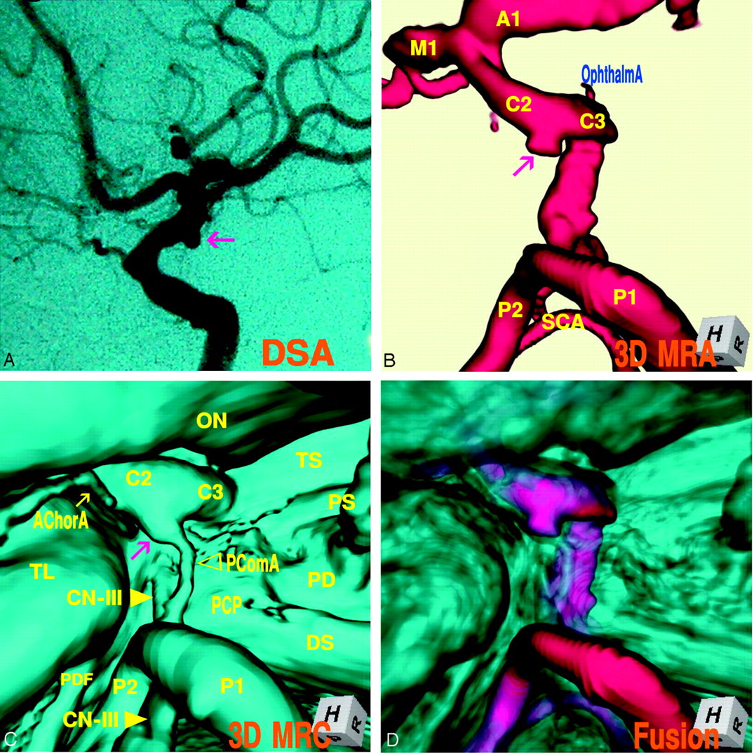
- Fig 1.
Case 1, Left infundibular dilation at the junction of the internal carotid artery–posterior communicating artery (adult type) in a 75-year-old woman.
A, Digital subtraction angiogram shows a round bulging (arrow) at the supraclinoid internal carotid artery.
B, 3D MR angiogram shows the trapezoid protrusion (arrow) at the supraclinoid internal carotid artery. A1, the first segment of the anterior cerebral artery; M1, the first segment of the middle cerebral artery; C2, the second segment of the internal carotid artery; C3, the third segment of the internal carotid artery; P1, the first segment of the posterior cerebral artery; P2, the second segment of the posterior cerebral artery; SCA, superior cerebellar artery.
C, 3D MR cisternogram, coordinated projection as to the 3D MR angiogram in B, shows an infundibular dilation (arrow) at the junction of the internal carotid artery-posterior communicating artery. A small posterior communicating artery arises at the apex. Intra- and juxtacisternal anatomic elements surrounding an infundibular dilation are visualized. ON, optic nerve; TS, tuberculum sellae; PS, pituitary stalk; PD, pituitary diaphragm; DS, dorsum sellae; PCP, posterior clinoid process; PComA, posterior communicating artery (▹); CN-III, oculomotor nerve (yellow arrow); PDF, petroclinoid dural fold.
D, Fusion image of the 3D MR angiography/cisternography shows the relationship of the bulging finding detected by the 3D MR angiography and its outer wall configuration depicted by the 3D MR cisternography.
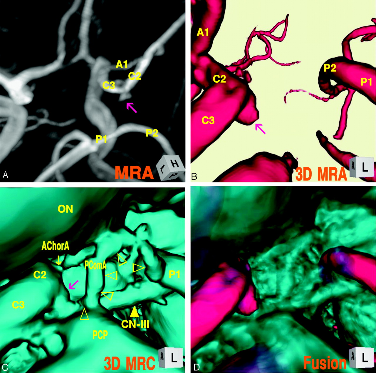
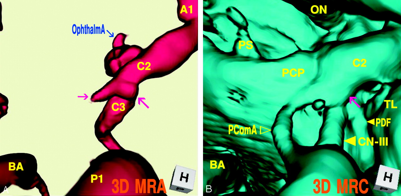
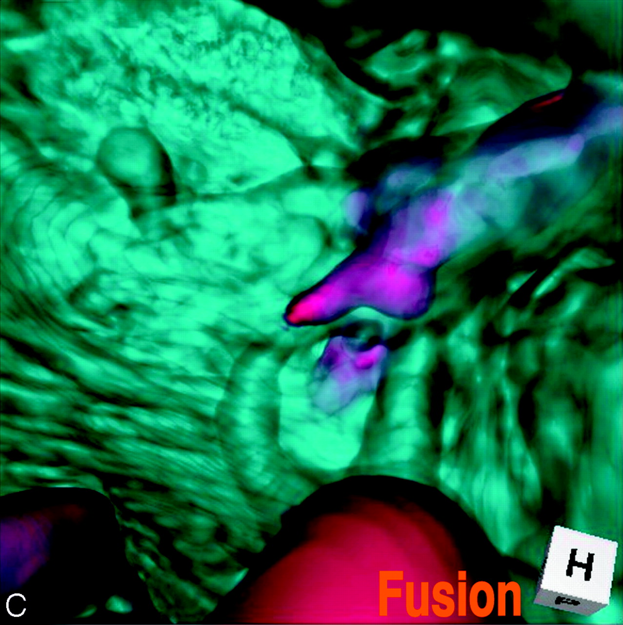
- Fig 2.
Case 2, Right infundibular dilation at the junction of the internal carotid artery–posterior communicating artery (fetal-type), in a 36-year-old woman.
A, Maximum intensity projection image of MR angiogram shows a trapezoidal bulging (arrow) at the posterior portion of the supraclinoid internal carotid artery.
B, 3D MR angiogram shows an aneurysm-like protrusion (arrow).
C, 3D MR cisternogram, coordinated projection as to the 3D MR angiogram B, shows an infundibular widening (large arrow) with a large posterior communicating artery (▹), bended at the posterior clinoid process and run tortuously to the posterior cerebral artery. ON, optic nerve; AChorA, anterior choroidal artery (small arrow); PComA (▹).
D, Fusion image of the 3D MR angiography/cisternography shows not an aneurysm but an infundibular dilation.
- Fig 3.
Case 3, Left infundibular dilation at the junction of the internal carotid artery–anterior choroidal artery, in a 69-year-old-woman.
A, 3D MR angiogram shows an aneurysm-like trapezoid bulging (arrow). C1, the first segment of the internal carotid artery; BA, basilar artery.
B, 3D MR cisternogram, coordinated projection as to the 3D MR angiogram in A, shows an infundibular dilation (large arrow) at the junction of the anterior choroidal artery (small arrow). PComA (▹); CN-III ([GRAPHIC]); TL, temporal lobe.
C, Fusion image of the 3D MR angiography/cisternography shows an infundibular dilation at the junction of an anterior choroidal artery.
- Fig 4.
Case 4, Right infundibular dilation at the junction of the internal carotid artery–posterior communicating artery (fetal type), misdiagnosed as an aneurysm and treated surgically, in a 44-year-old woman.
A, 3D MR angiogram shows an aneurysm-like protrusion (large arrow) and a conical bleblike elongation (small arrow).
B, 3D MR cisternogram, coordinated projection as to the 3D MR angiogram in A, shows an infundibular dilation (arrow) with a large posterior communicating artery. PDF (small arrow); CN-III (large arrow); PComA (▹).
C, Fusion image of the 3D MR angiography/cisternography shows the anatomic relationship of an aneurysm-like complex to a large posterior communicating artery, and indicating not an aneurysm but an infundibular dilation.
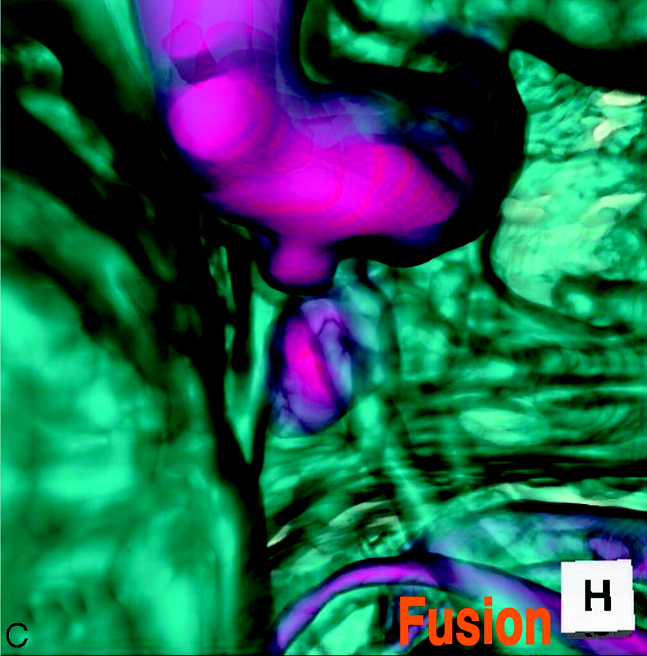
- Fig 5.
Case 5, Left internal carotid artery–posterior communicating artery aneurysm, in a 66-year-old woman.
A, 3D MR angiogram shows an aneurysm with an irregular dome (arrow).
B, 3D MR cisternogram, coordinated projection as to the 3D MR angiogram in A, shows an aneurysm (arrow) and a large posterior communicating artery. AChorA, anterior choroidal artery; CN-III (arrow).
C, Fusion image of the 3D MR angiography/cisternography shows the aneurysm complex in relation to its perianeurysmal environment.


In this issue


Jump to section
Related Articles
Cited By...
- Usefulness of high-resolution three-dimensional proton density-weighted turbo spin-echo MRI in distinguishing a junctional dilatation from an intracranial aneurysm of the posterior communicating artery: a pilot study
- Aneurysm outflow angle at MRA as discriminant for accurate diagnosis and differentiation between small sidewall cerebral aneurysms and infundibula
- Infundibular dilations of the posterior communicating arteries: pathogenesis, anatomical variants, aneurysm formation, and subarachnoid hemorrhage
- Identification of the Distal Dural Ring with Use of Fusion Images with 3D-MR Cisternography and MR Angiography: Application to Paraclinoid Aneurysms