Advertisement

Main menu

  • Home
  • Content
    • Current Issue
    • Accepted Manuscripts
    • Article Preview
    • Past Issue Archive
    • AJNR Case Collection
    • Case of the Week Archive
    • Classic Case Archive
    • Case of the Month Archive
  • Special Collections
    • Spinal CSF Leak Articles (Jan 2020-June 2024)
    • 2024 AJNR Journal Awards
    • Most Impactful AJNR Articles
  • Multimedia
    • AJNR Podcast
    • AJNR Scantastics
    • Video Articles
  • For Authors
    • Submit a Manuscript
    • Author Policies
    • Fast publishing of Accepted Manuscripts
    • Graphical Abstract Preparation
    • Manuscript Submission Guidelines
    • Imaging Protocol Submission
    • Submit a Case for the Case Collection
  • About Us
    • About AJNR
    • Editorial Board
  • More
    • Become a Reviewer/Academy of Reviewers
    • Subscribers
    • Permissions
    • Alerts
    • Feedback
    • Advertisers
    • ASNR Home
  • Other Publications
    • ajnr

User menu

  • Alerts
  • Log in

Search

  • Advanced search
American Journal of Neuroradiology

American Journal of Neuroradiology

ASHNR American Society of Functional Neuroradiology ASHNR American Society of Pediatric Neuroradiology ASSR
  • Alerts
  • Log in

Advanced Search

  • Home
  • Content
    • Current Issue
    • Accepted Manuscripts
    • Article Preview
    • Past Issue Archive
    • AJNR Case Collection
    • Case of the Week Archive
    • Classic Case Archive
    • Case of the Month Archive
  • Special Collections
    • Spinal CSF Leak Articles (Jan 2020-June 2024)
    • 2024 AJNR Journal Awards
    • Most Impactful AJNR Articles
  • Multimedia
    • AJNR Podcast
    • AJNR Scantastics
    • Video Articles
  • For Authors
    • Submit a Manuscript
    • Author Policies
    • Fast publishing of Accepted Manuscripts
    • Graphical Abstract Preparation
    • Manuscript Submission Guidelines
    • Imaging Protocol Submission
    • Submit a Case for the Case Collection
  • About Us
    • About AJNR
    • Editorial Board
  • More
    • Become a Reviewer/Academy of Reviewers
    • Subscribers
    • Permissions
    • Alerts
    • Feedback
    • Advertisers
    • ASNR Home
  • Follow AJNR on Twitter
  • Visit AJNR on Facebook
  • Follow AJNR on Instagram
  • Join AJNR on LinkedIn
  • RSS Feeds

Welcome to the new AJNR, Updated Hall of Fame, and more. Read the full announcements.


AJNR is seeking candidates for the position of Associate Section Editor, AJNR Case Collection. Read the full announcement.


Research ArticleSPINE

The Value of T2 Relaxation Times to Characterize Lumbar Intervertebral Disks: Preliminary Results

J. Perry, V. Haughton, P.A. Anderson, Y. Wu, J. Fine and C. Mistretta
American Journal of Neuroradiology February 2006, 27 (2) 337-342;
J. Perry
  • Find this author on Google Scholar
  • Find this author on PubMed
  • Search for this author on this site
V. Haughton
  • Find this author on Google Scholar
  • Find this author on PubMed
  • Search for this author on this site
P.A. Anderson
  • Find this author on Google Scholar
  • Find this author on PubMed
  • Search for this author on this site
Y. Wu
  • Find this author on Google Scholar
  • Find this author on PubMed
  • Search for this author on this site
J. Fine
  • Find this author on Google Scholar
  • Find this author on PubMed
  • Search for this author on this site
C. Mistretta
  • Find this author on Google Scholar
  • Find this author on PubMed
  • Search for this author on this site
  • Article
  • Figures & Data
  • Info & Metrics
  • Responses
  • References
  • PDF
Loading

Article Figures & Data

Figures

  • Tables
  • Fig 1.
    • Download figure
    • Open in new tab
    • Download powerpoint
    Fig 1.

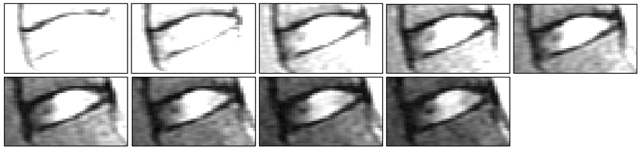
    Images of successive echoes in the FSE echo train of an L4/5 disk classified as stage II. The images progress from the first echo on the upper left to the ninth echo in the lower right.

  • Fig 2.
    • Download figure
    • Open in new tab
    • Download powerpoint
    Fig 2.

    3D contour plot of the disk shown in Fig 1. The anterior portion of the disk is to the reader’s left and superior portion toward the top of the page. The inclination of the disk in the 3D plot is due to the angulation of the intervertebral disk from the axial plane. Notice the intranuclear cleft is clearly distinguished from the more amorphous regions of the nucleus pulposus because of its lower T2 values.

  • Fig 3.
    • Download figure
    • Open in new tab
    • Download powerpoint
    Fig 3.

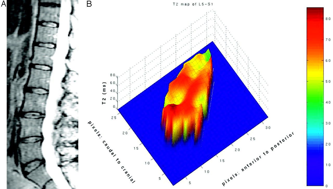
    Sagittal MR image (ninth echo; A) and contour plot (B) of the L5/S1 disk in volunteer 3. This volunteer had a less-conspicuous intranuclear cleft at L5-S1.

  • Fig 4.
    • Download figure
    • Open in new tab
    • Download powerpoint
    Fig 4.

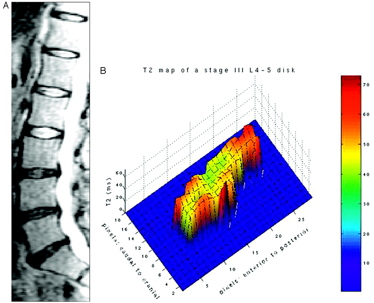
    Sagittal MR image (9th echo; A) and contour plot (B) of an L4–5 stage III intervertebral disk. The intranuclear cleft region is not distinguished in the image or the plot.
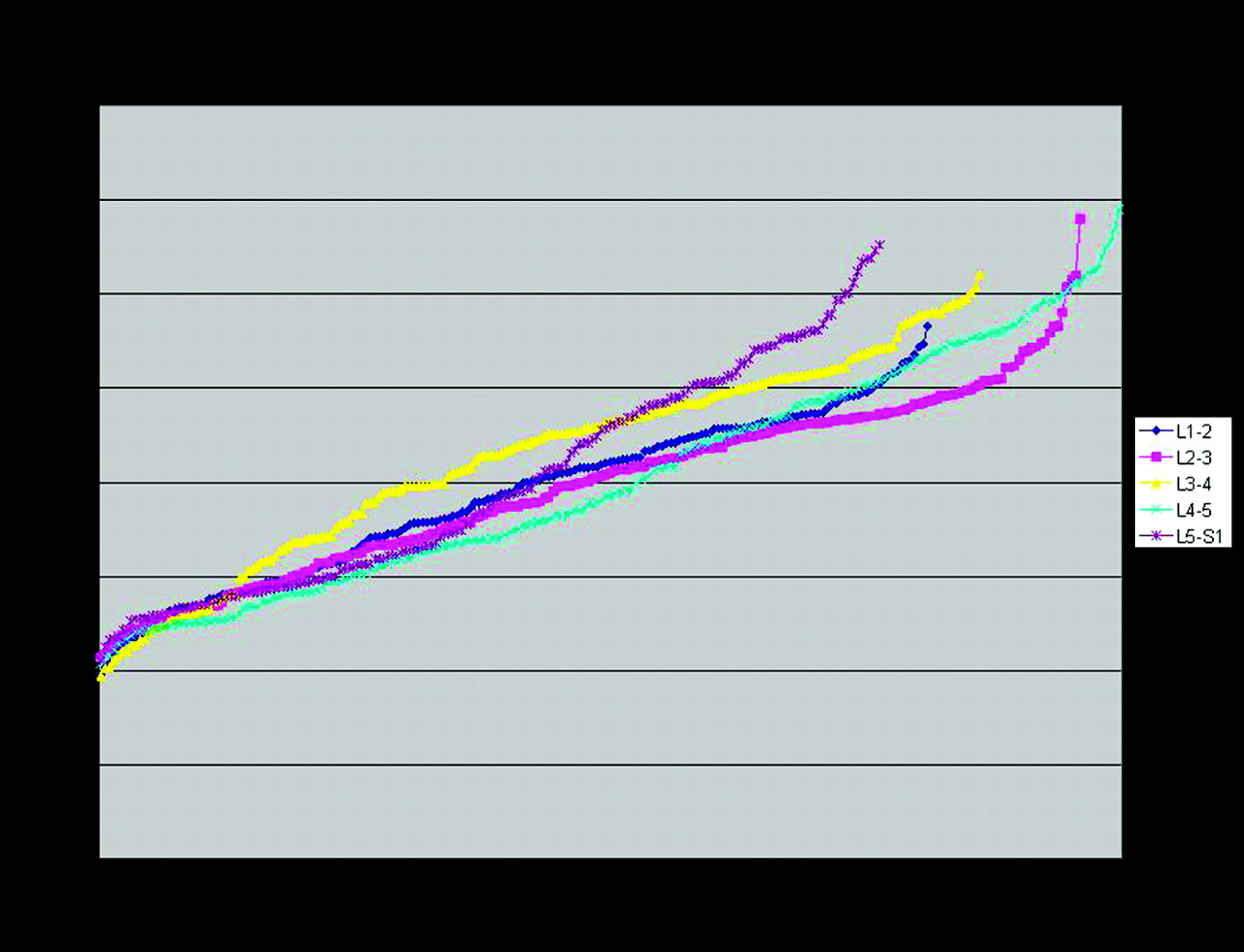
  • Fig 5.
    • Download figure
    • Open in new tab
    • Download powerpoint
    Fig 5.

    Rank plot of T2 values in a volunteer with 5 stage II disks. The T2 values are plotted in order of increasing value. Note that the plots for each level tend to coincide.

  • Fig 6.
    • Download figure
    • Open in new tab
    • Download powerpoint
    Fig 6.

    Rank plots of T2 values of disks in volunteer 3, who had a less-conspicuous intranuclear cleft at L5/S1. The rank plots show greater discrepancies than they did in the other 2 volunteers.
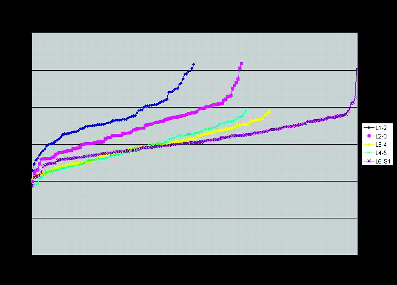
  • Fig 7.
    • Download figure
    • Open in new tab
    • Download powerpoint
    Fig 7.

    Rank plot of T2 values in a volunteer with stage II disks at L1–2 and L2–3 and stage III disks at L3–4, L4–5, and L5-S1. The stage II disks are clustered together, but the stage III disks are not.

Tables

  • Figures
  • Areas, means, percentiles, and slopes and intercepts for trendlines in a rank plot for T2 values in stage II and III disks in 5 volunteers

    ParameterDisk StageP value*
    IIIII,V
    No. of disks205
    Area (pixels)16165
    Mean T2 (ms)8453.0025
    SD of T2 (ms)228.014
    10th percentile (ms)55432.5E-05
    5th percentile (ms)6647.00026
    50th percentile (ms)8552.0032
    75th percentile (ms)10357.0035
    90th percentile (ms)11463.0041
    99th percentile (ms)13173.0033
    Slope of ranked T2 values−0.48−0.44.76
    Intercept of ranked T2 values12366.0055
    • * Differences in stage II and III disks.

PreviousNext
Back to top

In this issue

American Journal of Neuroradiology: 27 (2)
American Journal of Neuroradiology
Vol. 27, Issue 2
February, 2006
  • Table of Contents
  • Index by author
Advertisement
Print
Download PDF
Email Article

Thank you for your interest in spreading the word on American Journal of Neuroradiology.

NOTE: We only request your email address so that the person you are recommending the page to knows that you wanted them to see it, and that it is not junk mail. We do not capture any email address.

Enter multiple addresses on separate lines or separate them with commas.
The Value of T2 Relaxation Times to Characterize Lumbar Intervertebral Disks: Preliminary Results
(Your Name) has sent you a message from American Journal of Neuroradiology
(Your Name) thought you would like to see the American Journal of Neuroradiology web site.
CAPTCHA
This question is for testing whether or not you are a human visitor and to prevent automated spam submissions.
Cite this article
J. Perry, V. Haughton, P.A. Anderson, Y. Wu, J. Fine, C. Mistretta
The Value of T2 Relaxation Times to Characterize Lumbar Intervertebral Disks: Preliminary Results
American Journal of Neuroradiology Feb 2006, 27 (2) 337-342;

Citation Manager Formats

  • BibTeX
  • Bookends
  • EasyBib
  • EndNote (tagged)
  • EndNote 8 (xml)
  • Medlars
  • Mendeley
  • Papers
  • RefWorks Tagged
  • Ref Manager
  • RIS
  • Zotero
0 Responses
Respond to this article
Share
Bookmark this article
The Value of T2 Relaxation Times to Characterize Lumbar Intervertebral Disks: Preliminary Results
J. Perry, V. Haughton, P.A. Anderson, Y. Wu, J. Fine, C. Mistretta
American Journal of Neuroradiology Feb 2006, 27 (2) 337-342;
del.icio.us logo Twitter logo Facebook logo Mendeley logo
  • Tweet Widget
  • Facebook Like
  • Google Plus One

Jump to section

  • Article
    • Abstract
    • Methods
    • Results
    • Discussion
    • Conclusions
    • Acknowledgments
    • References
  • Figures & Data
  • Info & Metrics
  • Responses
  • References
  • PDF

Related Articles

  • No related articles found.
  • PubMed
  • Google Scholar

Cited By...

  • Clinical Utility of a Novel Ultrafast T2-Weighted Sequence for Spine Imaging
  • MR Imaging Assessment of Lumbar Intervertebral Disk Degeneration and Age-Related Changes: Apparent Diffusion Coefficient versus T2 Quantitation
  • Association between Annular Tears and Disk Degeneration: A Longitudinal Study
  • Crossref
  • Google Scholar

This article has not yet been cited by articles in journals that are participating in Crossref Cited-by Linking.

More in this TOC Section

  • Bern Score Validity for SIH
  • MP2RAGE 7T in MS Lesions of the Cervical Spine
  • Deep Learning for STIR Spine MRI Quality
Show more Spine

Similar Articles

Advertisement

Indexed Content

  • Current Issue
  • Accepted Manuscripts
  • Article Preview
  • Past Issues
  • Editorials
  • Editors Choice
  • Fellow Journal Club
  • Letters to the Editor

Cases

  • Case Collection
  • Archive - Case of the Week
  • Archive - Case of the Month
  • Archive - Classic Case

Special Collections

  • Special Collections

Resources

  • News and Updates
  • Turn around Times
  • Submit a Manuscript
  • Author Policies
  • Manuscript Submission Guidelines
  • Evidence-Based Medicine Level Guide
  • Publishing Checklists
  • Graphical Abstract Preparation
  • Imaging Protocol Submission
  • Submit a Case
  • Become a Reviewer/Academy of Reviewers
  • Get Peer Review Credit from Publons

Multimedia

  • AJNR Podcast
  • AJNR SCANtastic
  • Video Articles

About Us

  • About AJNR
  • Editorial Board
  • Not an AJNR Subscriber? Join Now
  • Alerts
  • Feedback
  • Advertise with us
  • Librarian Resources
  • Permissions
  • Terms and Conditions

American Society of Neuroradiology

  • Not an ASNR Member? Join Now

© 2025 by the American Society of Neuroradiology All rights, including for text and data mining, AI training, and similar technologies, are reserved.
Print ISSN: 0195-6108 Online ISSN: 1936-959X

Powered by HighWire