Advertisement

Main menu

  • Home
  • Content
    • Current Issue
    • Accepted Manuscripts
    • Article Preview
    • Past Issue Archive
    • AJNR Case Collection
    • Case of the Week Archive
    • Classic Case Archive
    • Case of the Month Archive
  • Special Collections
    • Spinal CSF Leak Articles (Jan 2020-June 2024)
    • 2024 AJNR Journal Awards
    • Most Impactful AJNR Articles
  • Multimedia
    • AJNR Podcast
    • AJNR Scantastics
    • Video Articles
  • For Authors
    • Submit a Manuscript
    • Author Policies
    • Fast publishing of Accepted Manuscripts
    • Graphical Abstract Preparation
    • Manuscript Submission Guidelines
    • Imaging Protocol Submission
    • Submit a Case for the Case Collection
  • About Us
    • About AJNR
    • Editorial Board
  • More
    • Become a Reviewer/Academy of Reviewers
    • Subscribers
    • Permissions
    • Alerts
    • Feedback
    • Advertisers
    • ASNR Home
  • Other Publications
    • ajnr

User menu

  • Alerts
  • Log in

Search

  • Advanced search
American Journal of Neuroradiology

American Journal of Neuroradiology

ASHNR American Society of Functional Neuroradiology ASHNR American Society of Pediatric Neuroradiology ASSR
  • Alerts
  • Log in

Advanced Search

  • Home
  • Content
    • Current Issue
    • Accepted Manuscripts
    • Article Preview
    • Past Issue Archive
    • AJNR Case Collection
    • Case of the Week Archive
    • Classic Case Archive
    • Case of the Month Archive
  • Special Collections
    • Spinal CSF Leak Articles (Jan 2020-June 2024)
    • 2024 AJNR Journal Awards
    • Most Impactful AJNR Articles
  • Multimedia
    • AJNR Podcast
    • AJNR Scantastics
    • Video Articles
  • For Authors
    • Submit a Manuscript
    • Author Policies
    • Fast publishing of Accepted Manuscripts
    • Graphical Abstract Preparation
    • Manuscript Submission Guidelines
    • Imaging Protocol Submission
    • Submit a Case for the Case Collection
  • About Us
    • About AJNR
    • Editorial Board
  • More
    • Become a Reviewer/Academy of Reviewers
    • Subscribers
    • Permissions
    • Alerts
    • Feedback
    • Advertisers
    • ASNR Home
  • Follow AJNR on Twitter
  • Visit AJNR on Facebook
  • Follow AJNR on Instagram
  • Join AJNR on LinkedIn
  • RSS Feeds

Welcome to the new AJNR, Updated Hall of Fame, and more. Read the full announcements.


AJNR is seeking candidates for the position of Associate Section Editor, AJNR Case Collection. Read the full announcement.


OtherSPINE

Recording CSF Pressure with a Transducer-Tipped Wire in an Animal Model of Chiari I

A. Turk, B.J. Iskandar, V. Haughton and D. Consigny
American Journal of Neuroradiology February 2006, 27 (2) 354-355;
A. Turk
  • Find this author on Google Scholar
  • Find this author on PubMed
  • Search for this author on this site
B.J. Iskandar
  • Find this author on Google Scholar
  • Find this author on PubMed
  • Search for this author on this site
V. Haughton
  • Find this author on Google Scholar
  • Find this author on PubMed
  • Search for this author on this site
D. Consigny
  • Find this author on Google Scholar
  • Find this author on PubMed
  • Search for this author on this site
  • Article
  • Figures & Data
  • Info & Metrics
  • Responses
  • References
  • PDF
Loading

Abstract

SUMMARY: In dogs, a wire with a pressure-sensitive transducer was inserted percutaneously into the subarachnoid space and manipulated under fluoroscopic monitoring in the posterior fossa or upper cervical spinal canal. Pressure recordings from the wire showed fluctuations in pressure corresponding to the cardiac cycle. When a balloon was distended in the foramen magnum, maximum and minimum pressures increased. Continuous monitoring of CSF pressure remote from the site of cannulation was feasible with a wire-based pressure transducer.

Apressure-tipped wire has been used to record the pressure in arteries and veins1,2 and within aneurysms.3 Fluctuations in pressure through the cardiac cycle are effectively demonstrated with this wire. We designed a test of this transducer-tipped wire to measure CSF pressures. We used an animal model in which the foramen magnum was partially obstructed, to simulate a Chiari I malformation.

Methods

Four adult mongrel dogs weighing 20–40 kg were used for the study. The dogs were anesthetized with ketamine and intubated in the dog laboratory. Oxygen levels and heart rate were monitored by means of an oximeter and electrocardiography (EKG) leads applied to the chest. The subarachnoid space was cannulated through the L3/4 neural foramen with an 18-gauge spinal needle as in a previous study.4 By using Seldinger technique the spinal needle was exchanged for a 6F sheath.

A microcatheter (Renegade Hi Flo; Target Therapeutics/Boston Scientific Industries, Fremont, Calif) with a Transcend EX guidewire (Target Therapeutics) was inserted through the sheath and manipulated to the level of foramen magnum under fluoroscopic monitoring. The guidewire was removed, and a 0.014-inch diameter wire equipped with a pressure sensor (Radi Medical Systems, Gothenburg, Sweden) was passed through the catheter. The wire was advanced under fluoroscopic monitoring until the pressure transducer mounted at the junction of the wire tip and core wire was positioned in the subarachnoid space at the level of cisterna magna or upper cervical spinal canal (Fig 1). A balloon catheter (Commodore 3.5 mm × 10 mm; Cordis Corporation, Miami Lakes, Fla) with the Transcend EX guidewire was then advanced through the sheath into the subarachnoid space. It was advanced, under fluoroscopic monitoring to a position in the foramen magnum (Fig 1). The pressure wire was connected to a recorder (Propaq Encore202, PROTOCOL Systems, Beaverton, Ore). The balloon was distended 3–5 times intermittently over a 10-minute time period by means of a hand injection of a dilute aqueous solution of water-soluble iodinated contrast medium. EKG was monitored for changes in cardiac rate. Before and after each balloon distension a paper trace of the pressure was obtained and maximal, minimal, and mean pressures were recorded from the values printed on the paper tracing.

Fig 1.
  • Download figure
  • Open in new tab
  • Download powerpoint
Fig 1.

Placement of the balloon catheter and Radi wire in the subarachnoid space in one dog.

The institution’s animal research supervisory committee approved the study.

Results

All 4 animals tolerated the procedures well. Heart rate varied little except during the positioning of the wire in the foramen magnum. In each animal, the wire was successfully manipulated into the inferior posterior fossa or the upper cervical spinal canal. Some repositioning of the wire was required to obtain a continuous pressure trace in some animals, but, once the wire was adequately positioned, the continuous pressure monitoring was feasible.

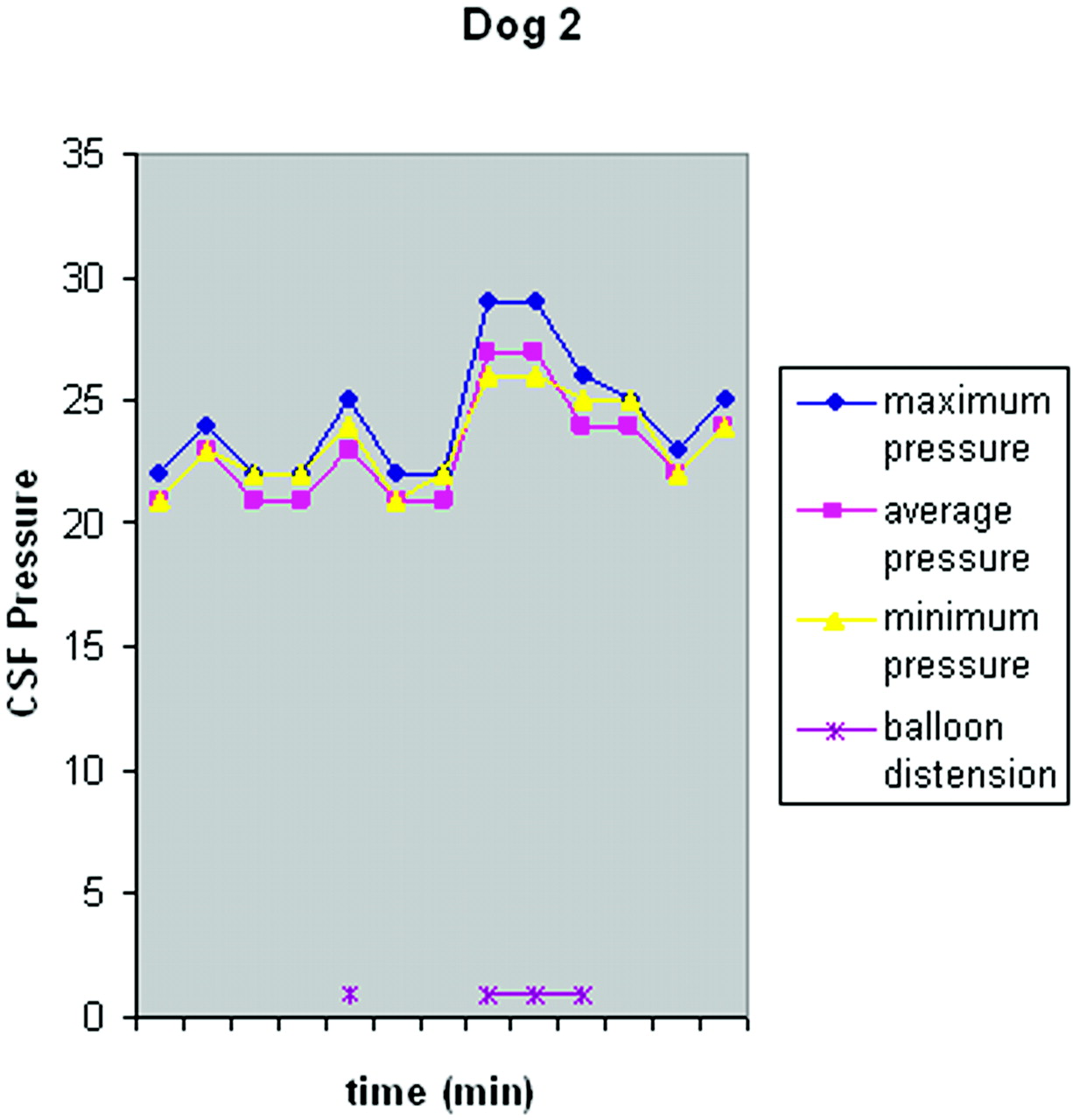
Pressure varied through the cardiac cycle (Fig 2). Except when the balloon catheter was expanded or collapsed, pressures varied little from one cardiac cycle to another.

Fig 2.
  • Download figure
  • Open in new tab
  • Download powerpoint
Fig 2.

CSF systolic, diastolic, mean, and pulse pressures in the CSF in dog 2 before and after inflation of the balloon catheter in the subarachnoid space.

With distension of the balloon, no changes in cardiac rate were noted. When the balloon was distended, it appeared to compromise the foramen magnum partially in each case.

Within seconds after each distension of the balloon, increases in CSF pressures were recorded. After deflation of the balloon, pressures dropped to preinflation levels.

Discussion

The study shows that CSF pressure can be recorded in the subarachnoid space with a transducer-tipped wire. Potentially, the wire can be placed anywhere in the spinal subarachnoid space and if appropriate techniques are employed, even in the cranial vault. The pressure recording showed variation in pressure through the cardiac cycle and little variation between cycles. In the experimental model used, significant pressure increases were recorded as the balloon in the foramen magnum was distended.

In other studies, the pressure measurements with the wire correlated exactly (r = 0.99) with the known values over a large range of pressures.1 The wire has been used intra-arterially.2–3 Applications in CSF pressure monitoring can be envisioned. With such a wire, fluctuations in CSF pressure below a block could be monitored during decompression of the spinal canal. The wire could theoretically be used to monitor CSF pressures as craniocervical decompression is performed in cases of Chiari I malformation or other foramen magnum abnormality. Specifically, one of the controversies of Chiari surgery is whether it is sufficient to remove the bone or whether duroplasty should be performed. We envision being able to insert a wire through the foramen magnum before the bony decompression and record pressures before and after the craniectomy and before and after duraplasty. Further, it might be useful to record the craniospinal pressure dissociation demonstrated with Chiari malformations by simultaneously recording pressures in the fourth ventricle and the upper cervical subarachnoid space.

The significance of this study is that CSF pressure can be measured (percutaneously) in the subarachnoid space with a small wire-mounted pressure transducer. With the transducer, elevated velocities have been documented secondary to partial obstruction of the foramen magnum in an animal model of a Chiari I malformation. Relatively safe invasive techniques such as this one would complement MR flow studies in helping us understand the CSF physiology at the foramen magnum. It is important to note that such studies would help elucidate the pathophysiology of obstructive foramen magnum lesions such as the Chiari I malformation.

References

  1. ↵
    Gorge G, Erbel R, Niessing S, et al. Miniaturized pressure guide-wire: evaluation in vitro and in isolated hearts. Cathet Cardiovasc Diagn 1993;30:341–47
    PubMed
  2. ↵
    Nisanci Y, Sezer M, Umman B, et al. Relationship between pressure-derived collateral blood flow and diabetes mellitus in patients with stable angina pectoris: a study based on coronary pressure measurement. J Invasive Cardiol 2002;14:118–22
    PubMed
  3. ↵
    Sorteberg A, Sorteberg W, Turk AS, et al. Effect of Guglielmi detachable coil placement on intraaneurysmal pressure: experimental study in canines. AJNR Am J Neuroradiol 2001;22:1750–56
    Abstract/FREE Full Text
  4. ↵
    Purdy PD, White CL 3rd, Baer DL, et al. Percutaneous translumbar spinal cord compression injury in dogs from an angioplasty balloon: MR and histopathologic changes with balloon sizes and compression times. AJNR Am J Neuroradiol 2004;25:1435–42
    Abstract/FREE Full Text
  • Received April 27, 2005.
  • Accepted after revision July 20, 2005.
  • Copyright © American Society of Neuroradiology
View Abstract
PreviousNext
Back to top

In this issue

American Journal of Neuroradiology: 27 (2)
American Journal of Neuroradiology
Vol. 27, Issue 2
February, 2006
  • Table of Contents
  • Index by author
Advertisement
Print
Download PDF
Email Article

Thank you for your interest in spreading the word on American Journal of Neuroradiology.

NOTE: We only request your email address so that the person you are recommending the page to knows that you wanted them to see it, and that it is not junk mail. We do not capture any email address.

Enter multiple addresses on separate lines or separate them with commas.
Recording CSF Pressure with a Transducer-Tipped Wire in an Animal Model of Chiari I
(Your Name) has sent you a message from American Journal of Neuroradiology
(Your Name) thought you would like to see the American Journal of Neuroradiology web site.
CAPTCHA
This question is for testing whether or not you are a human visitor and to prevent automated spam submissions.
Cite this article
A. Turk, B.J. Iskandar, V. Haughton, D. Consigny
Recording CSF Pressure with a Transducer-Tipped Wire in an Animal Model of Chiari I
American Journal of Neuroradiology Feb 2006, 27 (2) 354-355;

Citation Manager Formats

  • BibTeX
  • Bookends
  • EasyBib
  • EndNote (tagged)
  • EndNote 8 (xml)
  • Medlars
  • Mendeley
  • Papers
  • RefWorks Tagged
  • Ref Manager
  • RIS
  • Zotero
0 Responses
Respond to this article
Share
Bookmark this article
Recording CSF Pressure with a Transducer-Tipped Wire in an Animal Model of Chiari I
A. Turk, B.J. Iskandar, V. Haughton, D. Consigny
American Journal of Neuroradiology Feb 2006, 27 (2) 354-355;
del.icio.us logo Twitter logo Facebook logo Mendeley logo
  • Tweet Widget
  • Facebook Like
  • Google Plus One

Jump to section

  • Article
    • Abstract
    • Methods
    • Results
    • Discussion
    • References
  • Figures & Data
  • Info & Metrics
  • Responses
  • References
  • PDF

Related Articles

  • No related articles found.
  • PubMed
  • Google Scholar

Cited By...

  • Effect of Tonsillar Herniation on Cyclic CSF Flow Studied with Computational Flow Analysis
  • Crossref
  • Google Scholar

This article has not yet been cited by articles in journals that are participating in Crossref Cited-by Linking.

More in this TOC Section

  • Bern Score Validity for SIH
  • MP2RAGE 7T in MS Lesions of the Cervical Spine
  • Deep Learning for STIR Spine MRI Quality
Show more Spine

Similar Articles

Advertisement

Indexed Content

  • Current Issue
  • Accepted Manuscripts
  • Article Preview
  • Past Issues
  • Editorials
  • Editors Choice
  • Fellow Journal Club
  • Letters to the Editor

Cases

  • Case Collection
  • Archive - Case of the Week
  • Archive - Case of the Month
  • Archive - Classic Case

Special Collections

  • Special Collections

Resources

  • News and Updates
  • Turn around Times
  • Submit a Manuscript
  • Author Policies
  • Manuscript Submission Guidelines
  • Evidence-Based Medicine Level Guide
  • Publishing Checklists
  • Graphical Abstract Preparation
  • Imaging Protocol Submission
  • Submit a Case
  • Become a Reviewer/Academy of Reviewers
  • Get Peer Review Credit from Publons

Multimedia

  • AJNR Podcast
  • AJNR SCANtastic
  • Video Articles

About Us

  • About AJNR
  • Editorial Board
  • Not an AJNR Subscriber? Join Now
  • Alerts
  • Feedback
  • Advertise with us
  • Librarian Resources
  • Permissions
  • Terms and Conditions

American Society of Neuroradiology

  • Not an ASNR Member? Join Now

© 2025 by the American Society of Neuroradiology All rights, including for text and data mining, AI training, and similar technologies, are reserved.
Print ISSN: 0195-6108 Online ISSN: 1936-959X

Powered by HighWire