
Article Figures & Data
Figures
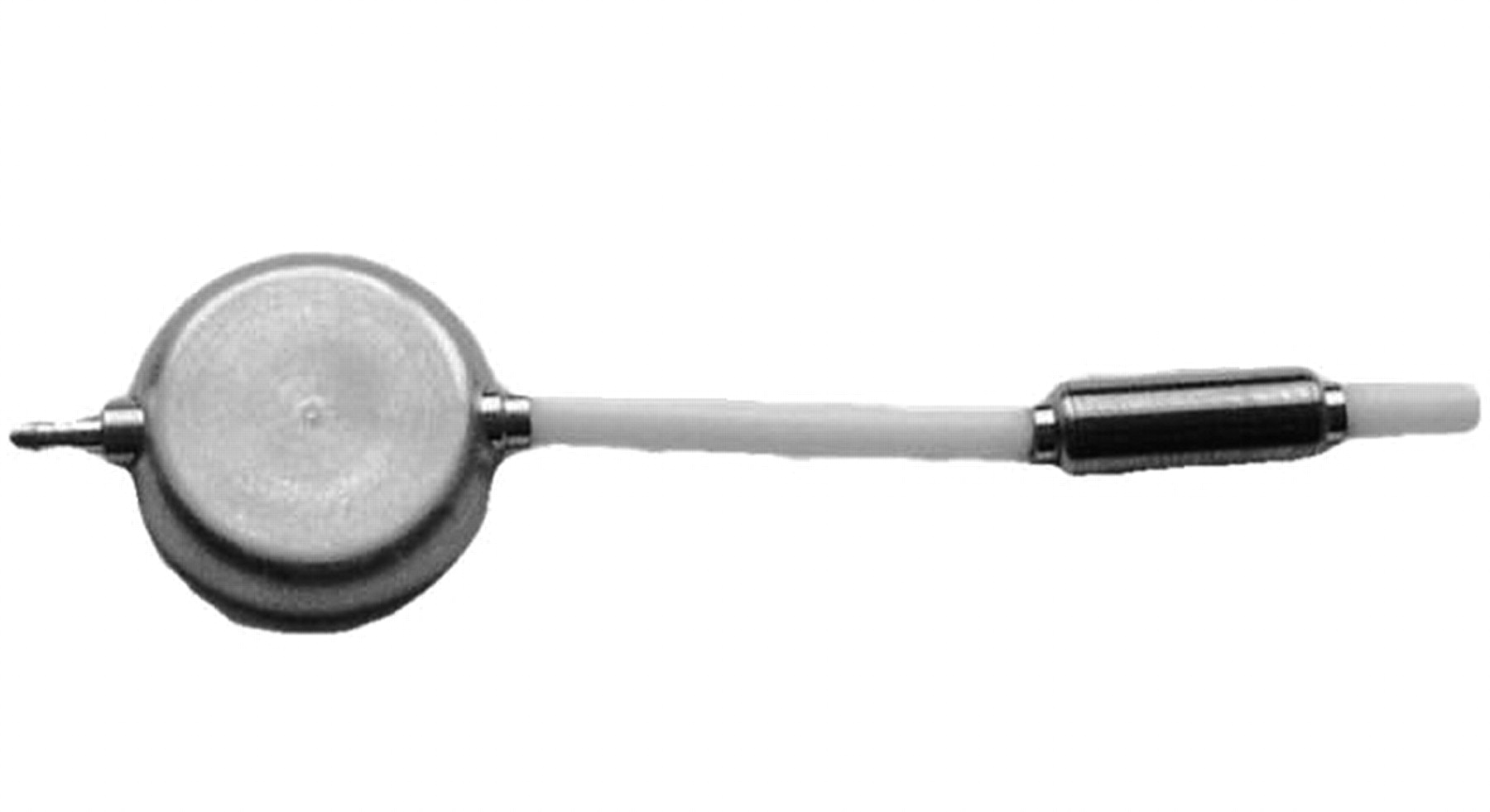
- Fig 1.
The programmable valve that underwent evaluation for MR imaging safety at 3T.
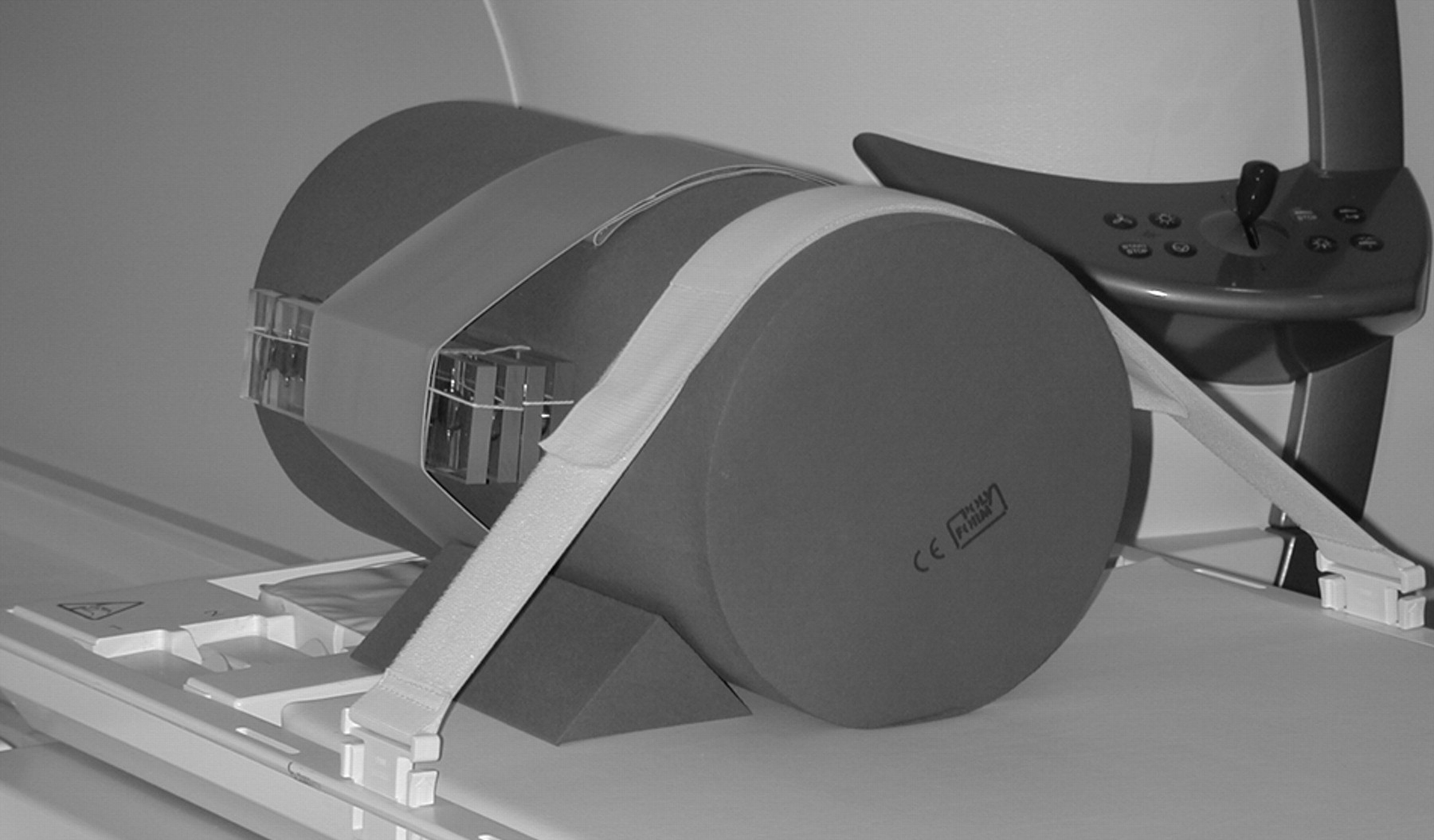
- Fig 2.
The experimental setup used to assess MR imaging–related heating for the programmable valve, showing the implant in position on the plastic frame placed on the bottom of the head/torso phantom. Note the cables going to the fluoroptic thermometry probes.
- Fig 3.
The 18 samples of the programmable valve shown attached to the cylinder-shaped phantom in preparation for the static magnetic field exposures (sagittal orientation).
- Fig 4.
The 18 samples of the programmable valve shown attached to the cylinder-shaped phantom in preparation for the static magnetic field exposures (simulated patient orientation).
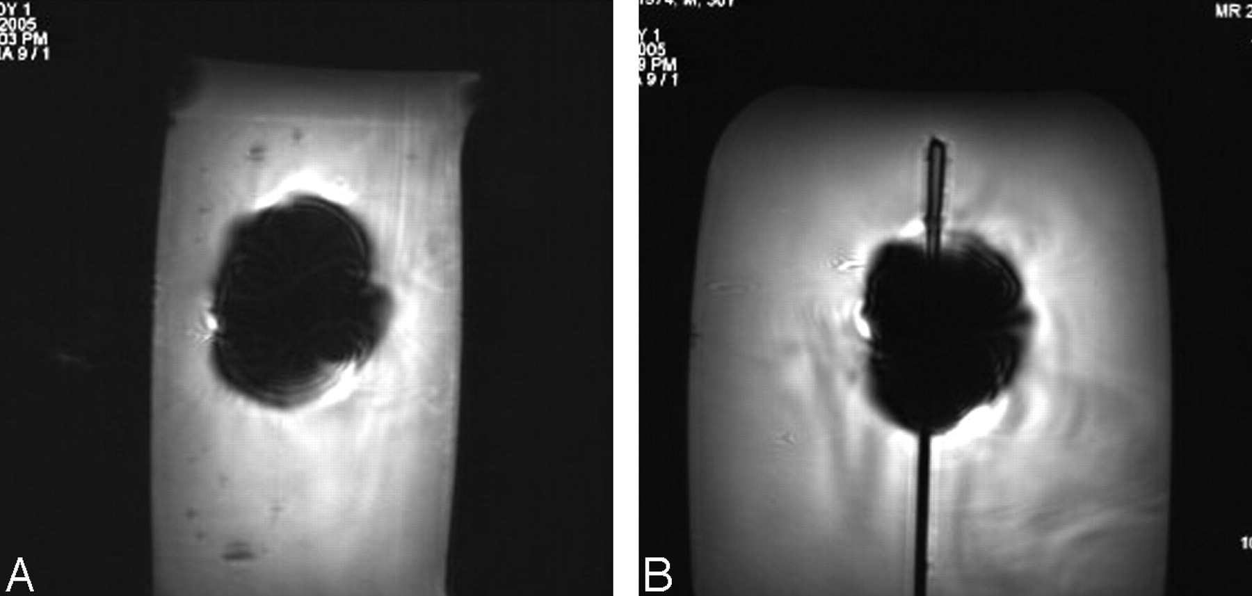
- Fig 5.
Examples of MR images showing artifacts for the programmable valve (gradient-echo pulse sequence; TR/TE, 100/15; flip angle, 30°; section thickness, 5 mm; field of view, 24 cm) at 3T. A, Section location oriented to the long axis of the programmable valve. B, Section location oriented to the short axis of the programmable valve.


Tables
Test Pressure Setting Orientation Arrow Direction 1 0 Sagittal Up 2 0 Sagittal Down 3 0 Axial Up 4 0 Axial Down 5 0 Coronal In 6 0 Coronal Out 7 8 Sagittal Up 8 8 Sagittal Down 9 8 Axial Up 10 8 Axial Down 11 8 Coronal In 12 8 Coronal Out 13 20 Sagittal Up 14 20 Sagittal Down 15 20 Axial Up 16 20 Axial Down 17 20 Coronal In 18 20 Coronal Out 19 5 Simulated patient In 20 5 Simulated patient Out Note:—All conditions were in-out × 5. There were no changes under any conditions.
- Table 2:
MR imaging parameters used in the assessment of the effects of MRI at 3T on the programmable valve
Pulse Sequence T1-SE T2-SE T1-FSE T2-FSE GRE, 3D GFRE, 3D GRE, MTC EPI TR (ms) 741 3000 700 5180 20 163 628 3400 TE (ms) 7 100 9 113 5 4 10 103 Flip angle N/A N/A N/A N/A 25 N/A 25 N/A Field of view (cm) 30 30 30 30 12 30 30 30 Matrix size 256 × 256 256 × 256 256 × 256 256 × 256 256 × 256 256 × 256 256 × 256 256 × 256 Section Thickness (mm) 10 10 10 10 3 3 10 10 Section gap (mm) 1 1 1 1 0.6 0.6 1 1 Imaging plane Axial Axial Axial Axial Volume Volume Axial Axial Imaging time 1:00 1:00 1:00 1:00 1:00 1:00 1:00 1:00 SAR, whole body (W/kg) 2.0 0.6 2.3 1.1 0.8 0.3 1.5 0.5 Note:—T1-SE indicates T1-weighted spin-echo; T2-SE, T2-weighted spin-echo; T1-FSE, T1-weighted fast spin-echo; T2-FSE, T2-weighted fast spin-echo; GRE, gradient echo; FGRE, fast gradient echo; MTC, magnetization transfer contrast; EPI, echo-planar imaging; N/A, not applicable; SAR, specific absorption rate.
Pulse Sequence Plane Orientation* Signal Void (mm2) T1-SE Long axis 1,359 Short axis 1,165 GRE Long axis 3,483 Short axis 3,156 Note:—T1-SE indicates T1-weighted spin-echo; GRE, gradient echo.
* Imaging plane relative to the programmable valve.


