Advertisement

Main menu

  • Home
  • Content
    • Current Issue
    • Accepted Manuscripts
    • Article Preview
    • Past Issue Archive
    • AJNR Case Collection
    • Case of the Week Archive
    • Classic Case Archive
    • Case of the Month Archive
  • Special Collections
    • Spinal CSF Leak Articles (Jan 2020-June 2024)
    • 2024 AJNR Journal Awards
    • Most Impactful AJNR Articles
  • Multimedia
    • AJNR Podcast
    • AJNR Scantastics
    • Video Articles
  • For Authors
    • Submit a Manuscript
    • Author Policies
    • Fast publishing of Accepted Manuscripts
    • Graphical Abstract Preparation
    • Manuscript Submission Guidelines
    • Imaging Protocol Submission
    • Submit a Case for the Case Collection
  • About Us
    • About AJNR
    • Editorial Board
  • More
    • Become a Reviewer/Academy of Reviewers
    • Subscribers
    • Permissions
    • Alerts
    • Feedback
    • Advertisers
    • ASNR Home
  • Other Publications
    • ajnr

User menu

  • Alerts
  • Log in

Search

  • Advanced search
American Journal of Neuroradiology

American Journal of Neuroradiology

ASHNR American Society of Functional Neuroradiology ASHNR American Society of Pediatric Neuroradiology ASSR
  • Alerts
  • Log in

Advanced Search

  • Home
  • Content
    • Current Issue
    • Accepted Manuscripts
    • Article Preview
    • Past Issue Archive
    • AJNR Case Collection
    • Case of the Week Archive
    • Classic Case Archive
    • Case of the Month Archive
  • Special Collections
    • Spinal CSF Leak Articles (Jan 2020-June 2024)
    • 2024 AJNR Journal Awards
    • Most Impactful AJNR Articles
  • Multimedia
    • AJNR Podcast
    • AJNR Scantastics
    • Video Articles
  • For Authors
    • Submit a Manuscript
    • Author Policies
    • Fast publishing of Accepted Manuscripts
    • Graphical Abstract Preparation
    • Manuscript Submission Guidelines
    • Imaging Protocol Submission
    • Submit a Case for the Case Collection
  • About Us
    • About AJNR
    • Editorial Board
  • More
    • Become a Reviewer/Academy of Reviewers
    • Subscribers
    • Permissions
    • Alerts
    • Feedback
    • Advertisers
    • ASNR Home
  • Follow AJNR on Twitter
  • Visit AJNR on Facebook
  • Follow AJNR on Instagram
  • Join AJNR on LinkedIn
  • RSS Feeds

Welcome to the new AJNR, Updated Hall of Fame, and more. Read the full announcements.


AJNR is seeking candidates for the position of Associate Section Editor, AJNR Case Collection. Read the full announcement.


Research ArticleBRAIN

Assessment of Ventricular Reconfiguration After Third Ventriculostomy: What Does Shape Analysis Provide in Addition to Volumetry?

C. Preul, T. Hübsch, D. Lindner and M. Tittgemeyer
American Journal of Neuroradiology March 2006, 27 (3) 689-693;
C. Preul
  • Find this author on Google Scholar
  • Find this author on PubMed
  • Search for this author on this site
T. Hübsch
  • Find this author on Google Scholar
  • Find this author on PubMed
  • Search for this author on this site
D. Lindner
  • Find this author on Google Scholar
  • Find this author on PubMed
  • Search for this author on this site
M. Tittgemeyer
  • Find this author on Google Scholar
  • Find this author on PubMed
  • Search for this author on this site
  • Article
  • Figures & Data
  • Info & Metrics
  • Responses
  • References
  • PDF
Loading

Article Figures & Data

Figures

  • Fig 1.
    • Download figure
    • Open in new tab
    • Download powerpoint
    Fig 1.

    3D shape decomposition with spheric harmonic-basis functions. The figure shows the reconstruction of the ventricular surface from order 1 (H1, left upper image) up to order 20 (H20, right lower image). The shape detail is effectively filtered as the number of harmonics is reduced.
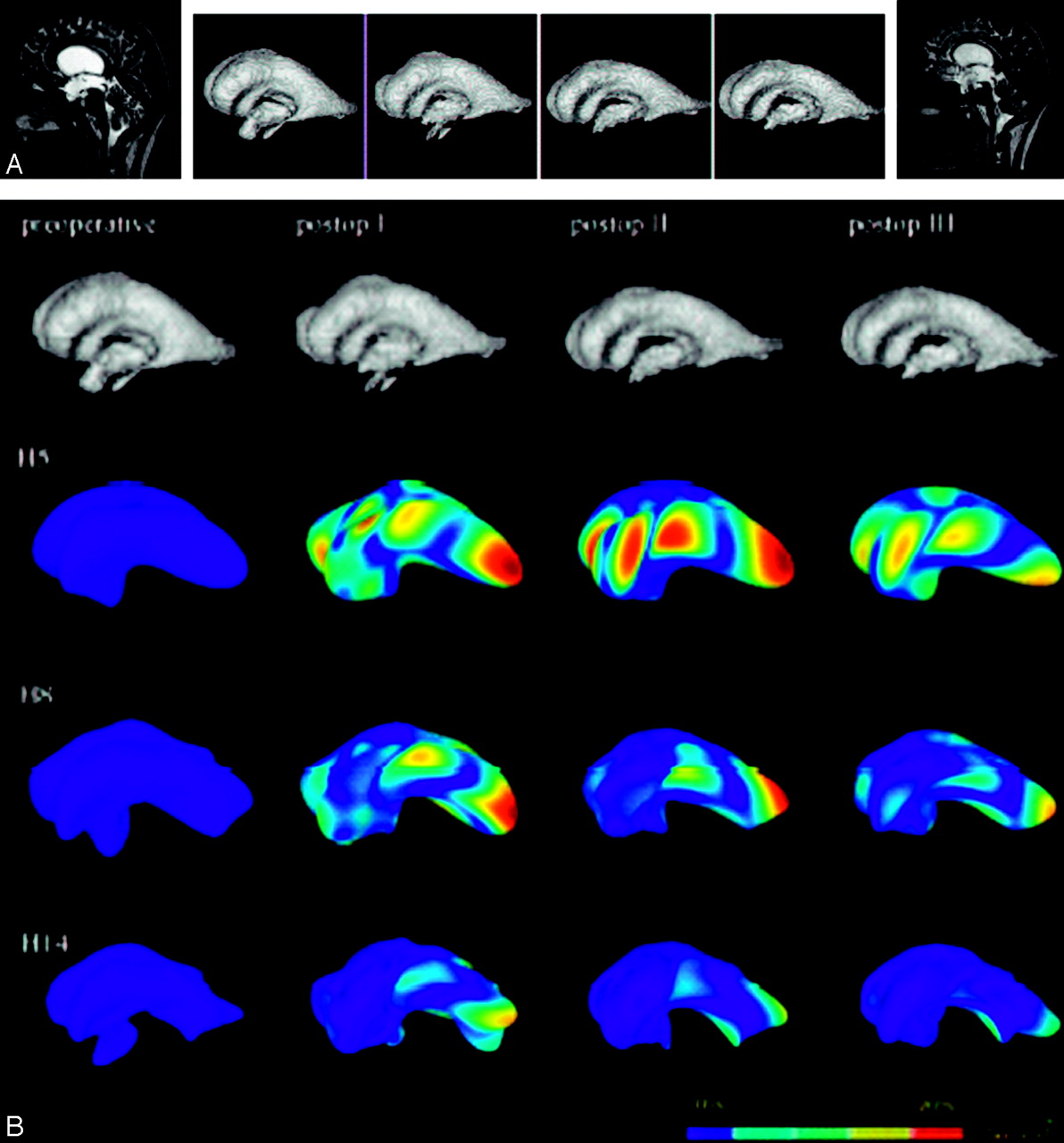
  • Fig 2.
    • Download figure
    • Open in new tab
    • Download powerpoint
    Fig 2.

    A, Pre- and second postoperative MR images of patient 1 (subacute hydrocephalus, left and right images) and segmented ventricular systems at preoperative and 3 postoperative time-steps (center). The segmented ventricular systems suggest a consecutive decrease in ventricular size (volumetric estimation, 218, 177, 132, 111 mL in the time course, respectively). Successful surgery is also documented by a flow void at the bottom of the third ventricle (right), indicating patency of the stoma.

    B, The upper row shows the segmented ventricular systems at the different pre- and postoperative time-steps. The 3 consecutive rows depict the models of the ventricular systems at harmonies 5, 8, and 14, respectively. Color-coding reveals differences in variance to the respective preoperative ventricle. Postoperative adaptational processes are most pronounced in the early postoperative course, whereas later adaptations are discrete. In the more complex harmony steps, the changes are less obvious than those in the simpler ones.
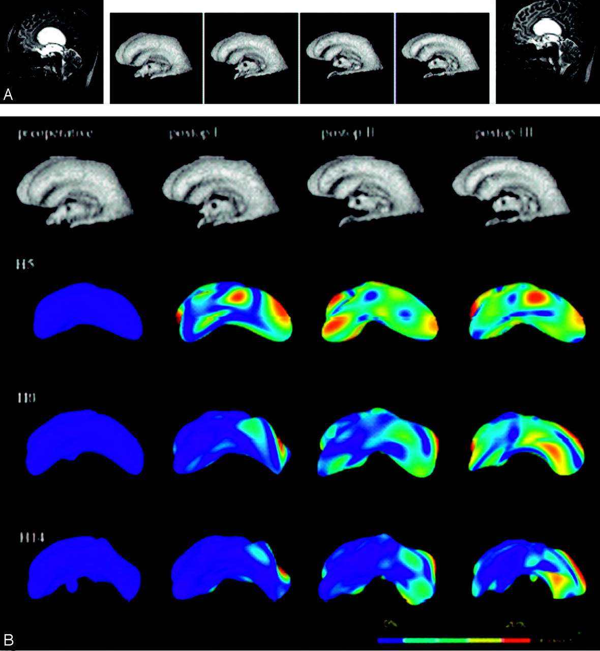
  • Fig 3.
    • Download figure
    • Open in new tab
    • Download powerpoint
    Fig 3.

    A, Left and right images show pre- and second postoperative MR images of patient 2 (chronic hydrocephalus). Segmented ventricular systems at preoperative and 3 postoperative time-steps are shown in the center. Although the segmented ventricular systems diminished in volume, the change in shape appears discrete (compared to T2-weighted midsagittal MR imaging scans [not shown]). (Volumetric estimation, 170, 128, 106, and 84 mL in the time course, respectively).

    B, The adaptational processes are more pronounced in the later postoperative course. The underlying simplified shapes of the ventricles (see harmony 5, Fig. 2B) differ substantially. Most changes occur in the posterior parts of the ventricles. As in the previous case, the changes are less impressive in the more complex harmonics.

PreviousNext
Back to top

In this issue

American Journal of Neuroradiology: 27 (3)
American Journal of Neuroradiology
Vol. 27, Issue 3
March, 2006
  • Table of Contents
  • Index by author
Advertisement
Print
Download PDF
Email Article

Thank you for your interest in spreading the word on American Journal of Neuroradiology.

NOTE: We only request your email address so that the person you are recommending the page to knows that you wanted them to see it, and that it is not junk mail. We do not capture any email address.

Enter multiple addresses on separate lines or separate them with commas.
Assessment of Ventricular Reconfiguration After Third Ventriculostomy: What Does Shape Analysis Provide in Addition to Volumetry?
(Your Name) has sent you a message from American Journal of Neuroradiology
(Your Name) thought you would like to see the American Journal of Neuroradiology web site.
CAPTCHA
This question is for testing whether or not you are a human visitor and to prevent automated spam submissions.
Cite this article
C. Preul, T. Hübsch, D. Lindner, M. Tittgemeyer
Assessment of Ventricular Reconfiguration After Third Ventriculostomy: What Does Shape Analysis Provide in Addition to Volumetry?
American Journal of Neuroradiology Mar 2006, 27 (3) 689-693;

Citation Manager Formats

  • BibTeX
  • Bookends
  • EasyBib
  • EndNote (tagged)
  • EndNote 8 (xml)
  • Medlars
  • Mendeley
  • Papers
  • RefWorks Tagged
  • Ref Manager
  • RIS
  • Zotero
0 Responses
Respond to this article
Share
Bookmark this article
Assessment of Ventricular Reconfiguration After Third Ventriculostomy: What Does Shape Analysis Provide in Addition to Volumetry?
C. Preul, T. Hübsch, D. Lindner, M. Tittgemeyer
American Journal of Neuroradiology Mar 2006, 27 (3) 689-693;
del.icio.us logo Twitter logo Facebook logo Mendeley logo
  • Tweet Widget
  • Facebook Like
  • Google Plus One

Jump to section

  • Article
    • Abstract
    • Methods
    • Results
    • Discussion
    • Footnotes
    • References
  • Figures & Data
  • Info & Metrics
  • Responses
  • References
  • PDF

Related Articles

  • No related articles found.
  • PubMed
  • Google Scholar

Cited By...

  • No citing articles found.
  • Crossref
  • Google Scholar

This article has not yet been cited by articles in journals that are participating in Crossref Cited-by Linking.

More in this TOC Section

  • Optimal MRI Sequence for Identifying Occlusion Location in Acute Stroke: Which Value of Time-Resolved Contrast-Enhanced MRA?
  • SWI or T2*: Which MRI Sequence to Use in the Detection of Cerebral Microbleeds? The Karolinska Imaging Dementia Study
  • Progression of Microstructural Damage in Spinocerebellar Ataxia Type 2: A Longitudinal DTI Study
Show more Brain

Similar Articles

Advertisement

Indexed Content

  • Current Issue
  • Accepted Manuscripts
  • Article Preview
  • Past Issues
  • Editorials
  • Editors Choice
  • Fellow Journal Club
  • Letters to the Editor

Cases

  • Case Collection
  • Archive - Case of the Week
  • Archive - Case of the Month
  • Archive - Classic Case

Special Collections

  • Special Collections

Resources

  • News and Updates
  • Turn around Times
  • Submit a Manuscript
  • Author Policies
  • Manuscript Submission Guidelines
  • Evidence-Based Medicine Level Guide
  • Publishing Checklists
  • Graphical Abstract Preparation
  • Imaging Protocol Submission
  • Submit a Case
  • Become a Reviewer/Academy of Reviewers
  • Get Peer Review Credit from Publons

Multimedia

  • AJNR Podcast
  • AJNR SCANtastic
  • Video Articles

About Us

  • About AJNR
  • Editorial Board
  • Not an AJNR Subscriber? Join Now
  • Alerts
  • Feedback
  • Advertise with us
  • Librarian Resources
  • Permissions
  • Terms and Conditions

American Society of Neuroradiology

  • Not an ASNR Member? Join Now

© 2025 by the American Society of Neuroradiology All rights, including for text and data mining, AI training, and similar technologies, are reserved.
Print ISSN: 0195-6108 Online ISSN: 1936-959X

Powered by HighWire