
Article Figures & Data
Figures
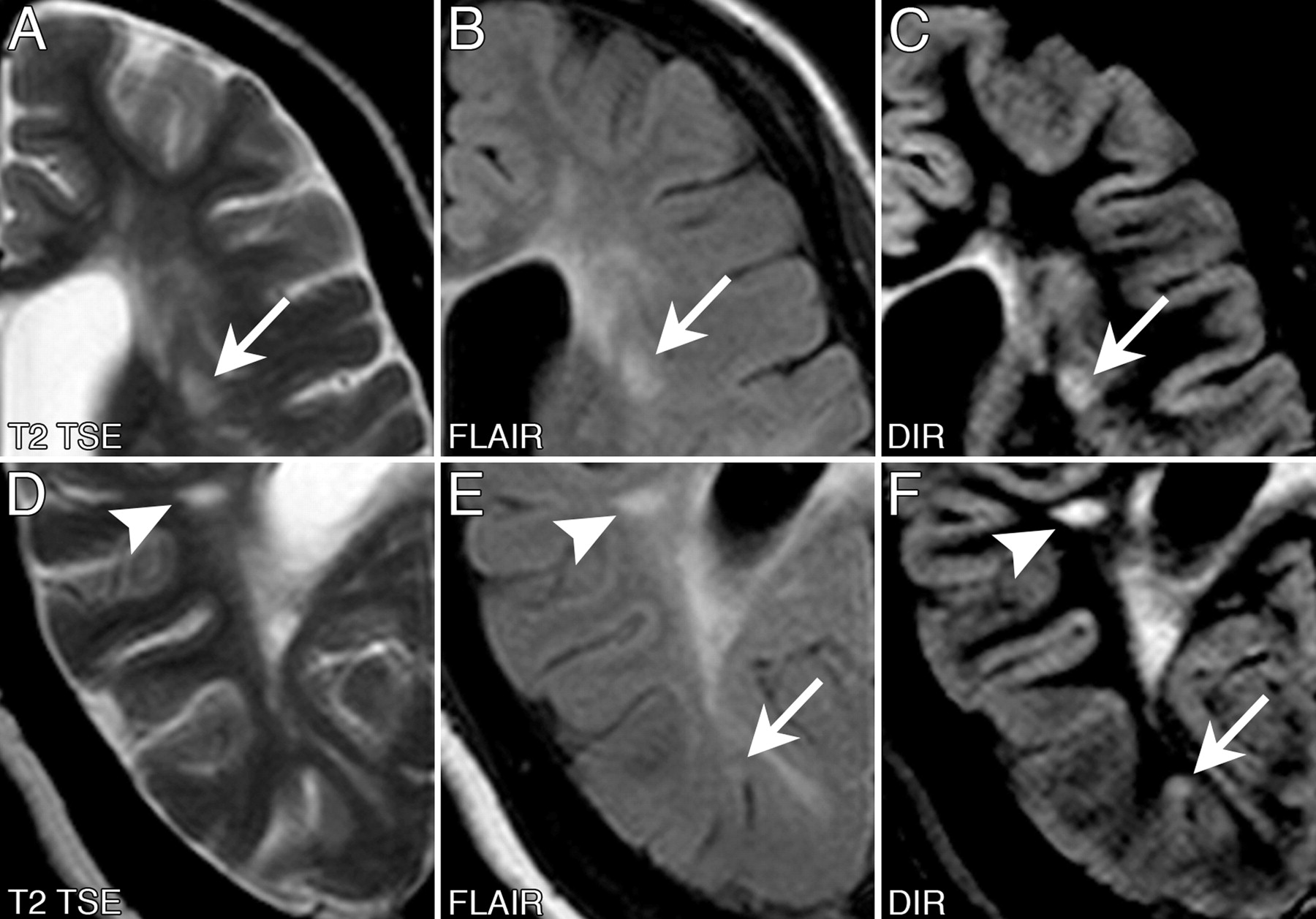
- Fig 1.
Transverse T2-weighted TSE, FLAIR, and DIR image examples documenting the higher sensitivity of DIR in the detection of inflammatory brain lesion in the infratentorial brain.
Top row (A–C), A 36-year-old woman presenting with a polysymptomatic CIS. A sharp delineated inflammatory lesion in the left pedunculus cerebelli (arrow) can be clearly identified on the DIR image but not on the corresponding sections of the T2 TSE and FLAIR sequences.
Bottom row (D–F), A 23-year-old man presenting with optic neuritis of the left eye. Compared with the T2 TSE and FLAIR images, more lesions in both hemispheres of the cerebellum (arrows) can be identified on the DIR image. Moreover, those lesions identified on all 3 sequences were better delineated on DIR compared with the corresponding T2 TSE and FLAIR images.
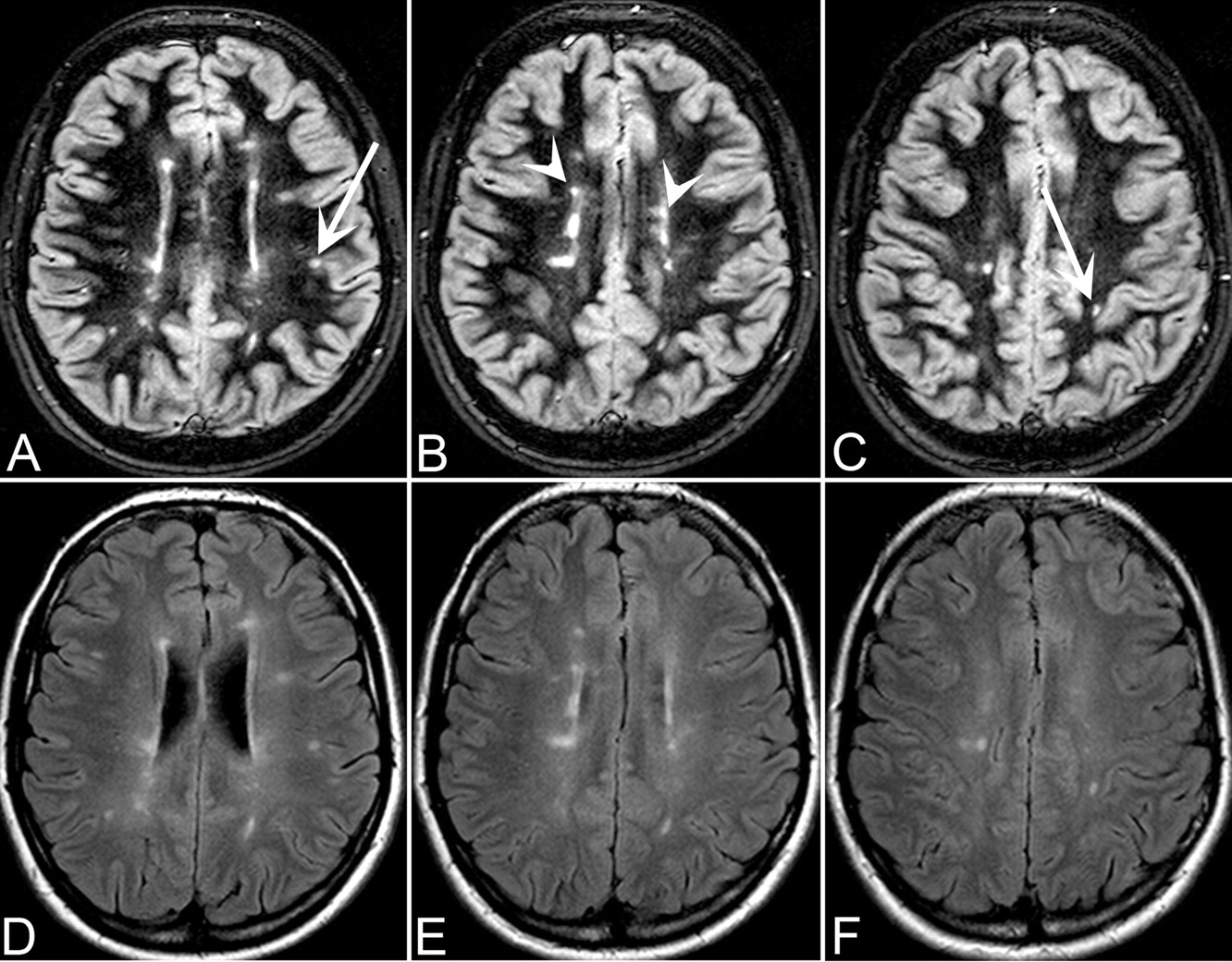
- Fig 2.
Transverse FLAIR (bottom row) and DIR (top row) sections of the supratentorial brain. The inflammatory lesions have a more sharp delineated appearance on the DIR compared with the corresponding FLAIR images. Despite a minor contrast between lesions and the normal-appearing gray matter, DIR showed a high sensitivity in the detection of juxtacortical and mixed white matter-gray matter lesions (arrows). A differentiation between juxtacortical and mixed white matter-gray matter lesions is much easier on the DIR than on the FLAIR images. Regarding the periventricular white matter, the lesions are easier to identify on the DIR compared with the FLAIR images (open arrows).
- Fig 3.
Image examples of the improved detection of mixed white matter-gray matter lesions on the DIR pulse sequence. These images were obtained from a 40-year-old woman with a relapsing-remitting course of MS (disease duration, 165 months; EDSS, 6) presenting with a high lesion load on the MR imaging, including mixed white matter-gray matter lesions. Top row, an example of different classifications of a lesion using different pulse sequences. A lesion (arrow) was prospectively classified as a juxtacortical lesion on the T2 TSE and FLAIR images. Because of the better delineation of the white and gray matter on the DIR image, this lesion had to reclassified into a mixed white matter-gray matter lesion. Bottom row, an example of the higher conspicuity of a cortical lesion (arrow) on the DIR image that was not prospectively identified on the corresponding T2 TSE and FLAIR images. Another lesion (open arrow) could be easily identified and categorized on the DIR image as a pure juxtacortical lesion without touching the cortex.


Tables
Parameter DIR FLAIR T2 TSE Field of view (mm) 230 230 230 Matrix 256 256 256 Section thickness (mm) 5 5 5 Measured voxel size (mm) 0.90/0.90/5 0.90/0.90/5 0.90/0.90/5 SENSE factor 1.5 1.6 Turbo factor 13 38 16 Repetition time (ms) 11000 12000 4100 Echo time (ms) 29 140 100 Inversion time (ms) 3400/325* 2850 Number of signals averaged 1 1 1 Bandwidth/pixel (Hz) 277.9 287 184.3 Acquisition time (minutes:seconds) 3:18 4:00 2:19 Note:—DIR indicates double inversion recovery; FLAIR, fluid-attenuated inversion recovery; T2 TSE, T2-weighted turbo spin-echo; SENSE sensitivity encoding.
* The long inversion time TI1 (3400 ms) is defined as the interval between the first 180° inversion pulse and the 90° excitation pulse. The short inversion time TI2 (325 ms) is defined as the interval between the second 180° inversion pulse and the 90° excitation pulse.
- Table 2:
Analysis of the lesion load measurement and relative comparisons of the DIR versus the FLAIR and T2-weighted TSE sequences
Region DIR* FLAIR* T2TSE* Relative Comparison (%)† DIR/FLAIR P Value‡ DIR/T2 TSE P Value‡ Infratentorial 39 25 27 56 0.02 44 0.02 Periventricular 85 81 76 5 0.16 12 0.03 Juxtacortical 54 57 49 −6 0.08 10 0.18 Mixed WM-GM 21 18 14 17 0.32 50 0.06 Deep WM 35 36 35 −3 0.32 0 1 Total 232 216 201 7 0.04 15 0.01 Note:—GM indicates gray matter; WM, white matter; DIR, double inversion recovery; FLAIR, fluid-attenuated inversion recovery; T2 TSE, T2-weighted turbo spin-echo.
* Data are numbers of detected lesions.
† Data are relative differences in the numbers of detected lesions expressed as percentages of lesion numbers identified with DIR imaging compared with the corresponding FLAIR and T2 TSE imaging.
‡ P value was obtained from the patient-wise analysis by Wilcoxon analysis for matched pairs indicating that more or fewer patients showed higher lesion load measurement with DIR imaging in comparison with the corresponding FLAIR or T2 TSE imaging.
Contrast* Contrast Ratio DIR FLAIR T2 TSE DIR vs FLAIR† DIR vs T2 TSE† Lesion/NAWM Infratentorial 0.460 ± 0.125 0.137 ± 0.060 0.181 ± 0.067 3.36 2.54 Periventricular 0.788 ± 0.098 0.253 ± 0.070 0.341 ± 0.085 3.11 2.31 Deep WM 0.720 ± 0.066 0.217 ± 0.047 0.298 ± 0.070 3.32 2.42 Juxtacortical 0.772 ± 0.110 0.253 ± 0.045 0.335 ± 0.047 3.05 2.30 Lesion/NAGM 0.190 ± 0.097 0.198 ± 0.064 0.213 ± 0.076 0.96 0.89 Lesion/CSF 0.832 ± 0.103 0.653 ± 0.243 0.201 ± 0.101 1.27 4.14 Note:—NAWM indicates normal-appearing white matter; WM, white matter; NAGM, normal-appearing grey matter; DIR, double inversion recovery; FLAIR, fluid-attenuated inversion recovery; T2 TSE, T2-weighted turbo spin-echo.
* Data are presented as means ± SD.
† Data are the contrast values of the DIR sequence in relative comparison with the corresponding contrast value of the FLAIR and T2 TSE sequence.
In this issue


Jump to section
Related Articles
Cited By...
- 3D Echo Planar Time-resolved Imaging (3D-EPTI) for ultrafast multi-parametric quantitative MRI
- Improving Detection of Multiple Sclerosis Lesions in the Posterior Fossa Using an Optimized 3D-FLAIR Sequence at 3T
- Pre- and Postcontrast 3D Double Inversion Recovery Sequence in Multiple Sclerosis: A Simple and Effective MR Imaging Protocol
- Current and Emerging Therapies in Multiple Sclerosis: Implications for the Radiologist, Part 1--Mechanisms, Efficacy, and Safety
- How Common Is Signal-Intensity Increase in Optic Nerve Segments on 3D Double Inversion Recovery Sequences in Visually Asymptomatic Patients with Multiple Sclerosis?
- Current and Emerging Therapies in Multiple Sclerosis: Implications for the Radiologist, Part 2--Surveillance for Treatment Complications and Disease Progression
- Synthetic MRI in the Detection of Multiple Sclerosis Plaques
- Juxtacortical Lesions and Cortical Thinning in Multiple Sclerosis
- Double Inversion Recovery Sequence of the Cervical Spinal Cord in Multiple Sclerosis and Related Inflammatory Diseases
- Multicontrast MR Imaging at 7T in Multiple Sclerosis: Highest Lesion Detection in Cortical Gray Matter with 3D-FLAIR
- Postmortem verification of MS cortical lesion detection with 3D DIR
- What you see depends on how you look: Gray matter lesions in multiple sclerosis
- In vivo imaging of cortical pathology in multiple sclerosis using ultra-high field MRI
- Tissue-Specific Imaging Is a Robust Methodology to Differentiate In Vivo T1 Black Holes with Advanced Multiple Sclerosis-Induced Damage