
Article Figures & Data
Figures
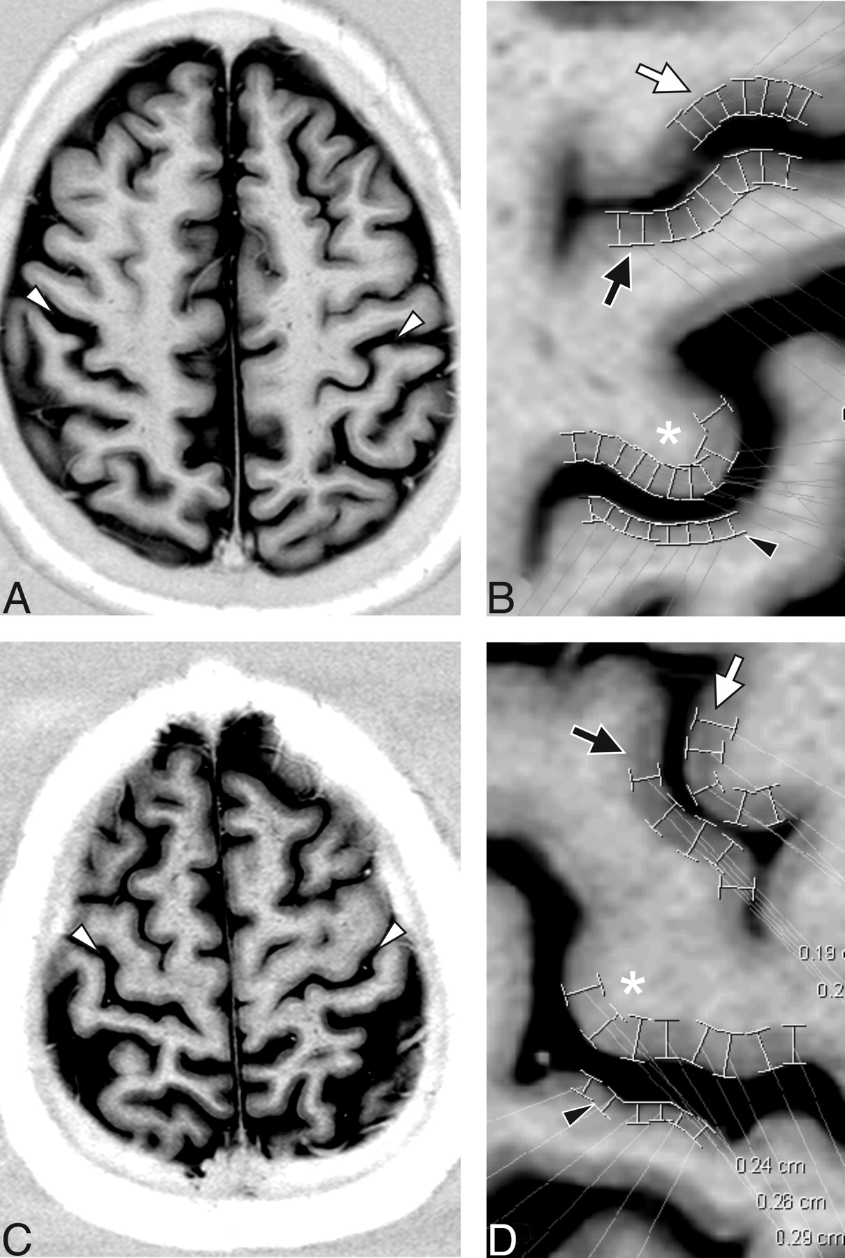
- Fig 1.
Cortical thickness measurements in a 56-year-old woman with primary lateral sclerosis (PLS) (A and B) and age- and sex-matched control subjects (C and D). Real reconstruction of axial 5-mm STIR MR imaging through M1 shows strong gray-white differentiation allowing for identification of the central sulcus (white arrowheads). The neighborhood of the central sulcus for patient (B) and control (D) is magnified from the corresponding images in A and C, respectively. Typical placements of digital calipers in the region of the hand knob (asterisks) for measurement of M1, as well as the measurements of S1 (black arrowheads), and the anterior (white arrows) and posterior (black arrows) banks of the precentral sulcus (pre-CS) are shown. M1 thickness for this patient was 2.16 and for the control subject was 2.63. The ratio of M1 to the pre-CS thickness was 0.92 for this patient and 1.14 for this control subject.
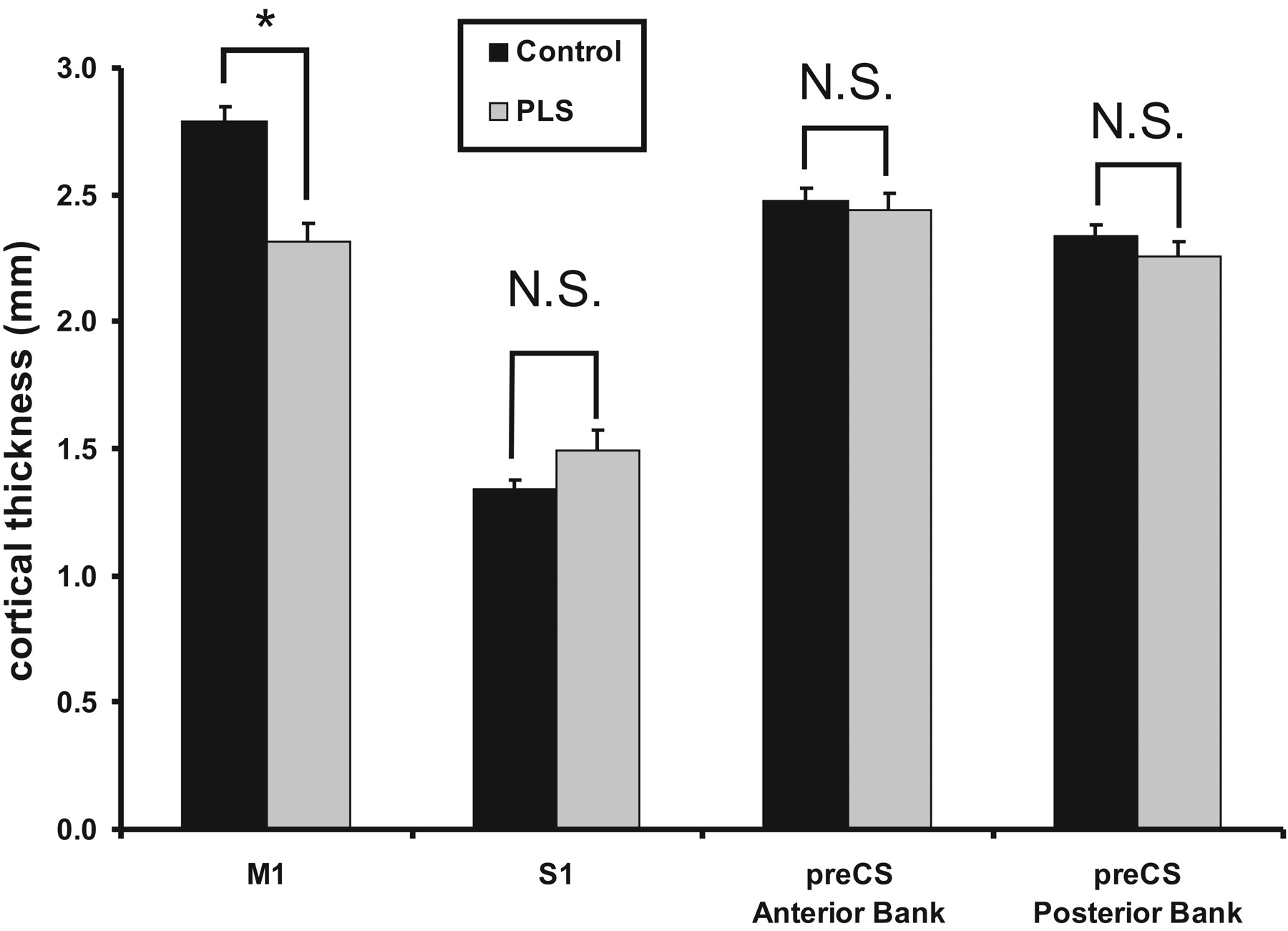
- Fig 2.
Cortical thickness in patients with primary lateral sclerosis (PLS) versus control subjects. There is a significant reduction in cortical thickness in primary motor cortex (M1), but no difference in the other 3 cortices measured. ∗, significant at P = .0008; N.S., not significant; preCS, precentral sulcus; S1, primary sensory cortex.
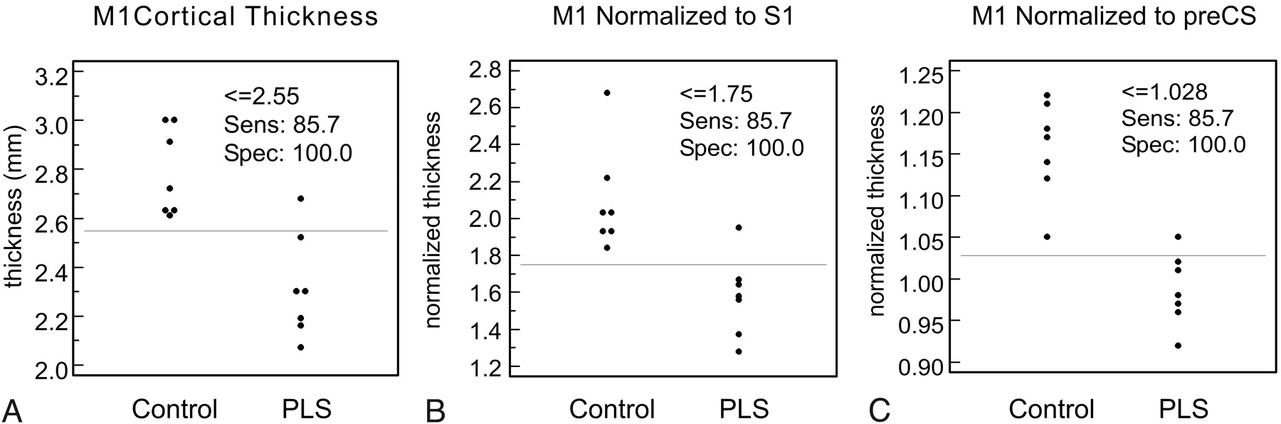
- Fig 3.
Cortical thickness distribution in control subjects and patients with primary lateral sclerosis (PLS) for M1 cortical thickness (A), M1 cortical thickness normalized to S1 cortical thickness (B), and M1 normalized to the thickness of the anterior and posterior banks of the precentral sulcus (C). Although thresholds with identical sensitivity and specificity can be identified, normalization to the precentral sulcus reduces the overlap between the 2 populations.


Tables
Subject Age (years) Disease Duration (years) Finger-Tapping Speed* (taps/s; R/L) Regions with UMN Signs 1 52 4 2.5/2.2 B, UE, LE 2 56 5 3.0/1.7 B, UE, LE 3 55 6 2.1/2.1 B, UE, LE 4 43 7 3.0/2.3 B, UE, LE 5 47 8 4.1/3.5 B, UE, LE 6 49 8 2.9/2.5 B, UE, LE 7 68 17 2.6/2.7 B, UE, LE Note:—R indicates right; L, left; B, bulbar; UE, upper extremities; LE, lower extremities; UMN, upper motor neuron.
* Control group tapping speed: R, 6.0 ± 0.6 taps/s; L, 5.5 ± 0.5 taps/s.
- Table 2:
Absolute and relative cortical thickness measurements in patients with PLS and control subjects
CS M1 Anterior (mm) Pre-CS S1 Posterior (mm) Anterior (mm) Posterior (mm) M1 Ratios M1:S1 M1:Pre-CS Control 2.79 ± 0.18 1.34 ± 0.11 2.48 ± 0.15 2.34 ± 0.12 2.10 ± 0.28 1.16 ± 0.06 PLS 2.32 ± 0.21 1.49 ± 0.23 2.44 ± 0.19 2.26 ± 0.18 1.58 ± 0.22 0.99 ± 0.04 P-value .0008 .14 .68 .33 .0023 .000057 Note:—CS indicates central sulcus; M1, primary motor cortex; pre-CS, precentral sulcus; S1, primary sensory cortex.
In this issue


Jump to section
Related Articles
Cited By...
- A fully synthetic three-dimensional human cerebrovascular model based on histological characteristics to investigate the hemodynamic fingerprint of the layer BOLD fMRI signal formation
- High T2 Signal in Primary Lateral Sclerosis Supports the Topographic Distribution of Fibers in the Corpus Callosum: Assessing Disease in the Primary Motor Segment