
Article Figures & Data
Figures
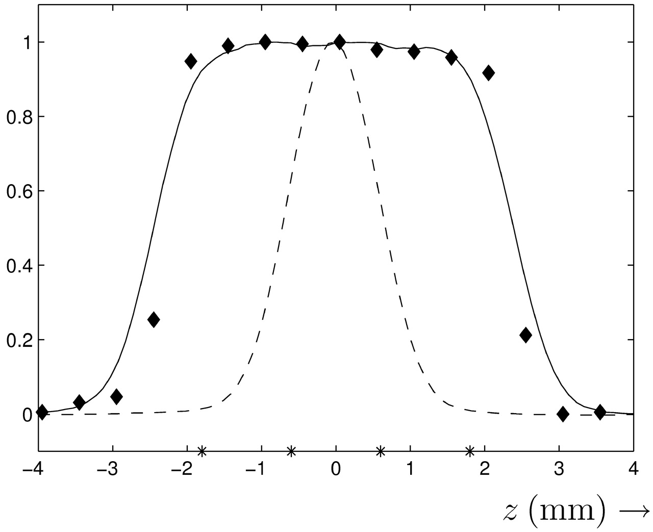
- Fig 1.
Section sensitivity profiles for sequential mode (diamonds) and spiral mode (dashed line). The continuous line depicts the profile of 4 combined spiral mode images with an increment of 1.2 mm (positions of each image given by asterisks on the z-axis).
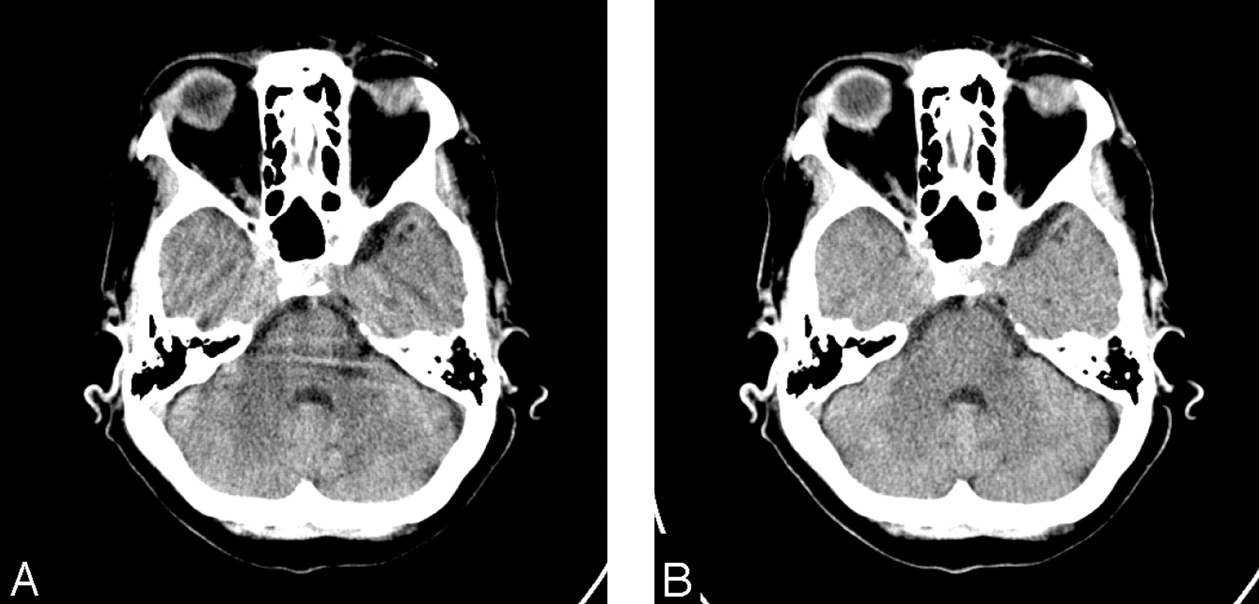
- Fig 2.
Cross-sectional images of the skull base.
A, Sequential technique. Severe streak artifacts are shown in the skull base.
B, Spiral technique.
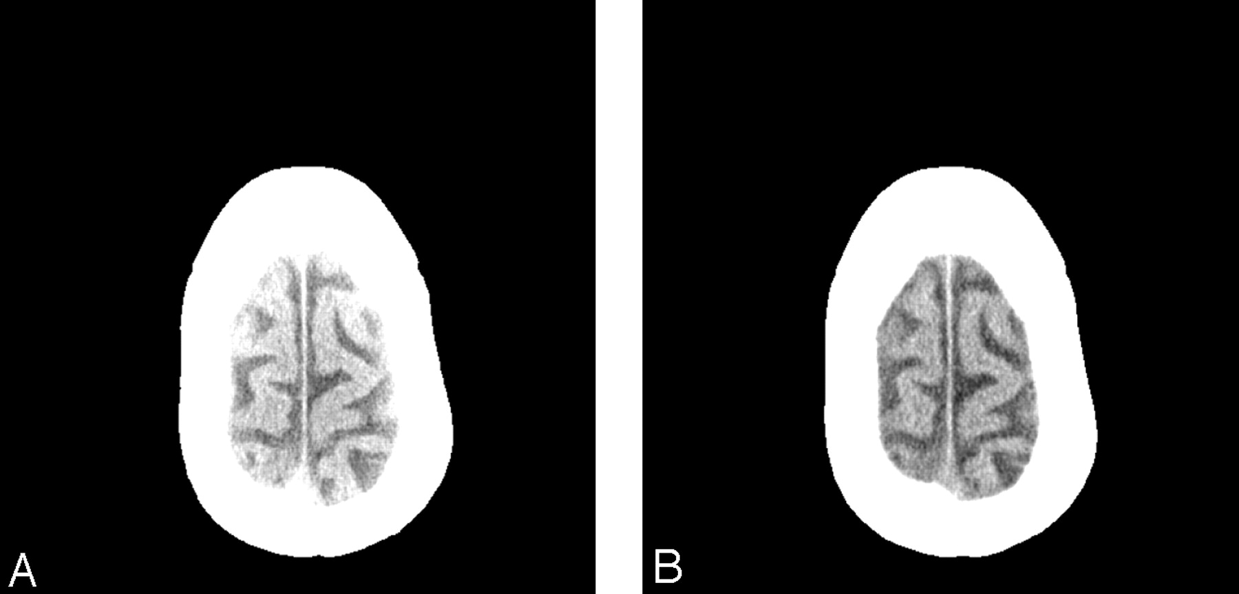
- Fig 3.
Cross-sectional images of the upper cranium.
A, Sequential technique. This image shows an increase of the CT values of the brain tissue, especially near the skull.
B, Spiral technique.
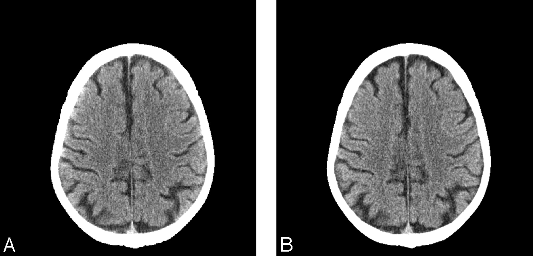
- Fig 4.
Cross-sectional images of the middle region of the brain. The spiral image has a higher contrast between the gyri and the CSF in the frontal region. Both observers had a slight preference for the spiral image with respect to “overall image quality,” “visualization of brain tissue near the skull,” and “image noise.”
A, Sequential technique.
B, Spiral technique.


Tables
Indication No. of Patients (Rule out) hydrocephalus 9 Stroke 6 Memory deficit 6 (Rule out) hematoma 4 Gait disturbance 3 Sensory deficit 2 Epilepsy 1 Vertigo 1 Note:—Some patients had multiple indications.
- Table 2:
Frequencies, mean, and SD of scores of observers in pairwise comparison of images (first part of observer study)
Aspects & Observers −2 −1 0 1 2 NA Mean SD P Streak artifacts 1 0 0 0 44 22 166 1.33 0.47 <.001 2 0 0 7 29 33 163 1.38 0.66 <.001 Visualization brain tissue near skull 1 0 2 105 106 19 0.61 0.65 <.001 2 1 10 75 104 42 0.76 0.81 <.001 Visualization hypodense lesion(s) 1 1 22 33 39 1 136 0.18 0.83 >.05 2 2 31 24 40 7 128 0.18 1.00 >.05 Gray/white matter differentiation 1 1 8 182 32 0 9 0.10 0.43 >.05 2 3 53 70 84 5 17 0.16 0.87 >.05 Image noise 1 1 49 92 90 0 0.17 0.77 >.05 2 1 52 52 115 12 0.37 0.90 <.001 Overall image quality 1 1 28 64 103 36 0.63 0.90 <.001 2 3 42 35 118 34 0.59 0.99 <.001 Note:—NA indicates not applicable. Scores range from −2 (preference for sequential technique) to +2 (preference for spiral technique). The tabulated P values are the those of the Wilcoxon signed-rank test multiplied by 24, which is the total number of significance tests performed (Bonferroni correction).
- Table 3:
Frequencies, mean, and SD of scores of observers in comparison of complete scans (second part of observer study)
Aspects & Observers −2 −1 0 1 2 NA Mean SD P Streak artifacts 1 0 0 0 18 5 0 1.22 0.41 <.001 2 0 0 0 3 20 0 1.87 0.34 <.001 Visualization brain tissue near skull 1 0 0 2 21 0 0.91 0.28 <.001 2 0 0 0 16 7 1.30 0.46 <.001 Visualization hypodense lesion(s) 1 0 0 10 5 1 7 0.44 0.61 >.05 2 0 1 6 8 0 8 0.47 0.62 >.05 Gray/white matter differentiation 1 0 0 22 1 0 0 0.04 0.20 >.05 2 0 0 3 20 0 0 0.87 0.34 <.001 Image noise 1 0 4 17 2 0 −0.09 0.50 >.05 2 0 0 0 4 19 1.83 0.38 <.001 Overall image quality 1 0 0 0 21 2 1.09 0.28 <.001 2 0 0 0 1 22 1.96 0.20 <.001 Note:—NA indicates not applicable. Scores range from −2 (preference for sequential technique) to +2 (preference for spiral technique). The tabulated P values are the those of the Wilcoxon signed-rank test multiplied by 24, which is the total number of significance tests performed (Bonferroni correction).
Aspect Observer Study: First Part Observer Study: Second Part N κ PABAK N κ PABAK Streak artifacts 60 0.00 0.88 23 0.00 1.00 Visualization brain tissue near skull 232 0.37 0.52 23 0.00 0.87 Visualization hypodense lesion(s) 71 0.09 0.11 15 −0.10 0.10 Gray/white matter differentiation 211 0.01 0.00 23 0.01 —* Image noise 232 0.14 0.16 23 0.00 —* Overall image quality 232 0.20 0.35 23 0.00 1.00 Note:—PABAK indicates prevalence- and bias-adjusted κ.
* No meaningful value could be determined because one or more of the concordant values of the cross-table became negative.


