
Article Figures & Data
Figures
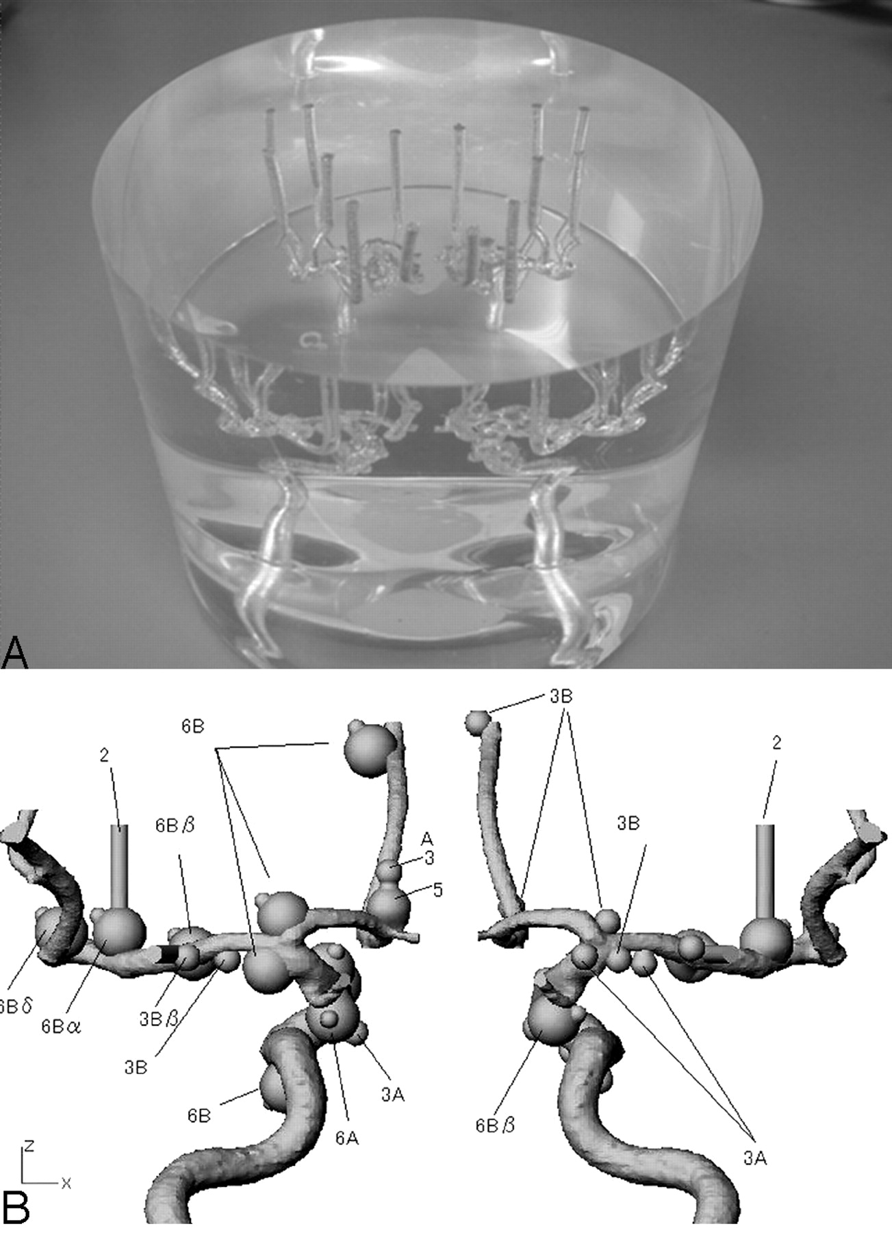
- Fig 1.
Photograph (A) and schematic drawing (B) of the anthropomorphic vascular phantom used in this study. The phantom was designed to simulate the intracranial arteries with a total of 32 aneurysms. Of 32 aneurysms, 15 had an aneurysmal bleb with diameter of 2 mm.
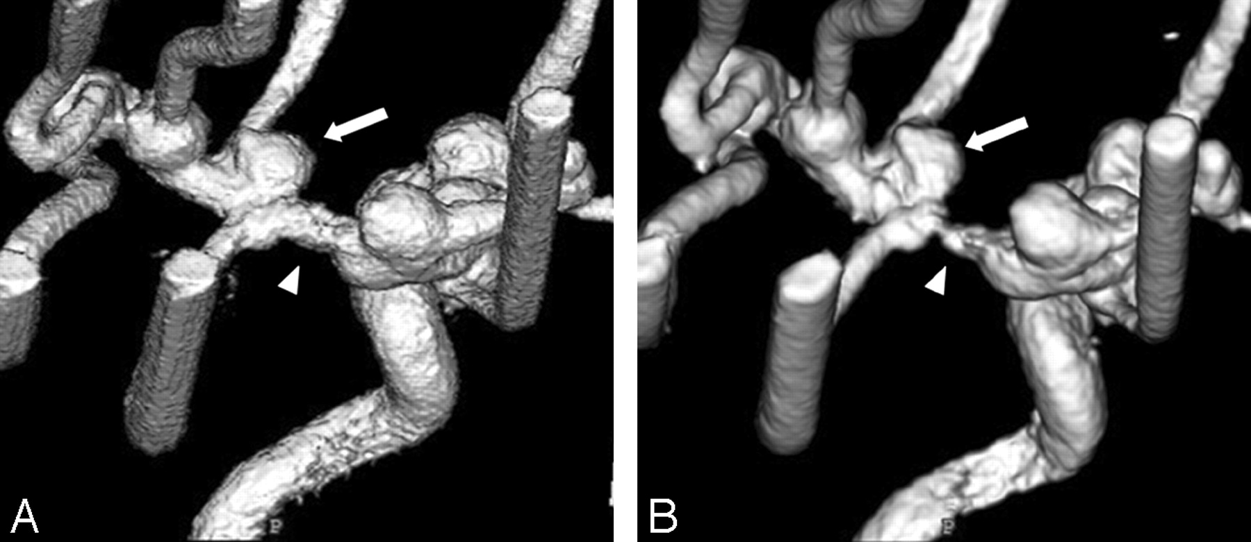
- Fig 2.
3D DSA from the superior and right posterior oblique view at 300 mg I/mL obtained with FPD system (A) and with I.I.-TV system (B). 3D DSA with FPD system shows mild stenosis at the M1 segment of the middle cerebral artery, whereas severe stenosis is observed on 3D DSA with I.I.-TV system (arrowheads). The pseudostenosis artifact caused the minor distortion of aneurysm on 3D DSA with FPD system, whereas the severe distortion of aneurysm on that with I.I.-TV system (arrows).
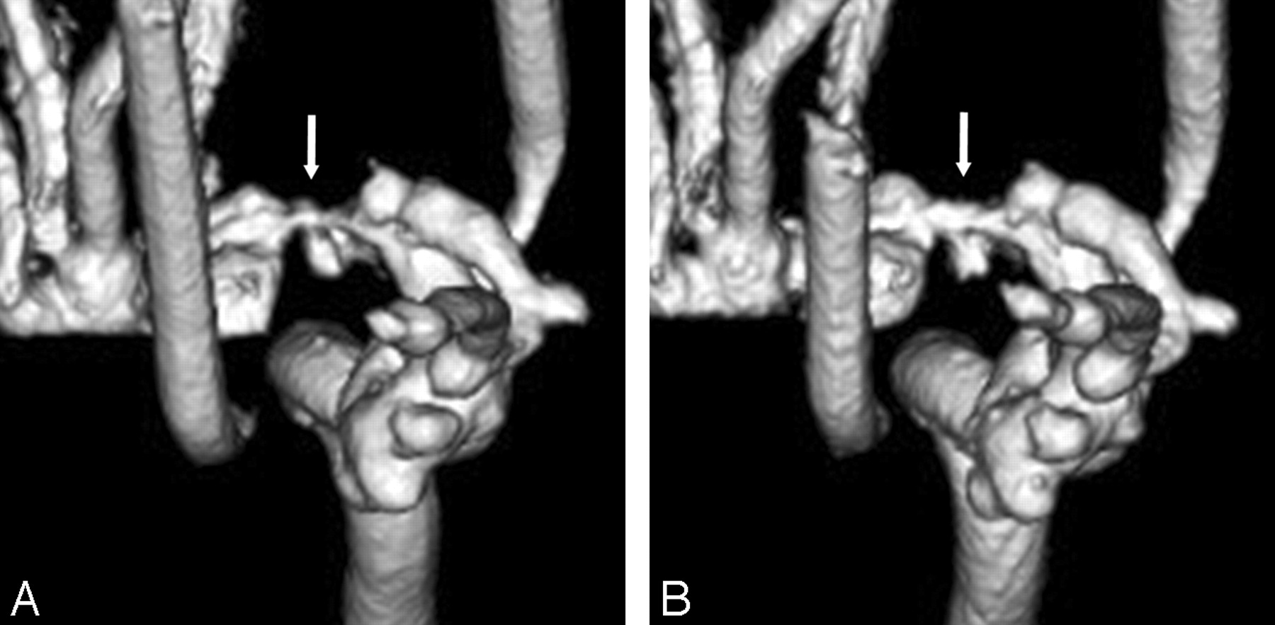
- Fig 3.
Anteroposterior 3D DSA obtained with I.I.-TV system at 300 (A) and at 150 mg I/mL (B) demonstrated that the pseudostenosis artifacts are more severe at 300 than at 150 mg I/mL (arrows).
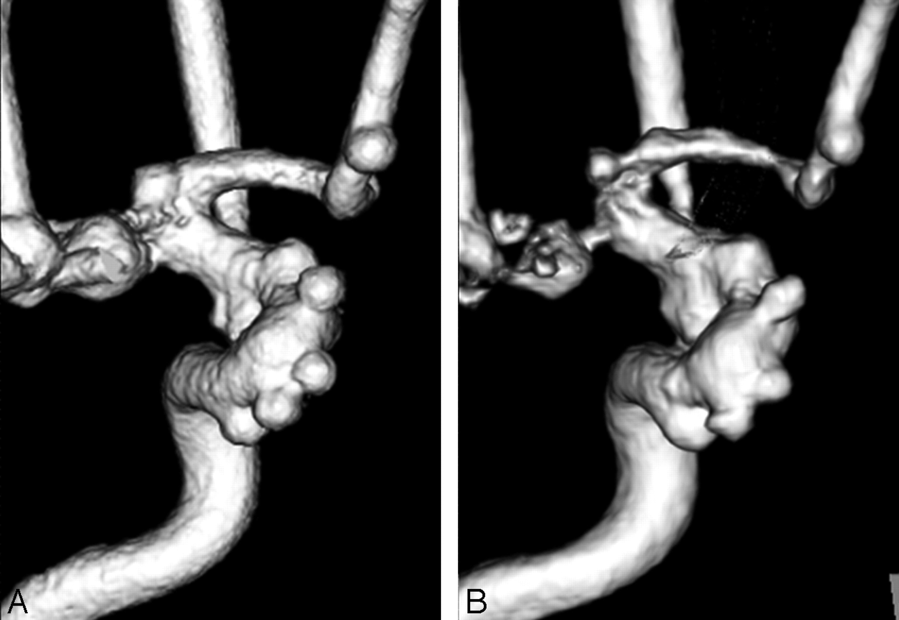
- Fig 4.
Anteroposterior 3D DSA (300 mg I/mL) obtained with the FPD system (A) and with the I.I.-TV system (B) show many aneurysms. For depiction of aneurysms such as the aneurysm neck and the shape of the aneurysm, the 3D DSA with FPD system is superior to that with the I.I.-TV system.
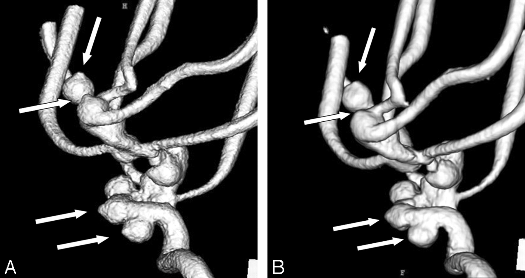
- Fig 5.
3D DSA (300 mg I/mL) from the right posterior oblique view obtained with FPD system (A) and with I.I.-TV system (B) show many aneurysmal blebs (arrows). For depiction of aneurysmal blebs, the 3D DSA with FPD system is superior to that with the I.I.-TV system.


Tables
Contrast Material Concentration & System Grade for Pseudostenosis Artifact Right M1 Left M1 300 mg I/mL FPD 1 1 I.I. 3 3 150 mg I/mL FPD 0 1 I.I. 2 2 Note:—FPD indicates flat panel detector; I.I., image intensifier; M1, sphenoidal segment of middle cerebral artery. Grades: 0, no stenosis; 1, mild stenosis less than 30%; 2, moderate stenosis of 30%–69%; 3, severe stenosis more than 70%.
Contrast Material Concentration & System Score for Image Quality Mean ± SD P* 4 3 2 1 300 mg I/mL FPD (n = 32) 24 5 3 0 3.66 ± 0.65 <.01 I.I. (n = 32) 20 2 5 5 3.16 ± 1.19 150 mg I/mL FPD (n = 32) 28 2 2 0 3.81 ± 0.54 <.01 I.I. (n = 32) 21 3 5 3 3.31 ± 1.06 Note:—FPD indicates flat panel detector; I.I., image intensifier. Scale: 4, no influence; 3, minor distortion of aneurysm; 2, marked distortion of aneurysm; 1, severe distortion of aneurysm.
* Wilcoxon signed rank test.
- Table 3:
Evaluation for the depiction of simulated aneurysms (I.I.-TV system vs FPD system)
Contrast Material Concentration & System Score for Image Quality Mean ± SD P* 5 4 3 2 1 300 mg I/mL FPD (n = 20) 10 3 5 2 0 4.05 ± 1.10 <.01 I.I. (n = 20) 3 4 6 4 3 3.00 ± 1.27 150 mg I/mL FPD (n = 21) 3 10 4 2 2 3.48 ± 1.14 <.01 I.I. (n = 21) 1 5 3 7 5 2.52 ± 1.20 Note:—FPD indicates flat panel detector; I.I., image intensifier. Scale: 5, excellent; 4, more than adequate; 3, adequate; 2, insufficient visualization; 1, not visible.
* Wilcoxon signed rank test.
Contrast Material Concentration & System Score for Image Quality Mean ± SD P* 5 4 3 2 1 300 mg I/mL FPD (n = 11) 4 2 3 2 0 3.73 ± 1.19 <.01 I.I. (n = 1) 0 3 1 3 4 2.27 ± 1.27 150 mg I/mL FPD (n = 1) 1 4 3 2 1 3.18 ± 1.17 <.01 I.I. (n = 1) 0 1 3 3 4 2.09 ± 1.04 Note:—FPD indicates flat panel detector; I.I., image intensifier. Scale: 5, excellent; 4, more than adequate; 3, adequate; 2, insufficient visualization; 1, not visible.
* Wilcoxon signed rank test.
In this issue


Jump to section
Related Articles
Cited By...
- No citing articles found.