
Article Figures & Data
Figures
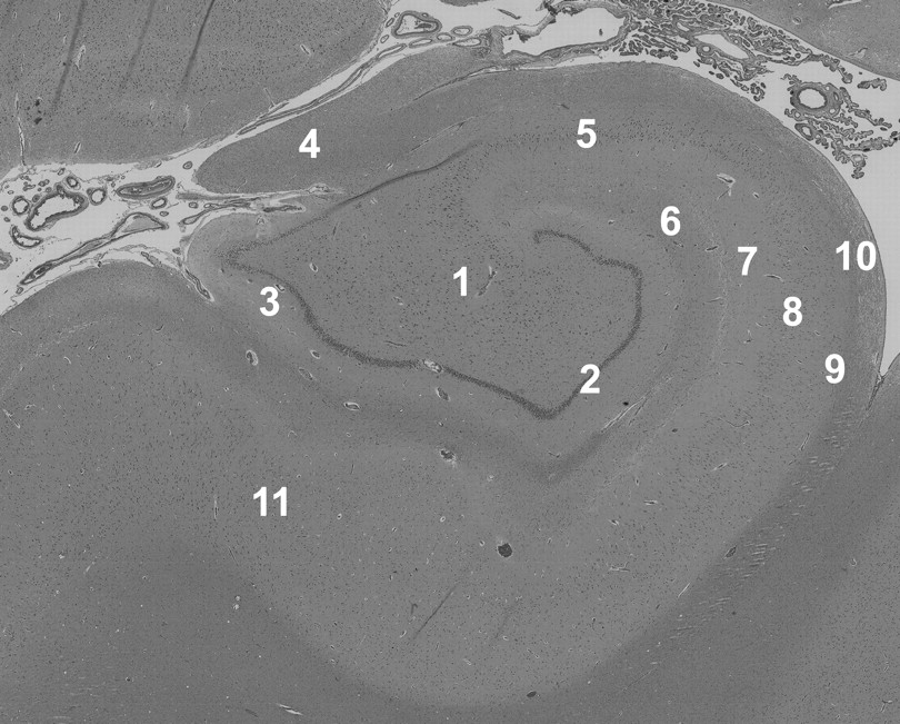
- Fig 1.
Histology demonstrates the laminar anatomy of the human hippocampus. This section was taken adjacent to one of the autopsy samples imaged in this study—no evidence of pathology was seen in any of the tissue samples. 1 indicates hilus; 2, granule cell layer; 3, molecular layer; 4, fimbria; 5, CA3 stratum pyramidale; 6, stratum lacunosum-moleculare; 7, stratum radiatum; 8, CA1 stratum pyramidale; 9, stratum oriens; 10, alveus; 11, subiculum (hematoxylin-eosin, original magnification ×4).
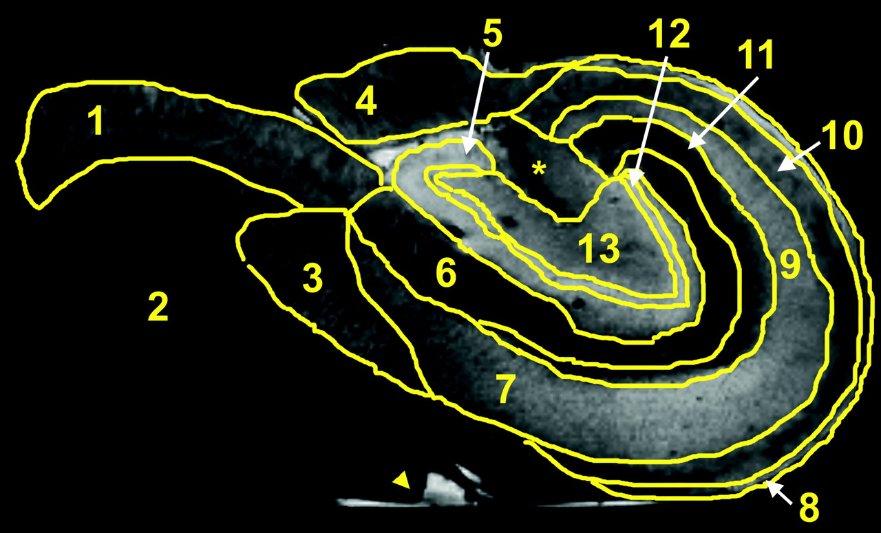
- Fig 2.
Manually segmented regions of interest on a non-diffusion-weighted image of a human hippocampus. These regions include the subiculum (1), the parahippocampal white matter (2), the presubiculum (3), the fimbria (4), the molecular layer (5), the stratum lacunosum-moleculare (6), the CA1 stratum pyramidale (7), the alveus (8), the CA3 stratum pyramidale (9), the stratum oriens (10), the stratum radiatum (11), the granule cell layer (12), and the hilus (13). The asterisk indicates a hippocampal area that could not be assigned unambiguously. The arrowhead indicates some water on the free surface of this particular sample that was not removed by immersion in Fluorinert.
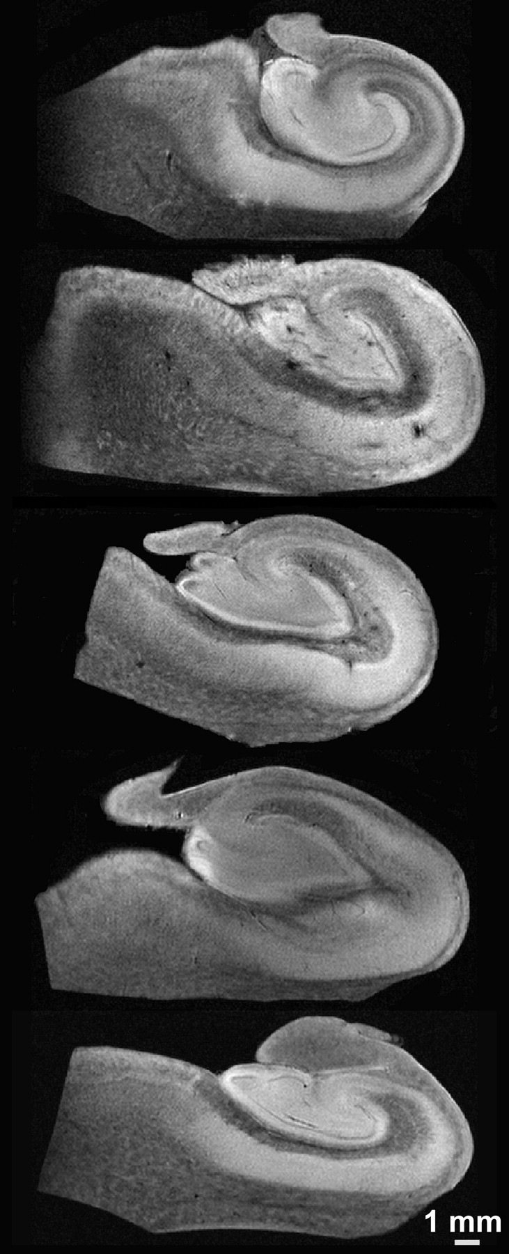
- Fig 3.
Representative 60-μm in-plane resolution diffusion-weighted images (b ∼ 1250 s/mm2) from each of the 5 human hippocampus autopsy specimens. Random diffusion gradient orientations (from the 21 directions acquired for each sample) are shown. The laminar anatomy of the hippocampal body is well resolved.
- Fig 4.
Images of the components of the diffusion tensor visualized on a section from one of the human hippocampus autopsy specimens. The diagonal elements of the tensor (Dxx, Dyy, Dzz) depict the water mobility along the 3 orthogonal directions, x, y, and z, respectively. The apparent differences between the images obtained from these diagonal elements are due to the anisotropy of water diffusion inside the sample, where the higher intensity voxels correspond to regions with higher (or less restricted) diffusion. These images are scaled between the values of zero and 8 × 10⁻⁴ mm2/s. The off-diagonal elements (Dxy, Dxz, Dyz) indicate the correlations of diffusional motion along 2 orthogonal directions. Because the off-diagonal elements can take negative values, these images are scaled between −2 × 10⁻⁴ and 2 × 10⁻⁴ mm2/s—hence the gray background.
- Fig 5.
Sixty-micrometer in-plane resolution images of no diffusion-weighting (S0), MD, and FA in the human hippocampus. In the S0 image, the layers of the hippocampus have different T2s and proton densities. In the MD image, water diffusion appeared highest (light) in regions of densely packed cells and lowest (dark) in coherent white matter such as the fimbria. Intermediate diffusivity was observed in the hilus, stratum radiatum, or subiculum. FA demonstrates excellent resolution of the hippocampal lamina. Diffusion anisotropy is highest in the fimbria, stratum oriens, alveus, and white matter from the parahippocampal gyrus. Anisotropy is lowest in the pyramidal and granule cell layers. Note also the division of the molecular layer into inner and outer layers on the basis of FA differences. 1 indicates molecular layer; 2, granule cell layer; 3, hilus; 4, fimbria; 5, CA3 stratum pyramidale; 6, alveus; 7, stratum lacunosum-moleculare; 8, stratum radiatum; 9, stratum oriens; 10, CA1 stratum pyramidale; 11, subiculum; 12, white matter.
- Fig 6.
Comparison of MD (×10−4 mm2/s) (A) and FA (no units) (B) for the different regions of the hippocampus (Mean ± standard error of the mean, 5 hippocampi). The MD and FA of the fimbria (asterisk) are statistically different from all other regions (Tukey multiple comparisons tests, P < .01). SUB indicates subiculum; SP, stratum pyramidale; SR/LM, stratum radiatum lacunosum-moleculare; SO, stratum oriens; GCL, granule cell layer; ML, molecular layer; HIL, hilus; FIM, fimbria.
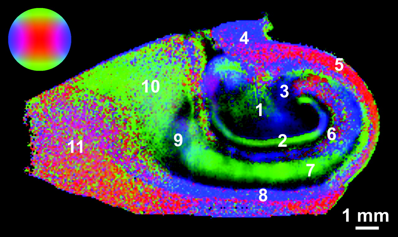
- Fig 7.
Color fiber-orientation maps of the human hippocampus derived from diffusion tensor microscopy data provide excellent contrast resolution for the different layers of the hippocampus. Color intensity is proportional to the FA within that pixel. The colorball indicates the direction of the principal eigenvector, which can be transverse (blue), vertical (green), or through the plane of the image (red). 1 indicates hilus; 2 , molecular layer; 3, stratum lucidum; 4, fimbria; 5, alveus, 6; stratum lacunosum-moleculare; 7, stratum radiatum/pyramidale; 8, stratum oriens; 9, presubiculum; 10, subiculum; 11, white matter.


In this issue


Jump to section
Related Articles
Cited By...
- Diffusion MRI of the Hippocampus
- Automated Surface-Based Segmentation of Deep Gray Matter Regions Based on Diffusion Tensor Images Reveals Unique Age Trajectories Over the Healthy Lifespan
- Mapping the Macrostructure and Microstructure of the in vivo Human Hippocampus using Diffusion MRI
- Differences in Microstructural Alterations of the Hippocampus in Alzheimer Disease and Idiopathic Normal Pressure Hydrocephalus: A Diffusion Tensor Imaging Study
- Hippocampal CA1 apical neuropil atrophy in mild Alzheimer disease visualized with 7-T MRI