Advertisement

Main menu

  • Home
  • Content
    • Current Issue
    • Accepted Manuscripts
    • Article Preview
    • Past Issue Archive
    • AJNR Case Collection
    • Case of the Week Archive
    • Classic Case Archive
    • Case of the Month Archive
  • Special Collections
    • Spinal CSF Leak Articles (Jan 2020-June 2024)
    • 2024 AJNR Journal Awards
    • Most Impactful AJNR Articles
  • Multimedia
    • AJNR Podcast
    • AJNR Scantastics
    • Video Articles
  • For Authors
    • Submit a Manuscript
    • Author Policies
    • Fast publishing of Accepted Manuscripts
    • Graphical Abstract Preparation
    • Manuscript Submission Guidelines
    • Imaging Protocol Submission
    • Submit a Case for the Case Collection
  • About Us
    • About AJNR
    • Editorial Board
  • More
    • Become a Reviewer/Academy of Reviewers
    • Subscribers
    • Permissions
    • Alerts
    • Feedback
    • Advertisers
    • ASNR Home
  • Other Publications
    • ajnr

User menu

  • Alerts
  • Log in

Search

  • Advanced search
American Journal of Neuroradiology

American Journal of Neuroradiology

ASHNR American Society of Functional Neuroradiology ASHNR American Society of Pediatric Neuroradiology ASSR
  • Alerts
  • Log in

Advanced Search

  • Home
  • Content
    • Current Issue
    • Accepted Manuscripts
    • Article Preview
    • Past Issue Archive
    • AJNR Case Collection
    • Case of the Week Archive
    • Classic Case Archive
    • Case of the Month Archive
  • Special Collections
    • Spinal CSF Leak Articles (Jan 2020-June 2024)
    • 2024 AJNR Journal Awards
    • Most Impactful AJNR Articles
  • Multimedia
    • AJNR Podcast
    • AJNR Scantastics
    • Video Articles
  • For Authors
    • Submit a Manuscript
    • Author Policies
    • Fast publishing of Accepted Manuscripts
    • Graphical Abstract Preparation
    • Manuscript Submission Guidelines
    • Imaging Protocol Submission
    • Submit a Case for the Case Collection
  • About Us
    • About AJNR
    • Editorial Board
  • More
    • Become a Reviewer/Academy of Reviewers
    • Subscribers
    • Permissions
    • Alerts
    • Feedback
    • Advertisers
    • ASNR Home
  • Follow AJNR on Twitter
  • Visit AJNR on Facebook
  • Follow AJNR on Instagram
  • Join AJNR on LinkedIn
  • RSS Feeds

Welcome to the new AJNR, Updated Hall of Fame, and more. Read the full announcements.


AJNR is seeking candidates for the position of Associate Section Editor, AJNR Case Collection. Read the full announcement.


OtherHEAD & NECK

Severe Diffuse Systemic Amyloidosis with Involvement of the Pharynx, Larynx, and Trachea: CT and MR Findings

R. Gilad, P. Milillo and P.M. Som
American Journal of Neuroradiology September 2007, 28 (8) 1557-1558; DOI: https://doi.org/10.3174/ajnr.A0604
R. Gilad
  • Find this author on Google Scholar
  • Find this author on PubMed
  • Search for this author on this site
P. Milillo
  • Find this author on Google Scholar
  • Find this author on PubMed
  • Search for this author on this site
P.M. Som
  • Find this author on Google Scholar
  • Find this author on PubMed
  • Search for this author on this site
  • Article
  • Figures & Data
  • Info & Metrics
  • Responses
  • References
  • PDF
Loading

Abstract

SUMMARY: Amyloidosis is a term applied to a diverse group of disorders that share the deposition of amyloid protein in various extracellular tissues. Systemic amyloidosis may involve almost any organ system in the body including regions in the head and neck; however, pharyngeal involvement is rare, with only 12 cases having been previously reported. Ten of these cases were localized disease, and only 2 cases were systemic amyloidosis. We present the case of a patient with severe diffuse systemic amyloidosis with extensive involvement of the pharynx, larynx, trachea, lungs, eyelids, and breasts. We also review the imaging characteristics and pertinent literature.

Amyloidosis is a rare, infiltrative condition characterized by deposition of abnormal protein in various tissues. When systemic amyloidosis involves regions of the head and neck, the larynx and trachea are the most common sites. To date, there have been only 2 prior reported cases of systemic amyloidosis involving the pharynx. We present a case of severe diffuse systemic amyloidosis in the chest, head and neck, with extensive involvement of the pharynx and we review the literature.

Case Report

The patient was a 64-year-old woman with a history of myasthenia gravis for 30 years, type 1 diabetes mellitus, and amyloidosis for 4 years. In the past, the patient was found to have amyloid deposits in the eyelids and breasts. A biopsy confirmed findings consistent with the AL type (amyloid light chain) of amyloidosis. The patient presented with increased shortness of breath and hoarseness on the most recent hospital admission. There was no past medical history of renal failure or multiple myeloma. The patient was being treated for myasthenia gravis with pyridostigmine (Mestinon) and high-dose steroids and had received plasmapheresis therapy in the past. Physical examination was significant for bilateral ptoses and indurations of the eyelids, pharyngeal fullness, shortness of breath, and a decreased range of motion of the neck. Her urine was negative for Bence-Jones protein, and the results of serum protein and urine protein electrophoreses were unremarkable.

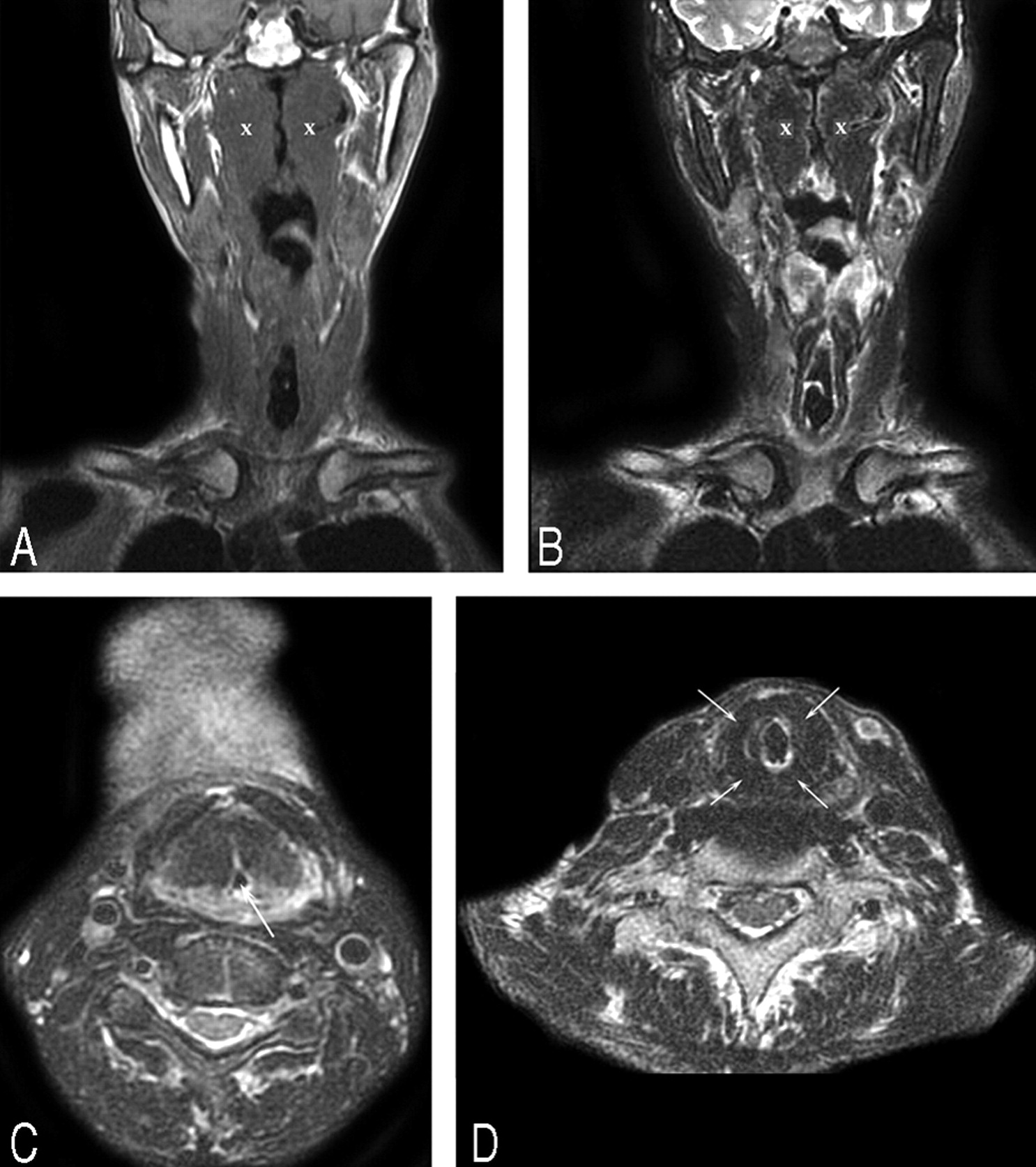
A tracheostomy was performed, and a high-resolution chest CT study showed extensive interstitial lung disease, abnormally attenuated breast tissue (Fig 1) for the patient's stated age, and small nodules in the tracheal mucosa with marked thickening of the tracheal wall with significant stenosis (not shown). An endoscopic examination of the trachea revealed nodular deposits and plaques on the mucosal surface along its entire length. Noncontrast MR imaging of the neck was performed on a 1.5T magnet. Contrast was not administered because of a reported allergy to gadolinium. The images showed large submucosal, nodular masses in the pharynx and supraglottic larynx, and thickening of the tracheal wall with stenosis. These masses had intermediate T1-weighted signal intensity (Figs 2A) and low T2-weighted signal intensity (Figs 2B, C, D). The large submucosal supraglottic laryngeal deposits were the primary cause of her airway obstruction. These findings on imaging were consistent with systemic amyloidosis. Biopsy specimens of the tracheal wall deposits on the mucosal surface confirmed the diagnosis of AL-type amyloidosis.

Fig 1.
  • Download figure
  • Open in new tab
  • Download powerpoint
Fig 1.

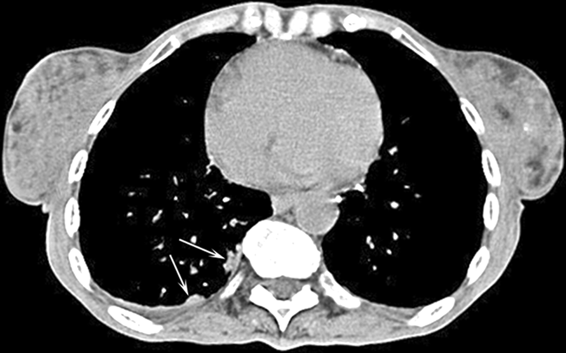
Axial noncontrast CT of the chest shows abnormally attenuated nodular breast tissue bilaterally and focal nodularity (arrows) along the right posterior pleura.

Fig 2.
  • Download figure
  • Open in new tab
  • Download powerpoint
Fig 2.

Noncontrast MR images show large nodular, submucosal masses along the pharyngeal walls, supraglottic larynx, and thickening of the tracheal wall with stenosis. Note the extensive involvement of the nasopharynx and oropharynx. A, Coronal T1-weighted MR image shows the pharyngeal masses (x) to have intermediate signal intensity. B, Coronal T2-weighted MR image shows the masses (x) to have low signal intensity. The signal intensities of the masses parallel those of skeletal muscle. C, Axial T2-weighted MR image shows the masses to have low signal intensity. There is marked narrowing of the laryngeal airway (arrow). D, Axial T2-weighted MR image shows marked low signal intensity thickening (arrows) of the tracheal walls.

Discussion

Amyloidosis is a condition characterized by infiltration and extracellular deposition of homogeneous, eosinophilic proteinaceous fibrils in a beta-pleated sheet configuration. It classically has an apple-green birefringence when stained with Congo red and viewed under polarized light. From a clinical standpoint, it may be classified as a localized or systemic disease. The localized disease involves only 1 organ, typically the larynx or brain (where it has been associated with aging and Alzheimer disease). Systemic amyloidosis involves many organ systems and is typically divided into 3 separate categories. Primary amyloidosis (AL type or amyloid light chain) is the result of spontaneous overproduction of the antibody light chain in the bone marrow plasma cell. Secondary amyloidosis (AA type or amyloid A protein) occurs as a result of an underlying condition such as multiple myeloma; chronic infections (tuberculosis or osteomyelitis); and chronic inflammatory diseases such as rheumatoid arthritis, Sjögren disease, and ankylosing spondylitis. The familial type (ATTR type, or amyloid transthyretin protein) is rare and exhibits an autosomal dominant pattern of inheritance, and the amyloid deposits are composed of the protein transthyretin, which is made in the liver. There are 12 other protein subtypes that have been identified but are far less common.1–4

From a radiographic standpoint, MR imaging is the technique of choice to demonstrate the most specific features of amyloidosis. Typically, the amyloid deposits have intermediate T1-weighted signal intensity and low T2-weighted signal intensity, similar to skeletal muscle. This is secondary to the beta-pleated structure of the amyloid deposits that occurs as protein fibrils in a parallel sheetlike configuration, similar to the organization of skeletal muscle fibers. Because of these unique imaging features, MR imaging is considered to be a more specific technique than CT to establish the diagnosis of amyloidosis.1,5

In systemic primary amyloidosis, deposits in the region of the head and neck are uncommon. However, the most common head and neck sites are the supraglottic and subglottic larynx as well as the thyroid gland.4,6,7 There were 12 previous cases that date back to 1980.8 Of these 12 cases, 10 were of localized disease of the nasopharynx. In the cases of localized disease, systemic amyloidosis was ruled out by either abdominal or rectal biopsy.2–10 There were only 2 previous cases in the literature of systemic amyloidosis (both AL type) that involved the pharynx.11,12 The current case is an example of a patient with severe systemic amyloidosis with diffuse involvement of the pharynx.

Amyloidosis of the breasts is also a rare entity and has been described in only 35 cases since 1973.13 Ten of these cases were of the AL systemic type and were not associated with an underlying condition.13 In systemic amyloidosis, involvement of the eyelids can occur, but orbital involvement is uncommon.14 It is usually localized, and the imaging features are similar to those described above.14

Amyloidosis, either systemic or localized, may affect the upper and lower respiratory tract.15 In amyloidosis of the lung, the imaging characteristics are diverse and therefore may mimic a variety of pathologic processes in the lung.16 Tracheobronchial amyloidosis is characterized by amyloid deposits in the trachea and large bronchi, with extension at times into segmental bronchi.17 Tracheal stenosis often results from thickening of the tracheal wall. Any inflammatory or granulomatous process demonstrates similar features as amyloid on CT.18,19 However, on MR imaging, the signal intensities mentioned above should suggest the diagnosis.13,20

Systemic amyloidosis carries a poor prognosis because accumulation of amyloid fibrils in the tissues destroys normal structure and function. However, the localized form of amyloidosis carries a much better prognosis. Therefore, it is important to identify the presence of amyloid on imaging by using the characteristics described and then determine whether this is localized or systemic disease.

Conclusion

Amyloidosis is a rare, infiltrative condition characterized by deposition of abnormal protein in various tissues. When systemic amyloidosis involves regions of the head and neck, the larynx and trachea are the most common sites. To date, there have been only 2 previous reported cases of systemic amyloidosis involving the pharynx. We described the case of a patient with severe systemic amyloidosis who presented with airway obstruction as a result of extensive involvement of the larynx, trachea, nasopharynx, and oropharynx. We also reported the findings on imaging. There was also a review of the literature of amyloid deposits in the pharynx.

References

  1. ↵
    Som PM, Curtin HD, eds. Head and Neck Imaging. 3rd ed. St. Louis: Mosby;1996
  2. ↵
    Ghekiere O, Desuter G, Weynand B, et al. Hypopharyngeal amyloidoma. AJR Am J Roentgenol 2003;181:1720–21
    PubMed
  3. Takahashi M, Matsuda H, Ito K, et al. Pediatric localized amyloidosis in mesopharynx. Int J Pediatr Otorhinolaryngol 2002;66:181–84
    CrossRefPubMed
  4. ↵
    Patel A, Pambuccian S, Maisel R. Nasopharyngeal amyloidosis. Am J Otolaryngol 2002;23:308–11
    CrossRefPubMed
  5. ↵
    Hegarty JL, Rao VM. Amyloidoma of the nasopharynx: CT and MR findings. AJNR Am J Neuroradiol 1993;14:215–18
    Abstract/FREE Full Text
  6. ↵
    Penner CR, Muller S. Head and neck amyloidosis: a clinicopathologic study of 15 cases. Oral Oncol 2006;42:421–29
    CrossRefPubMed
  7. ↵
    Zhuang YL, Tsai TL, Lin CZ. Localized amyloid deposition in the nasopharynx and neck, mimicking nasopharyngeal carcinoma with neck metastasis. J Chin Med Assoc 2005;68:142–45
    PubMed
  8. ↵
    Panda NK, Sharma SC, Mann SB, et al. Localized amyloidosis of the nasopharynx. Ear Nose Throat J 1994;73:335–36
    PubMed
  9. Pitkaranta A, Malmberg H. Localized amyloid tumor of the nasopharynx. Otolaryngol Head Neck Surg 2000;122:309–10
    CrossRefPubMed
  10. ↵
    Dominguez S, Weinberg P, Claros P, et al. Primary localized nasopharyngeal amyloidosis. A case report. Int J Pediatr Otorhinolaryngol 1996;36:61–67
    CrossRefPubMed
  11. ↵
    Chadwick MA, Buckland JR, Mason P, et al. A rare case of dysphagia: hypopharyngeal amyloidosis masquerading as a post-cricoid tumour. J Laryngol Otol 2002;116:54–56
    CrossRefPubMed
  12. ↵
    Lim JS, Lebowitz RA, Jacobs JB. Primary amyloidosis presenting as a nasopharyngeal mass. Am J Rhinol 1999;13:209–12
    CrossRefPubMed
  13. ↵
    Röcken C, Kronsbein H, Sletten K, et al. Amyloidosis of the breast. Virchows Arch 2002;440:527–35
    CrossRefPubMed
  14. ↵
    Holmstrom GE, Nyman KG. Primary orbital amyloidosis localised to an extraocular muscle. Br J Ophthalmol 1987;71:32–33
    Abstract/FREE Full Text
  15. ↵
    Murdoch IE, Sullivan TJ, Moseley I, et al. Primary localised amyloidosis of the orbit. Br J Ophthalmol 1996;80:1083–86
    Abstract/FREE Full Text
  16. ↵
    Utz JP, Swensen SJ, Gertz MA. Pulmonary amyloidosis. The Mayo Clinic experience from 1980 to 1993. Ann Intern Med 1996;124:407–13
    PubMed
  17. ↵
    Lachmann HJ, Hawkins PN. Amyloidosis and the lung. Chron Respir Dis 2006;3:203–14
    CrossRefPubMed
  18. ↵
    Thompson PJ, Citron KM. Amyloid and the lower respiratory tract. Thorax 1983;38:84–87
    FREE Full Text
  19. ↵
    Webb EM, Elicker BM, Webb WR. Using CT to diagnose nonneoplastic tracheal abnormalities: appearance of the tracheal wall. AJR Am J Roentgenol 2000;174:1315–21
    CrossRefPubMed
  20. ↵
    Cordier JF, Loire R, Brune J. Amyloidosis of the lower respiratory tract. Clinical and pathologic features in a series of 21 patients. Chest 1986;90:827–31
    CrossRefPubMed
  • Received November 30, 2006.
  • Accepted after revision January 20, 2007.
  • Copyright © American Society of Neuroradiology
View Abstract
PreviousNext
Back to top

In this issue

American Journal of Neuroradiology: 28 (8)
American Journal of Neuroradiology
Vol. 28, Issue 8
September 2007
  • Table of Contents
  • Index by author
Advertisement
Print
Download PDF
Email Article

Thank you for your interest in spreading the word on American Journal of Neuroradiology.

NOTE: We only request your email address so that the person you are recommending the page to knows that you wanted them to see it, and that it is not junk mail. We do not capture any email address.

Enter multiple addresses on separate lines or separate them with commas.
Severe Diffuse Systemic Amyloidosis with Involvement of the Pharynx, Larynx, and Trachea: CT and MR Findings
(Your Name) has sent you a message from American Journal of Neuroradiology
(Your Name) thought you would like to see the American Journal of Neuroradiology web site.
CAPTCHA
This question is for testing whether or not you are a human visitor and to prevent automated spam submissions.
Cite this article
R. Gilad, P. Milillo, P.M. Som
Severe Diffuse Systemic Amyloidosis with Involvement of the Pharynx, Larynx, and Trachea: CT and MR Findings
American Journal of Neuroradiology Sep 2007, 28 (8) 1557-1558; DOI: 10.3174/ajnr.A0604

Citation Manager Formats

  • BibTeX
  • Bookends
  • EasyBib
  • EndNote (tagged)
  • EndNote 8 (xml)
  • Medlars
  • Mendeley
  • Papers
  • RefWorks Tagged
  • Ref Manager
  • RIS
  • Zotero
0 Responses
Respond to this article
Share
Bookmark this article
Severe Diffuse Systemic Amyloidosis with Involvement of the Pharynx, Larynx, and Trachea: CT and MR Findings
R. Gilad, P. Milillo, P.M. Som
American Journal of Neuroradiology Sep 2007, 28 (8) 1557-1558; DOI: 10.3174/ajnr.A0604
del.icio.us logo Twitter logo Facebook logo Mendeley logo
  • Tweet Widget
  • Facebook Like
  • Google Plus One
Purchase

Jump to section

  • Article
    • Abstract
    • Case Report
    • Discussion
    • Conclusion
    • References
  • Figures & Data
  • Info & Metrics
  • Responses
  • References
  • PDF

Related Articles

  • No related articles found.
  • PubMed
  • Google Scholar

Cited By...

  • Localised laryngotracheal amyloidosis: a differential diagnosis not to forget
  • Crossref (43)
  • Google Scholar

This article has been cited by the following articles in journals that are participating in Crossref Cited-by Linking.

  • Imaging appearance of granulomatous lesions of head and neck
    Ahmed Abdel Khalek Abdel Razek, Mauricio Castillo
    European Journal of Radiology 2010 76 1
  • CT of Diffuse Tracheal Diseases
    Jonathan H. Chung, Jeffrey P. Kanne, Matthew D. Gilman
    American Journal of Roentgenology 2011 196 3
  • Larynx
    Benjamin Y. Huang, Michael Solle, Mark C. Weissler
    Otolaryngologic Clinics of North America 2012 45 6
  • Long-term follow-up after surgery in localized laryngeal amyloidosis
    Aldert J. C. Hazenberg, Bouke P. C. Hazenberg, Frederik G. Dikkers
    European Archives of Oto-Rhino-Laryngology 2016 273 9
  • Laryngeal amyloidosis: diagnosis, pathophysiology and management
    N M Phillips, E Matthews, C Altmann, J Agnew, H Burns
    The Journal of Laryngology & Otology 2017 131 S2
  • Clinical characteristics of laryngeal versus nonlaryngeal amyloidosis
    Shannon F. Rudy, Caroline C. Jeffery, Edward J. Damrose
    The Laryngoscope 2018 128 3
  • MDCT of Trachea and Main Bronchi
    Cylen Javidan-Nejad
    Radiologic Clinics of North America 2010 48 1
  • Easier to Swallow: Pictorial Review of Structural Findings of the Pharynx at Barium Pharyngography
    Ting Y. Tao, Christine O. Menias, Thomas E. Herman, William H. McAlister, Dennis M. Balfe
    RadioGraphics 2013 33 7
  • Amyloidosis in the head and neck: CT findings with clinicopathological correlation
    Koji Takumi, Pedro V. Staziaki, Rania Hito, Rohini N. Nadgir, John L. Berk, V. Carlota Andreu-Arasa, Wilson Chavez, Osamu Sakai
    European Journal of Radiology 2020 128
  • Intensity-modulated radiotherapy for localized nasopharyngeal amyloidosis
    Ming Luo, Gang Peng, Liangliang Shi, Xing Ming, Zhenyu Li, Shijiang Fei, Qian Ding, Jing Cheng
    Strahlentherapie und Onkologie 2016 192 12

More in this TOC Section

  • WHO Classification Update: Nasal&Skull Base Tumors
  • Peritumoral Signal in Vestibular Schwannomas
  • Chondrosarcoma vs Synovial Chondromatosis: Imaging
Show more HEAD & NECK

Similar Articles

Advertisement

Indexed Content

  • Current Issue
  • Accepted Manuscripts
  • Article Preview
  • Past Issues
  • Editorials
  • Editors Choice
  • Fellow Journal Club
  • Letters to the Editor

Cases

  • Case Collection
  • Archive - Case of the Week
  • Archive - Case of the Month
  • Archive - Classic Case

Special Collections

  • Special Collections

Resources

  • News and Updates
  • Turn around Times
  • Submit a Manuscript
  • Author Policies
  • Manuscript Submission Guidelines
  • Evidence-Based Medicine Level Guide
  • Publishing Checklists
  • Graphical Abstract Preparation
  • Imaging Protocol Submission
  • Submit a Case
  • Become a Reviewer/Academy of Reviewers
  • Get Peer Review Credit from Publons

Multimedia

  • AJNR Podcast
  • AJNR SCANtastic
  • Video Articles

About Us

  • About AJNR
  • Editorial Board
  • Not an AJNR Subscriber? Join Now
  • Alerts
  • Feedback
  • Advertise with us
  • Librarian Resources
  • Permissions
  • Terms and Conditions

American Society of Neuroradiology

  • Not an ASNR Member? Join Now

© 2025 by the American Society of Neuroradiology All rights, including for text and data mining, AI training, and similar technologies, are reserved.
Print ISSN: 0195-6108 Online ISSN: 1936-959X

Powered by HighWire