Advertisement

Main menu

  • Home
  • Content
    • Current Issue
    • Accepted Manuscripts
    • Article Preview
    • Past Issue Archive
    • AJNR Case Collection
    • Case of the Week Archive
    • Classic Case Archive
    • Case of the Month Archive
  • Special Collections
    • Spinal CSF Leak Articles (Jan 2020-June 2024)
    • 2024 AJNR Journal Awards
    • Most Impactful AJNR Articles
  • Multimedia
    • AJNR Podcast
    • AJNR Scantastics
    • Video Articles
  • For Authors
    • Submit a Manuscript
    • Author Policies
    • Fast publishing of Accepted Manuscripts
    • Graphical Abstract Preparation
    • Manuscript Submission Guidelines
    • Imaging Protocol Submission
    • Submit a Case for the Case Collection
  • About Us
    • About AJNR
    • Editorial Board
  • More
    • Become a Reviewer/Academy of Reviewers
    • Subscribers
    • Permissions
    • Alerts
    • Feedback
    • Advertisers
    • ASNR Home
  • Other Publications
    • ajnr

User menu

  • Alerts
  • Log in

Search

  • Advanced search
American Journal of Neuroradiology

American Journal of Neuroradiology

ASHNR American Society of Functional Neuroradiology ASHNR American Society of Pediatric Neuroradiology ASSR
  • Alerts
  • Log in

Advanced Search

  • Home
  • Content
    • Current Issue
    • Accepted Manuscripts
    • Article Preview
    • Past Issue Archive
    • AJNR Case Collection
    • Case of the Week Archive
    • Classic Case Archive
    • Case of the Month Archive
  • Special Collections
    • Spinal CSF Leak Articles (Jan 2020-June 2024)
    • 2024 AJNR Journal Awards
    • Most Impactful AJNR Articles
  • Multimedia
    • AJNR Podcast
    • AJNR Scantastics
    • Video Articles
  • For Authors
    • Submit a Manuscript
    • Author Policies
    • Fast publishing of Accepted Manuscripts
    • Graphical Abstract Preparation
    • Manuscript Submission Guidelines
    • Imaging Protocol Submission
    • Submit a Case for the Case Collection
  • About Us
    • About AJNR
    • Editorial Board
  • More
    • Become a Reviewer/Academy of Reviewers
    • Subscribers
    • Permissions
    • Alerts
    • Feedback
    • Advertisers
    • ASNR Home
  • Follow AJNR on Twitter
  • Visit AJNR on Facebook
  • Follow AJNR on Instagram
  • Join AJNR on LinkedIn
  • RSS Feeds

Welcome to the new AJNR, Updated Hall of Fame, and more. Read the full announcements.


AJNR is seeking candidates for the position of Associate Section Editor, AJNR Case Collection. Read the full announcement.


Research ArticleHead and Neck

Intra- and Extracranial Solitary Fibrous Tumor of the Trigeminal Nerve: CT and MR Imaging Appearance

K. Motoori, T. Hanazawa, I. Yamakami, A. Sugimoto and H. Ito
American Journal of Neuroradiology February 2010, 31 (2) 280-281; DOI: https://doi.org/10.3174/ajnr.A1702
K. Motoori
  • Find this author on Google Scholar
  • Find this author on PubMed
  • Search for this author on this site
T. Hanazawa
  • Find this author on Google Scholar
  • Find this author on PubMed
  • Search for this author on this site
I. Yamakami
  • Find this author on Google Scholar
  • Find this author on PubMed
  • Search for this author on this site
A. Sugimoto
  • Find this author on Google Scholar
  • Find this author on PubMed
  • Search for this author on this site
H. Ito
  • Find this author on Google Scholar
  • Find this author on PubMed
  • Search for this author on this site
  • Article
  • Figures & Data
  • Info & Metrics
  • Responses
  • References
  • PDF
Loading

Abstract

SUMMARY: We describe a rare case of SFT existing along the mandibular division of the trigeminal nerve and extending down into the infratemporal fossa through the foramen ovale. The tumor showed heterogeneous hypointensity on T2-weighted images and marked enhancement on CT and MR images.

Abbreviations

EMA
epithelial membrane antigen
SFT
solitary fibrous tumor
SMA
smooth muscle antigen.

SFT is an uncommon neoplasm and has been reported with increasing frequency in various anatomic sites, including the central nervous system.1–7 We present an SFT arising along the fifth cranial nerve.

Case Report

The patient was a 49-year-old woman with a several-month history of left-sided mandibular hypoesthesia and trismus. Contrast-enhanced CT demonstrated a large dumbbell-shaped mass extending from the left middle temporal fossa into the infratemporal fossa via an enlarged foramen ovale. The tumor had a well-defined margin and showed heterogeneous enhancement. The mass pushed against the lateral wall of the left maxillary sinus and the pterygoid process without destruction (Fig 1). MR imaging was performed to provide delineation of the location and size of the tumor and the relationship to the surrounding structures. It revealed a solid tumor, 55 × 32 × 84 mm, arising from the middle temporal fossa and extending into the infratemporal fossa. The temporal lobe was pushed aside by intracranial extension and showed hyperintensities on T2-weighted images. The tumor was isointense on T1-weighted imaging with hypointense foci, suggesting flow voids. On T2-weighted images, the tumor showed heterogeneous hypointensity. Most of the tumor showed isointense on T1-weighted imaging with hypointense foci, suggesting flow voids, and heterogeneous hypointensity on T2-weighted imaging. In contrast, the signal intensities of the lateral side of the intracranial component were higher than those of the rest of the tumor on T1- and T2-weighted images. This site showed marked enhancement and early washout on a dynamic contrast study. The rest of the tumor showed early enhancement and poor washout (Fig 2).

Fig 1.
  • Download figure
  • Open in new tab
  • Download powerpoint
Fig 1.

A, Axial contrast-enhanced CT scan shows an enlarged left foramen ovale (arrows) by the enhanced tumor. B, Axial contrast-enhanced CT scan of the caudal side of A shows the tumor compressing the maxillary bone, left pterygoid plate, and mandible (arrows), without destruction.

Fig 2.
  • Download figure
  • Open in new tab
  • Download powerpoint
Fig 2.

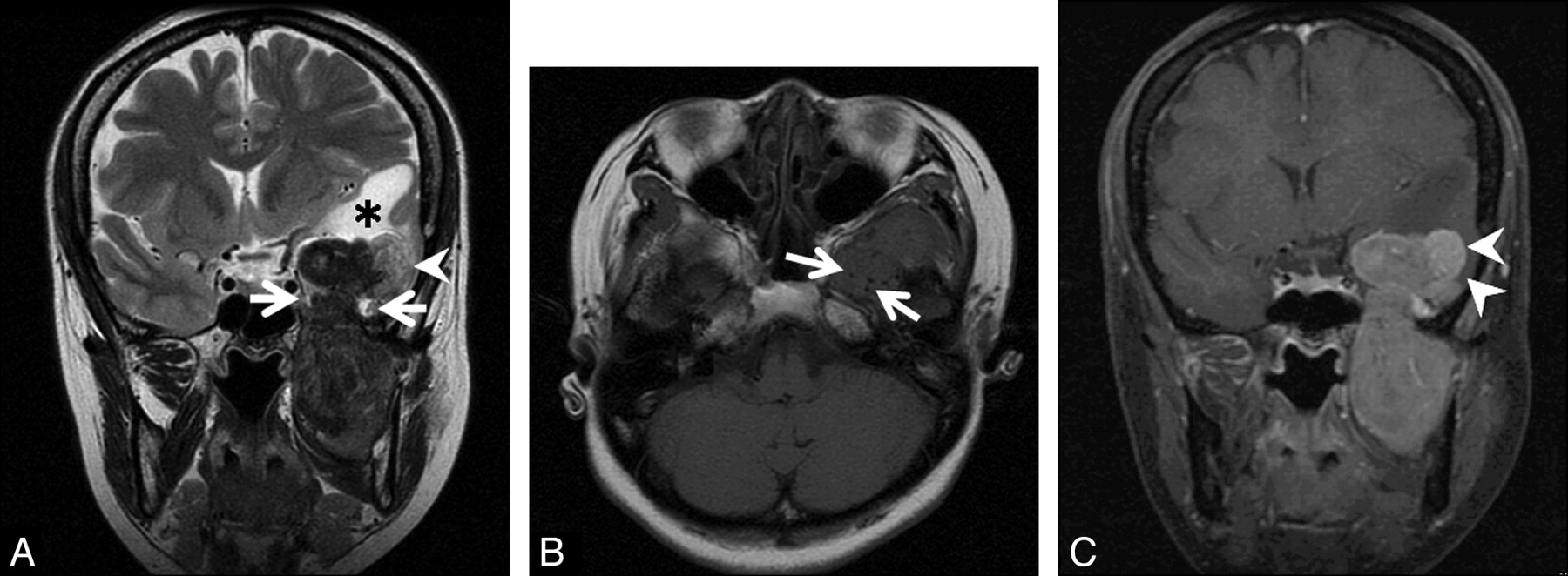
A, Coronal T2-weighted image shows a dumbbell-shaped tumor passing through left foramen ovale (arrows). The tumor is mainly hypointense to white matter. The lateral part of the intracranial component (arrowhead) shows higher intensities than other parts of the tumor. White matter of the left temporal lobe shows high intensities (asterisk). B, Axial T1-weighted image shows isointensity of the tumor to the brain. The tumor has low-intensity foci, suggesting a flow-void condition (arrows). C, On a coronal contrast-enhanced fat-suppressed T1-weighted image, the tumor shows heterogeneous enhancement. The lateral part of the intracranial component shows different strong enhancement (arrowheads).

Biopsy of the tumor was performed via the left maxillary sinus. The microscopic appearance with hematoxylin-eosin staining revealed spindle cells in a characteristic “patternless pattern,” with amorphous areas of collagen and haphazardly arranged cells. Immunohistochemical testing showed the neoplastic cells to be positive for CD34 and vimentin and negative for SMA, S100, EMA, desmin, and c-kit. On the basis of these findings, a diagnosis of SFT was made. Complete surgical excision was achieved via left frontotemporal craniotomy with zygomectomy. There was no cerebral invasion or adhesion.

Discussion

In 1931, Klemperer and Rabin8 reported SFT as a localized fibrous tumor arising in the pleura. The further development of electron microscopy and immunohistochemistry clarified that this tumor was not from mesothelial but rather from mesenchymal origin, and it is now referred to as a SFT, different from a mesothelial tumor. Although initially most SFTs were reported to arise within the thoracic cavity, recently they have been more frequently reported in other sites and organs. The diagnosis of SFT rests on a combination of histopathologic and immunohistochemical features. Because SFT shows such a variety of patterns on radiologic imaging, it would be difficult to make a correct presurgical diagnosis.9 Most SFTs present as well-circumscribed and often encapsuled lesions of 1–25 cm. On sections, they frequently show a multinodular, whitish, and firm appearance. In addition, myxoid and hemorrhagic changes are occasionally observed. Histopathologically, SFTs typically exhibit a patternless architecture characterized by the coexistence of hypo- and hypercellular areas, which are separated by fibrous stroma having hemangiopericytoma-like branching blood vessels. Immunohistochemically, SFTs tend to be positive for CD34, CD99, and BCL2. They are usually negative for S100 protein, cytokeratin, and/or desmin.9

On CT scans, SFTs usually appear as well-circumscribed, round, or lobulated masses with attenuation of soft tissue.9,10 On T1-weighted MR images, they are usually isointense to the muscle.9 On T2-weighted images, SFTs are mainly hypointense, corresponding to fibrous stroma intermixed with prominent vascular channels.2,9,10 In our case, SFT had rich collagenous stroma with hyalinization. After intravenous contrast administration, the tumor usually shows homogeneous enhancement but also sometimes heterogeneous enhancement, corresponding to the cellularity, stromal components, and degeneration. The enhancement can be from moderate to strong, presumably depending on the cellularity, stromal component, and vascularity. In a tumor with strong enhancement, the vascular bed may be seen as serpiginous signal-intensity voids on MR imaging.10–12

The typical presentation of intracranial SFT strongly mimics that of a dural-based meningioma, on the basis of the likely mesenchymal origin of SFT. When located in the spine or in close relationship with the central nervous system, it may be difficult to distinguish from a schwannoma.3 Meningioma usually appears isointense relative to brain parenchyma on both T1- and T2-weighted images, and schwannoma usually appears hyperintense on T2-weighted images. On T2-weighted and postcontrast T1-weighted images, the intracranial component of jugular foramen meningioma has higher intensities and stronger enhancement than its extracranial component.13 These findings represent a clue to the possibility of a jugular foraminal tumor being a meningioma.13 In our case of SFT, different signal intensities and contrast enhancements were noted between intra- and extracranial components. Although SFT could show variable intensities on T2-weighted images, many SFTs are reported as mainly hypointense on T2-weighted images.

Conclusions

This is the first report of an intra- and extracranial SFT in the foramen ovale arising from a branch of the trigeminal nerve. SFT should be considered in the differential diagnosis of hypervascular and hypointense soft-tissue tumors on T2-weighted images.

References

  1. 1.↵
    1. Carneiro SS,
    2. Scheithauer BW,
    3. Nascimento AG,
    4. et al
    . Solitary fibrous tumor of the meninges: a lesion distinct from fibrous meningioma—a clinicopathologic and immunohistochemical study. Am J Clin Pathol 1996;106:217–24
    PubMed
  2. 2.↵
    1. Bohinski RJ,
    2. Mendel E,
    3. Aldape KD,
    4. et al
    . Intramedullary and extramedullary solitary fibrous tumor of the cervical spine: case report and review of the literature. J Neurosurg 2004;100:358–63
    PubMed
  3. 3.↵
    1. Endo K,
    2. Komagata M,
    3. Ikegami H,
    4. et al
    . Dumbbell-type solitary fibrous tumor in the cervical spine. J Orthop Sci 2003;8:428–31
    CrossRefPubMed
  4. 4.↵
    1. Kocak A,
    2. Cayli SR,
    3. Sarac K,
    4. et al
    . Intraventricular solitary fibrous tumor: an unusual tumor with radiological, ultrastructural, and immunohistochemical evaluation—case report. Neurosurgery 2004;54:213–17
    PubMed
  5. 5.↵
    1. Waldron JS,
    2. Tihan T,
    3. Parsa AT
    . Solitary fibrous tumor arising from cranial nerve VI in the prepontine cistern: case report and review of a tumor subpopulation mimicking schwannoma. Neurosurgery 2006;59:E939–40
    CrossRefPubMed
  6. 6.↵
    1. Badion ML,
    2. Lim CC,
    3. Teo J,
    4. et al
    . Solitary fibrous tumor of the hypoglossal nerve. AJNR Am J Neuroradiol; 24:343–45
  7. 7.↵
    1. Kim KA,
    2. Gonzalez I,
    3. McComb JG,
    4. et al
    . Unusual presentations of cerebral solitary fibrous tumors: report of four cases. Neurosurgery 2004;54:1004–09
    PubMed
  8. 8.↵
    1. Klemperer P,
    2. Rabin CG
    . Primary neoplasms of the pleura. Arch Pathol 1931;11:385–412
  9. 9.↵
    1. Ganly I,
    2. Petel SG,
    3. Stambuk HE,
    4. et al
    . Solitary fibrous tumors of the head and neck: a clinicopathologic and radiologic review. Arch Otolaryngol Head Neck Surg 2006;132:517–25
    CrossRefPubMed
  10. 10.↵
    1. Kim TA,
    2. Brunberg JA,
    3. Pearson JP,
    4. et al
    . Solitary fibrous tumor of the paranasal sinuses: CT and MR appearance. AJNR Am J Neuroradiol 1996;17:1767–72
    Abstract
  11. 11.↵
    1. Jeong AK,
    2. Lee HK,
    3. Kim SY,
    4. et al
    . Solitary fibrous tumor of the parapharyngeal space: MR imaging findings. AJNR Am J Neuroradiol 2002;23:473–75
    Abstract/FREE Full Text
  12. 12.↵
    1. Cardillo G,
    2. Facciolo F,
    3. Cavazzana AO,
    4. et al
    . Localized (solitary) fibrous tumors of the pleura: an analysis of 55 patients. Ann Thorac Surg 2000;70:1808–12
    CrossRefPubMed
  13. 13.↵
    1. Shimono T,
    2. Akai F,
    3. Yamamoto A,
    4. et al
    . Different signal intensities between intra- and extracranial components in jugular foramen meningioma: an enigma. AJNR Am J Neuroradiol 2005;26:1122–27
    Abstract/FREE Full Text
  • Received April 22, 2009.
  • Accepted after revision April 28, 2009.
  • Copyright © American Society of Neuroradiology
View Abstract
PreviousNext
Back to top

In this issue

American Journal of Neuroradiology: 31 (2)
American Journal of Neuroradiology
Vol. 31, Issue 2
1 Feb 2010
  • Table of Contents
  • Index by author
Advertisement
Print
Download PDF
Email Article

Thank you for your interest in spreading the word on American Journal of Neuroradiology.

NOTE: We only request your email address so that the person you are recommending the page to knows that you wanted them to see it, and that it is not junk mail. We do not capture any email address.

Enter multiple addresses on separate lines or separate them with commas.
Intra- and Extracranial Solitary Fibrous Tumor of the Trigeminal Nerve: CT and MR Imaging Appearance
(Your Name) has sent you a message from American Journal of Neuroradiology
(Your Name) thought you would like to see the American Journal of Neuroradiology web site.
CAPTCHA
This question is for testing whether or not you are a human visitor and to prevent automated spam submissions.
Cite this article
K. Motoori, T. Hanazawa, I. Yamakami, A. Sugimoto, H. Ito
Intra- and Extracranial Solitary Fibrous Tumor of the Trigeminal Nerve: CT and MR Imaging Appearance
American Journal of Neuroradiology Feb 2010, 31 (2) 280-281; DOI: 10.3174/ajnr.A1702

Citation Manager Formats

  • BibTeX
  • Bookends
  • EasyBib
  • EndNote (tagged)
  • EndNote 8 (xml)
  • Medlars
  • Mendeley
  • Papers
  • RefWorks Tagged
  • Ref Manager
  • RIS
  • Zotero
0 Responses
Respond to this article
Share
Bookmark this article
Intra- and Extracranial Solitary Fibrous Tumor of the Trigeminal Nerve: CT and MR Imaging Appearance
K. Motoori, T. Hanazawa, I. Yamakami, A. Sugimoto, H. Ito
American Journal of Neuroradiology Feb 2010, 31 (2) 280-281; DOI: 10.3174/ajnr.A1702
del.icio.us logo Twitter logo Facebook logo Mendeley logo
  • Tweet Widget
  • Facebook Like
  • Google Plus One
Purchase

Jump to section

  • Article
    • Abstract
    • Abbreviations
    • Case Report
    • Discussion
    • Conclusions
    • References
  • Figures & Data
  • Info & Metrics
  • Responses
  • References
  • PDF

Related Articles

  • No related articles found.
  • PubMed
  • Google Scholar

Cited By...

  • Value of MR Imaging in Differentiation between Solitary Fibrous Tumor and Schwannoma in the Orbit
  • Crossref (6)
  • Google Scholar

This article has been cited by the following articles in journals that are participating in Crossref Cited-by Linking.

  • Spinal Solitary Fibrous Tumor/Hemangiopericytoma: A Clinicopathologic and Radiologic Analysis of Eleven Cases
    Xiaoping Yi, Desheng Xiao, Yujiao He, Hongling Yin, Guanghui Gong, Xueying Long, Weihua Liao, Xuejun Li, Lunquan Sun, Youming Zhang, Bo Zhang
    World Neurosurgery 2017 104
  • Value of MR Imaging in Differentiation between Solitary Fibrous Tumor and Schwannoma in the Orbit
    Z. Zhang, J. Shi, J. Guo, F. Yan, L. Fu, J. Xian
    American Journal of Neuroradiology 2013 34 5
  • High-Resolution Isotropic Three-Dimensional MR Imaging of the Extraforaminal Segments of the Cranial Nerves
    Jessica Wen, Naman S. Desai, Dean Jeffery, Nafi Aygun, Ari Blitz
    Magnetic Resonance Imaging Clinics of North America 2018 26 1
  • Lung cancer presenting with trigeminal neuropathy
    Alfonso Cerase, Leopoldo Brindisi, Luigi Lazzeretti, Emanuela Pepponi, Carlo Venturi
    Neurological Sciences 2011 32 5
  • Head and Neck Sarcoma Tumor Board Survival Guide for Neuroradiologists: Imaging Findings, History, and Pathology
    Daniel Warren, Cameron Koch, Matthew S. Parsons, Gloria J. Guzmán Pérez-Carrillo, Rami W. Eldaya
    Current Problems in Diagnostic Radiology 2023 52 4
  • Solitary fibrous tumor of the kidney
    Zongyu Xie, Guanghui Zhu, Liuquan Cheng, Jinhong Liu, Huiyi Ye, Haiyi Wang
    Medicine 2018 97 34

More in this TOC Section

  • Correlation of Apparent Diffusion Coefficient at 3T with Prognostic Parameters of Retinoblastoma
  • Parathyroid Lesions: Characterization with Dual-Phase Arterial and Venous Enhanced CT of the Neck
  • MR Diagnosis of Facial Neuritis: Diagnostic Performance of Contrast-Enhanced 3D-FLAIR Technique Compared with Contrast-Enhanced 3D-T1-Fast-Field Echo with Fat Suppression
Show more Head and Neck

Similar Articles

Advertisement

Indexed Content

  • Current Issue
  • Accepted Manuscripts
  • Article Preview
  • Past Issues
  • Editorials
  • Editors Choice
  • Fellow Journal Club
  • Letters to the Editor

Cases

  • Case Collection
  • Archive - Case of the Week
  • Archive - Case of the Month
  • Archive - Classic Case

Special Collections

  • Special Collections

Resources

  • News and Updates
  • Turn around Times
  • Submit a Manuscript
  • Author Policies
  • Manuscript Submission Guidelines
  • Evidence-Based Medicine Level Guide
  • Publishing Checklists
  • Graphical Abstract Preparation
  • Imaging Protocol Submission
  • Submit a Case
  • Become a Reviewer/Academy of Reviewers
  • Get Peer Review Credit from Publons

Multimedia

  • AJNR Podcast
  • AJNR SCANtastic
  • Video Articles

About Us

  • About AJNR
  • Editorial Board
  • Not an AJNR Subscriber? Join Now
  • Alerts
  • Feedback
  • Advertise with us
  • Librarian Resources
  • Permissions
  • Terms and Conditions

American Society of Neuroradiology

  • Not an ASNR Member? Join Now

© 2025 by the American Society of Neuroradiology All rights, including for text and data mining, AI training, and similar technologies, are reserved.
Print ISSN: 0195-6108 Online ISSN: 1936-959X

Powered by HighWire