Advertisement

Main menu

  • Home
  • Content
    • Current Issue
    • Accepted Manuscripts
    • Article Preview
    • Past Issue Archive
    • AJNR Case Collection
    • Case of the Week Archive
    • Classic Case Archive
    • Case of the Month Archive
  • Special Collections
    • Spinal CSF Leak Articles (Jan 2020-June 2024)
    • 2024 AJNR Journal Awards
    • Most Impactful AJNR Articles
  • Multimedia
    • AJNR Podcast
    • AJNR Scantastics
    • Video Articles
  • For Authors
    • Submit a Manuscript
    • Author Policies
    • Fast publishing of Accepted Manuscripts
    • Graphical Abstract Preparation
    • Manuscript Submission Guidelines
    • Imaging Protocol Submission
    • Submit a Case for the Case Collection
  • About Us
    • About AJNR
    • Editorial Board
  • More
    • Become a Reviewer/Academy of Reviewers
    • Subscribers
    • Permissions
    • Alerts
    • Feedback
    • Advertisers
    • ASNR Home
  • Other Publications
    • ajnr

User menu

  • Alerts
  • Log in

Search

  • Advanced search
American Journal of Neuroradiology

American Journal of Neuroradiology

ASHNR American Society of Functional Neuroradiology ASHNR American Society of Pediatric Neuroradiology ASSR
  • Alerts
  • Log in

Advanced Search

  • Home
  • Content
    • Current Issue
    • Accepted Manuscripts
    • Article Preview
    • Past Issue Archive
    • AJNR Case Collection
    • Case of the Week Archive
    • Classic Case Archive
    • Case of the Month Archive
  • Special Collections
    • Spinal CSF Leak Articles (Jan 2020-June 2024)
    • 2024 AJNR Journal Awards
    • Most Impactful AJNR Articles
  • Multimedia
    • AJNR Podcast
    • AJNR Scantastics
    • Video Articles
  • For Authors
    • Submit a Manuscript
    • Author Policies
    • Fast publishing of Accepted Manuscripts
    • Graphical Abstract Preparation
    • Manuscript Submission Guidelines
    • Imaging Protocol Submission
    • Submit a Case for the Case Collection
  • About Us
    • About AJNR
    • Editorial Board
  • More
    • Become a Reviewer/Academy of Reviewers
    • Subscribers
    • Permissions
    • Alerts
    • Feedback
    • Advertisers
    • ASNR Home
  • Follow AJNR on Twitter
  • Visit AJNR on Facebook
  • Follow AJNR on Instagram
  • Join AJNR on LinkedIn
  • RSS Feeds

Welcome to the new AJNR, Updated Hall of Fame, and more. Read the full announcements.


AJNR is seeking candidates for the position of Associate Section Editor, AJNR Case Collection. Read the full announcement.


Research ArticleHead and Neck

Intra- and Extracranial Solitary Fibrous Tumor of the Trigeminal Nerve: CT and MR Imaging Appearance

K. Motoori, T. Hanazawa, I. Yamakami, A. Sugimoto and H. Ito
American Journal of Neuroradiology February 2010, 31 (2) 280-281; DOI: https://doi.org/10.3174/ajnr.A1702
K. Motoori
  • Find this author on Google Scholar
  • Find this author on PubMed
  • Search for this author on this site
T. Hanazawa
  • Find this author on Google Scholar
  • Find this author on PubMed
  • Search for this author on this site
I. Yamakami
  • Find this author on Google Scholar
  • Find this author on PubMed
  • Search for this author on this site
A. Sugimoto
  • Find this author on Google Scholar
  • Find this author on PubMed
  • Search for this author on this site
H. Ito
  • Find this author on Google Scholar
  • Find this author on PubMed
  • Search for this author on this site
  • Article
  • Figures & Data
  • Info & Metrics
  • Responses
  • References
  • PDF
Loading

Article Figures & Data

Figures

  • Fig 1.
    • Download figure
    • Open in new tab
    • Download powerpoint
    Fig 1.

    A, Axial contrast-enhanced CT scan shows an enlarged left foramen ovale (arrows) by the enhanced tumor. B, Axial contrast-enhanced CT scan of the caudal side of A shows the tumor compressing the maxillary bone, left pterygoid plate, and mandible (arrows), without destruction.
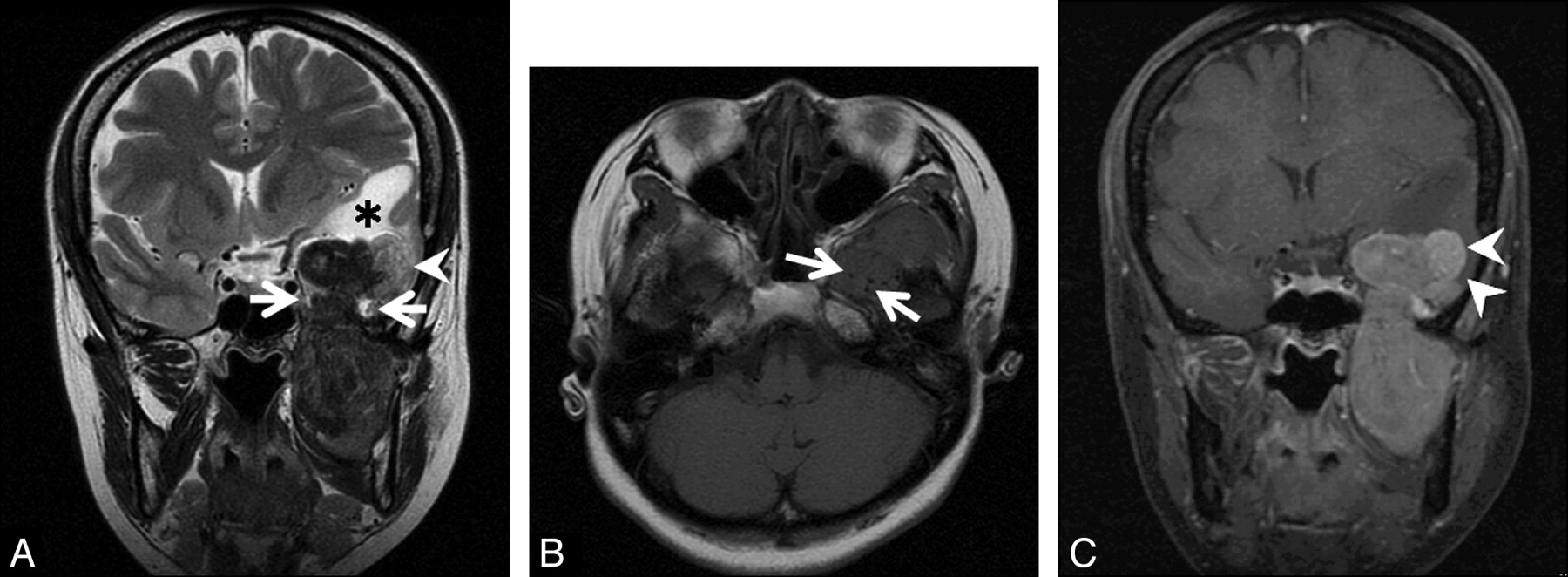
  • Fig 2.
    • Download figure
    • Open in new tab
    • Download powerpoint
    Fig 2.

    A, Coronal T2-weighted image shows a dumbbell-shaped tumor passing through left foramen ovale (arrows). The tumor is mainly hypointense to white matter. The lateral part of the intracranial component (arrowhead) shows higher intensities than other parts of the tumor. White matter of the left temporal lobe shows high intensities (asterisk). B, Axial T1-weighted image shows isointensity of the tumor to the brain. The tumor has low-intensity foci, suggesting a flow-void condition (arrows). C, On a coronal contrast-enhanced fat-suppressed T1-weighted image, the tumor shows heterogeneous enhancement. The lateral part of the intracranial component shows different strong enhancement (arrowheads).

PreviousNext
Back to top

In this issue

American Journal of Neuroradiology: 31 (2)
American Journal of Neuroradiology
Vol. 31, Issue 2
1 Feb 2010
  • Table of Contents
  • Index by author
Advertisement
Print
Download PDF
Email Article

Thank you for your interest in spreading the word on American Journal of Neuroradiology.

NOTE: We only request your email address so that the person you are recommending the page to knows that you wanted them to see it, and that it is not junk mail. We do not capture any email address.

Enter multiple addresses on separate lines or separate them with commas.
Intra- and Extracranial Solitary Fibrous Tumor of the Trigeminal Nerve: CT and MR Imaging Appearance
(Your Name) has sent you a message from American Journal of Neuroradiology
(Your Name) thought you would like to see the American Journal of Neuroradiology web site.
CAPTCHA
This question is for testing whether or not you are a human visitor and to prevent automated spam submissions.
Cite this article
K. Motoori, T. Hanazawa, I. Yamakami, A. Sugimoto, H. Ito
Intra- and Extracranial Solitary Fibrous Tumor of the Trigeminal Nerve: CT and MR Imaging Appearance
American Journal of Neuroradiology Feb 2010, 31 (2) 280-281; DOI: 10.3174/ajnr.A1702

Citation Manager Formats

  • BibTeX
  • Bookends
  • EasyBib
  • EndNote (tagged)
  • EndNote 8 (xml)
  • Medlars
  • Mendeley
  • Papers
  • RefWorks Tagged
  • Ref Manager
  • RIS
  • Zotero
0 Responses
Respond to this article
Share
Bookmark this article
Intra- and Extracranial Solitary Fibrous Tumor of the Trigeminal Nerve: CT and MR Imaging Appearance
K. Motoori, T. Hanazawa, I. Yamakami, A. Sugimoto, H. Ito
American Journal of Neuroradiology Feb 2010, 31 (2) 280-281; DOI: 10.3174/ajnr.A1702
del.icio.us logo Twitter logo Facebook logo Mendeley logo
  • Tweet Widget
  • Facebook Like
  • Google Plus One
Purchase

Jump to section

  • Article
    • Abstract
    • Abbreviations
    • Case Report
    • Discussion
    • Conclusions
    • References
  • Figures & Data
  • Info & Metrics
  • Responses
  • References
  • PDF

Related Articles

  • No related articles found.
  • PubMed
  • Google Scholar

Cited By...

  • Value of MR Imaging in Differentiation between Solitary Fibrous Tumor and Schwannoma in the Orbit
  • Crossref (6)
  • Google Scholar

This article has been cited by the following articles in journals that are participating in Crossref Cited-by Linking.

  • Spinal Solitary Fibrous Tumor/Hemangiopericytoma: A Clinicopathologic and Radiologic Analysis of Eleven Cases
    Xiaoping Yi, Desheng Xiao, Yujiao He, Hongling Yin, Guanghui Gong, Xueying Long, Weihua Liao, Xuejun Li, Lunquan Sun, Youming Zhang, Bo Zhang
    World Neurosurgery 2017 104
  • Value of MR Imaging in Differentiation between Solitary Fibrous Tumor and Schwannoma in the Orbit
    Z. Zhang, J. Shi, J. Guo, F. Yan, L. Fu, J. Xian
    American Journal of Neuroradiology 2013 34 5
  • High-Resolution Isotropic Three-Dimensional MR Imaging of the Extraforaminal Segments of the Cranial Nerves
    Jessica Wen, Naman S. Desai, Dean Jeffery, Nafi Aygun, Ari Blitz
    Magnetic Resonance Imaging Clinics of North America 2018 26 1
  • Lung cancer presenting with trigeminal neuropathy
    Alfonso Cerase, Leopoldo Brindisi, Luigi Lazzeretti, Emanuela Pepponi, Carlo Venturi
    Neurological Sciences 2011 32 5
  • Head and Neck Sarcoma Tumor Board Survival Guide for Neuroradiologists: Imaging Findings, History, and Pathology
    Daniel Warren, Cameron Koch, Matthew S. Parsons, Gloria J. Guzmán Pérez-Carrillo, Rami W. Eldaya
    Current Problems in Diagnostic Radiology 2023 52 4
  • Solitary fibrous tumor of the kidney
    Zongyu Xie, Guanghui Zhu, Liuquan Cheng, Jinhong Liu, Huiyi Ye, Haiyi Wang
    Medicine 2018 97 34

More in this TOC Section

  • Correlation of Apparent Diffusion Coefficient at 3T with Prognostic Parameters of Retinoblastoma
  • Parathyroid Lesions: Characterization with Dual-Phase Arterial and Venous Enhanced CT of the Neck
  • Comparison of Contrast Effect on the Cochlear Perilymph after Intratympanic and Intravenous Gadolinium Injection
Show more Head and Neck

Similar Articles

Advertisement

Indexed Content

  • Current Issue
  • Accepted Manuscripts
  • Article Preview
  • Past Issues
  • Editorials
  • Editors Choice
  • Fellow Journal Club
  • Letters to the Editor

Cases

  • Case Collection
  • Archive - Case of the Week
  • Archive - Case of the Month
  • Archive - Classic Case

Special Collections

  • Special Collections

Resources

  • News and Updates
  • Turn around Times
  • Submit a Manuscript
  • Author Policies
  • Manuscript Submission Guidelines
  • Evidence-Based Medicine Level Guide
  • Publishing Checklists
  • Graphical Abstract Preparation
  • Imaging Protocol Submission
  • Submit a Case
  • Become a Reviewer/Academy of Reviewers
  • Get Peer Review Credit from Publons

Multimedia

  • AJNR Podcast
  • AJNR SCANtastic
  • Video Articles

About Us

  • About AJNR
  • Editorial Board
  • Not an AJNR Subscriber? Join Now
  • Alerts
  • Feedback
  • Advertise with us
  • Librarian Resources
  • Permissions
  • Terms and Conditions

American Society of Neuroradiology

  • Not an ASNR Member? Join Now

© 2025 by the American Society of Neuroradiology All rights, including for text and data mining, AI training, and similar technologies, are reserved.
Print ISSN: 0195-6108 Online ISSN: 1936-959X

Powered by HighWire