Advertisement

Main menu

  • Home
  • Content
    • Current Issue
    • Accepted Manuscripts
    • Article Preview
    • Past Issue Archive
    • AJNR Case Collection
    • Case of the Week Archive
    • Classic Case Archive
    • Case of the Month Archive
  • Special Collections
    • Spinal CSF Leak Articles (Jan 2020-June 2024)
    • 2024 AJNR Journal Awards
    • Most Impactful AJNR Articles
  • Multimedia
    • AJNR Podcast
    • AJNR Scantastics
    • Video Articles
  • For Authors
    • Submit a Manuscript
    • Author Policies
    • Fast publishing of Accepted Manuscripts
    • Graphical Abstract Preparation
    • Manuscript Submission Guidelines
    • Imaging Protocol Submission
    • Submit a Case for the Case Collection
  • About Us
    • About AJNR
    • Editorial Board
  • More
    • Become a Reviewer/Academy of Reviewers
    • Subscribers
    • Permissions
    • Alerts
    • Feedback
    • Advertisers
    • ASNR Home
  • Other Publications
    • ajnr

User menu

  • Alerts
  • Log in

Search

  • Advanced search
American Journal of Neuroradiology

American Journal of Neuroradiology

ASHNR American Society of Functional Neuroradiology ASHNR American Society of Pediatric Neuroradiology ASSR
  • Alerts
  • Log in

Advanced Search

  • Home
  • Content
    • Current Issue
    • Accepted Manuscripts
    • Article Preview
    • Past Issue Archive
    • AJNR Case Collection
    • Case of the Week Archive
    • Classic Case Archive
    • Case of the Month Archive
  • Special Collections
    • Spinal CSF Leak Articles (Jan 2020-June 2024)
    • 2024 AJNR Journal Awards
    • Most Impactful AJNR Articles
  • Multimedia
    • AJNR Podcast
    • AJNR Scantastics
    • Video Articles
  • For Authors
    • Submit a Manuscript
    • Author Policies
    • Fast publishing of Accepted Manuscripts
    • Graphical Abstract Preparation
    • Manuscript Submission Guidelines
    • Imaging Protocol Submission
    • Submit a Case for the Case Collection
  • About Us
    • About AJNR
    • Editorial Board
  • More
    • Become a Reviewer/Academy of Reviewers
    • Subscribers
    • Permissions
    • Alerts
    • Feedback
    • Advertisers
    • ASNR Home
  • Follow AJNR on Twitter
  • Visit AJNR on Facebook
  • Follow AJNR on Instagram
  • Join AJNR on LinkedIn
  • RSS Feeds

Welcome to the new AJNR, Updated Hall of Fame, and more. Read the full announcements.


AJNR is seeking candidates for the position of Associate Section Editor, AJNR Case Collection. Read the full announcement.


Research ArticleBrain
Open Access

Evaluation of Traumatic Subarachnoid Hemorrhage Using Susceptibility-Weighted Imaging

Z. Wu, S. Li, J. Lei, D. An and E.M. Haacke
American Journal of Neuroradiology August 2010, 31 (7) 1302-1310; DOI: https://doi.org/10.3174/ajnr.A2022
Z. Wu
  • Find this author on Google Scholar
  • Find this author on PubMed
  • Search for this author on this site
S. Li
  • Find this author on Google Scholar
  • Find this author on PubMed
  • Search for this author on this site
J. Lei
  • Find this author on Google Scholar
  • Find this author on PubMed
  • Search for this author on this site
D. An
  • Find this author on Google Scholar
  • Find this author on PubMed
  • Search for this author on this site
E.M. Haacke
  • Find this author on Google Scholar
  • Find this author on PubMed
  • Search for this author on this site
  • Article
  • Figures & Data
  • Info & Metrics
  • Responses
  • References
  • PDF
Loading

Article Figures & Data

Figures

  • Tables
  • Fig 1.
    • Download figure
    • Open in new tab
    • Download powerpoint
    Fig 1.

    Two cases with both convexity and interhemispheric fissure SAH. Case 1: CT (A) shows high attenuation in the sulci of the convexity (arrows) and the paramedium sulci (along the centerline). SWI (B) shows SAH with a thick and rough boundary (short arrows and sulci inside the circle), while the vein is smooth and thin (long white arrow). SWI phase image (C) shows aliasing in the paramedian sulci (inside circle). Case 2: CT (D) shows high attenuation in 1 sulcus (white arrow) and falx high attenuation. SWI (E) shows SAH in the same sulcus (black arrow) and hemorrhage in the paramedian sulci (inside circle). SWI phase image (F) shows aliasing in the sulci (inside circle).

  • Fig 2.
    • Download figure
    • Open in new tab
    • Download powerpoint
    Fig 2.

    Two cases with Sylvian fissure SAH. Case 1: CT (A) shows a large amount of hemorrhage accumulating in the lateral part of the left Sylvian fissure (white arrow). SWI (B) shows only a small amount of hemorrhage deposited along the Sylvian fissure. SWI phase image (C) shows a similar pattern as in B without obvious aliasing. Case 2: CT (D) does not show high attenuation at the right Sylvian fissure (white arrow). SWI (E) shows a small amount of hemorrhage (black arrow). Phase image (F) shows aliasing effect (white >arrow).

  • Fig 3.
    • Download figure
    • Open in new tab
    • Download powerpoint
    Fig 3.

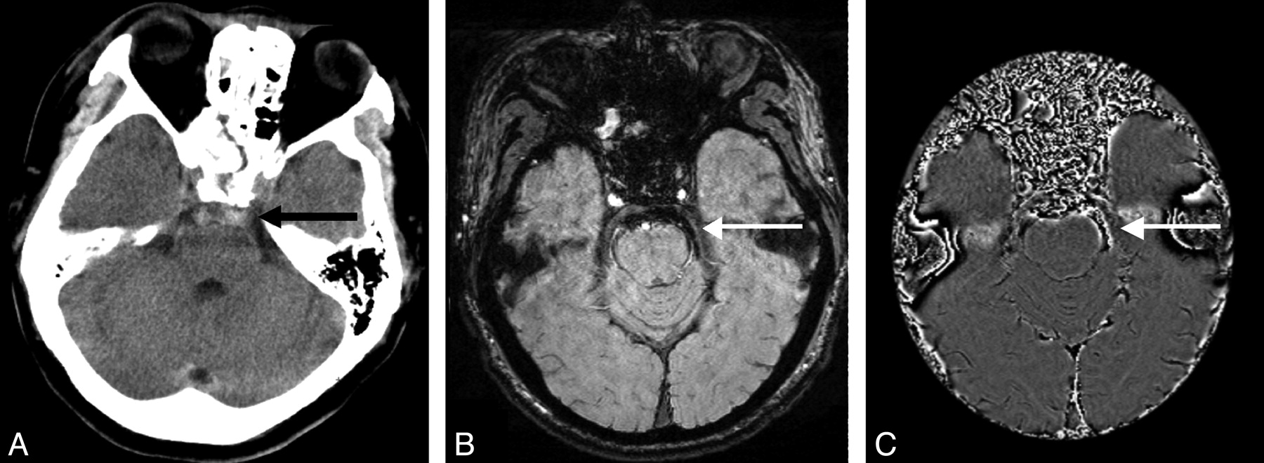
    SAH of the prepontine cistern: CT (A) shows high attenuation in the pontine cistern (black arrow). SWI (B) shows very dark signal intensity at the same location with CSF signal intensity surrounding (white arrow). SWI phase image (C) shows aliasing (white arrow).

  • Fig 4.
    • Download figure
    • Open in new tab
    • Download powerpoint
    Fig 4.

    High attenuation of the tentorium on CT with different signals on SWI on 3 different patients. Case 1: CT (A) shows high attenuation of the tentorium (white arrow). SWI (B) shows irregular hemorrhagic signal intensity on top of the tentorium and in between the cortex of vermis of the cerebellum (black arrow). Phase image (C) shows aliasing effect (white arrow). Case 2: CT (D) shows high attenuation of the tentorium (white arrow). SWI (E) shows bulging dark signal intensity along the tentorium. SWI magnitude image (F) shows 2 smooth lines at the edge of the hemorrhage, indicating subdural hematoma. Case 3: CT (G) shows high attenuation of the tentorium (white arrow). SWI (H) and phase image (I) show no hemorrhage at the tentorial area at all. (The small foci of dark signal intensity near the tentorium in H are microhemorrhages.)

  • Fig 5.
    • Download figure
    • Open in new tab
    • Download powerpoint
    Fig 5.

    Two cases with intraventricular hemorrhage. Case 1: CT (A) shows a calcified choroid plexus (long arrow) and hemorrhage (short arrow) in the left posterior horn of the lateral ventricle. SWI (B) shows hemorrhage in both sides of the posterior horn with distinct fluid levels (black arrows) below that are blood. Case 2: CT (C) does not show intraventricular hemorrhage, but only 2 calcified choroid plexus. SWI (D) shows a very small amount of hemorrhage in both posterior horns of the lateral ventricle (arrows).

  • Fig 6.
    • Download figure
    • Open in new tab
    • Download powerpoint
    Fig 6.

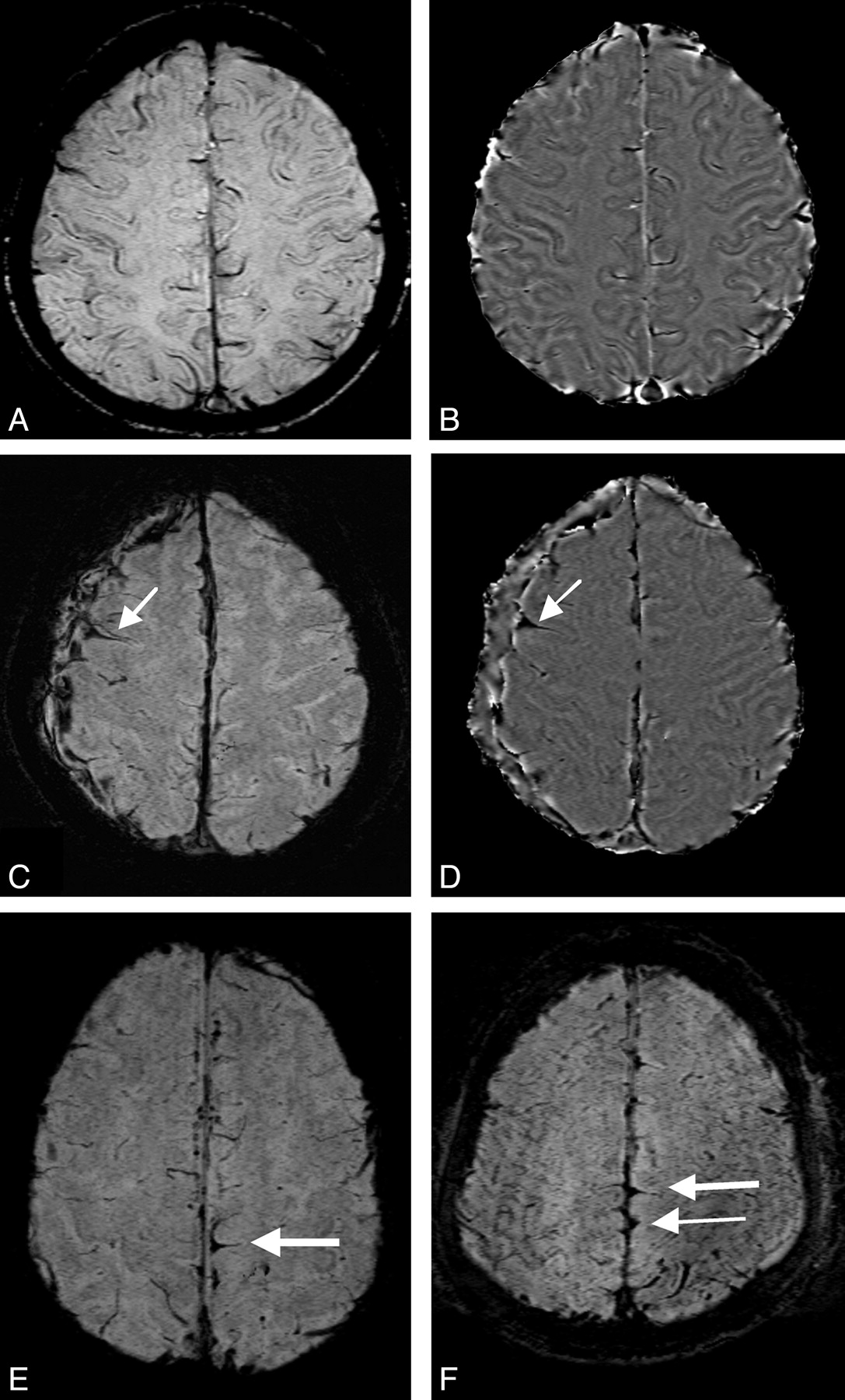
    A and B are the SWI and phase image from a healthy volunteer, which show the normal sulci, veins, and falx. Note the interhemispheric fissure has CSF and therefore appears slightly brighter than WM in the phase image. C and D represent a TBI patient showing 1 sulcus filled with blood, creating a typical triangle sign. Additionally, a crescent-shaped subdural hematoma along the right convexity is seen. E and F are 2 other TBI cases showing a similar triangle sign in the paramedian sulci.

  • Fig 7.
    • Download figure
    • Open in new tab
    • Download powerpoint
    Fig 7.

    A normal volunteer's tentorium appears as isointense on SWI (A) and slightly hyperintense (diamagnetic) on the SWI phase image (B).

  • Fig 8.
    • Download figure
    • Open in new tab
    • Download powerpoint
    Fig 8.

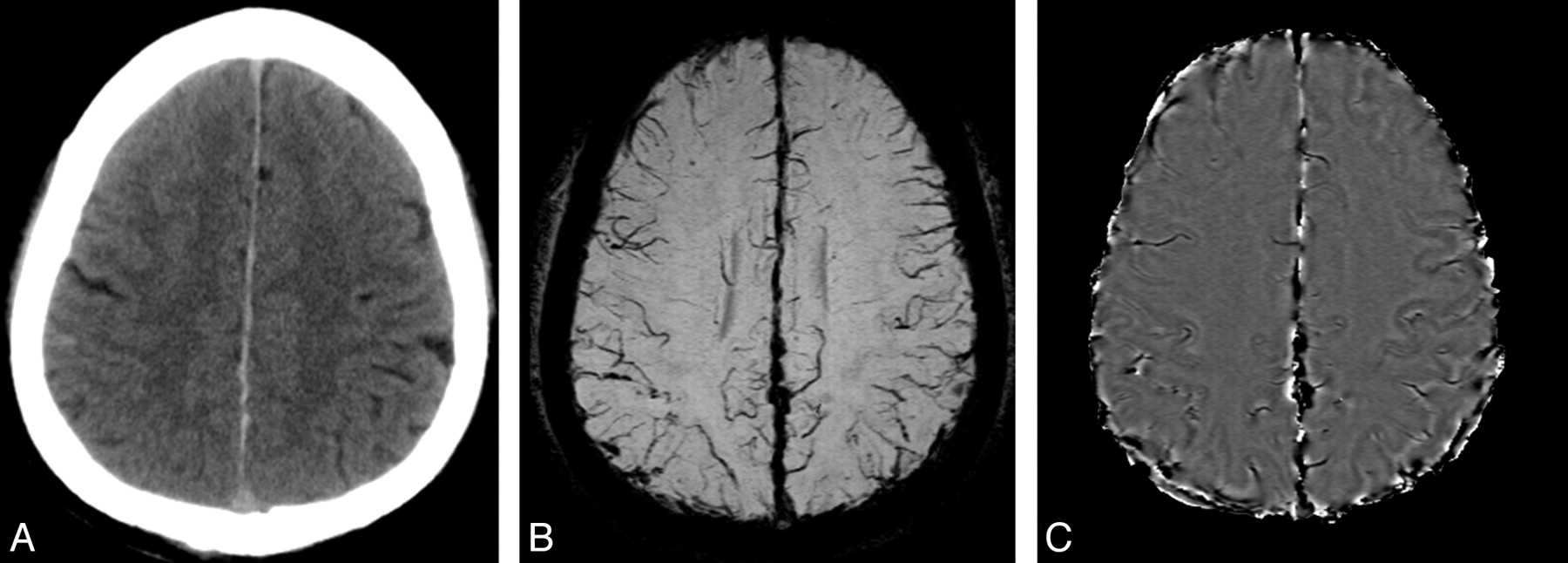
    CT (A) shows high attenuation of the falx, while SWI (B) and phase image (C) do not show the triangle sign, but rather show a bulging effect indicating subdural hemorrhage.

Tables

  • Figures
  • Number of SAH areas identified by CT and SWI

    Identified by Method ofFPCTOCIHFSVFPMCPFCTNCIVHTotal
    Both CT and SWI112137418555
    Only by CT1212810
    Only by SWI422513
    Total153171043161078
PreviousNext
Back to top

In this issue

American Journal of Neuroradiology: 31 (7)
American Journal of Neuroradiology
Vol. 31, Issue 7
1 Aug 2010
  • Table of Contents
  • Index by author
Advertisement
Print
Download PDF
Email Article

Thank you for your interest in spreading the word on American Journal of Neuroradiology.

NOTE: We only request your email address so that the person you are recommending the page to knows that you wanted them to see it, and that it is not junk mail. We do not capture any email address.

Enter multiple addresses on separate lines or separate them with commas.
Evaluation of Traumatic Subarachnoid Hemorrhage Using Susceptibility-Weighted Imaging
(Your Name) has sent you a message from American Journal of Neuroradiology
(Your Name) thought you would like to see the American Journal of Neuroradiology web site.
CAPTCHA
This question is for testing whether or not you are a human visitor and to prevent automated spam submissions.
Cite this article
Z. Wu, S. Li, J. Lei, D. An, E.M. Haacke
Evaluation of Traumatic Subarachnoid Hemorrhage Using Susceptibility-Weighted Imaging
American Journal of Neuroradiology Aug 2010, 31 (7) 1302-1310; DOI: 10.3174/ajnr.A2022

Citation Manager Formats

  • BibTeX
  • Bookends
  • EasyBib
  • EndNote (tagged)
  • EndNote 8 (xml)
  • Medlars
  • Mendeley
  • Papers
  • RefWorks Tagged
  • Ref Manager
  • RIS
  • Zotero
0 Responses
Respond to this article
Share
Bookmark this article
Evaluation of Traumatic Subarachnoid Hemorrhage Using Susceptibility-Weighted Imaging
Z. Wu, S. Li, J. Lei, D. An, E.M. Haacke
American Journal of Neuroradiology Aug 2010, 31 (7) 1302-1310; DOI: 10.3174/ajnr.A2022
del.icio.us logo Twitter logo Facebook logo Mendeley logo
  • Tweet Widget
  • Facebook Like
  • Google Plus One
Purchase

Jump to section

  • Article
    • Abstract
    • Abbreviations
    • Materials and Methods
    • Results
    • Discussion
    • Conclusions
    • Footnotes
    • References
  • Figures & Data
  • Info & Metrics
  • Responses
  • References
  • PDF

Related Articles

  • No related articles found.
  • PubMed
  • Google Scholar

Cited By...

  • Traumatic subarachnoid hemorrhage: a scoping review
  • Detection of aneurysmal subarachnoid hemorrhage 3 months after initial bleeding: evaluation of T2* and FLAIR MR sequences at 3 T in comparison with initial non-enhanced CT as a gold standard
  • Double Inversion Recovery MR Sequence for the Detection of Subacute Subarachnoid Hemorrhage
  • Detection of Microhemorrhage in Posterior Reversible Encephalopathy Syndrome Using Susceptibility-Weighted Imaging
  • Crossref (81)
  • Google Scholar

This article has been cited by the following articles in journals that are participating in Crossref Cited-by Linking.

  • Susceptibility-weighted imaging and quantitative susceptibility mapping in the brain
    Chunlei Liu, Wei Li, Karen A. Tong, Kristen W. Yeom, Samuel Kuzminski
    Journal of Magnetic Resonance Imaging 2015 42 1
  • Detection of Microhemorrhage in Posterior Reversible Encephalopathy Syndrome Using Susceptibility-Weighted Imaging
    A.M. McKinney, B. Sarikaya, C. Gustafson, C.L. Truwit
    American Journal of Neuroradiology 2012 33 5
  • Detecting Traumatic Brain Lesions in Children: CT versus MRI versus Susceptibility Weighted Imaging (SWI)
    Miriam H. Beauchamp, Michael Ditchfield, Franz E. Babl, Michael Kean, Cathy Catroppa, Keith O. Yeates, Vicki Anderson
    Journal of Neurotrauma 2011 28 6
  • Detecting subarachnoid hemorrhage: Comparison of combined FLAIR/SWI versus CT
    Rajeev Kumar Verma, Raimund Kottke, Lukas Andereggen, Christian Weisstanner, Christoph Zubler, Jan Gralla, Claus Kiefer, Johannes Slotboom, Roland Wiest, Gerhard Schroth, Christoph Ozdoba, Marwan El-Koussy
    European Journal of Radiology 2013 82 9
  • Emerging Imaging Tools for Use with Traumatic Brain Injury Research
    Jill V. Hunter, Elisabeth A. Wilde, Karen A. Tong, Barbara A. Holshouser
    Journal of Neurotrauma 2012 29 4
  • ACR Appropriateness Criteria ® Cerebrovascular Disease
    Michael B. Salmela, Shabnam Mortazavi, Bharathi D. Jagadeesan, Daniel F. Broderick, Judah Burns, Tejaswini K. Deshmukh, H. Benjamin Harvey, Jenny Hoang, Christopher H. Hunt, Tabassum A. Kennedy, Alexander A. Khalessi, William Mack, Nandini D. Patel, Joel S. Perlmutter, Bruno Policeni, Jason W. Schroeder, Gavin Setzen, Matthew T. Whitehead, Rebecca S. Cornelius, Amanda S. Corey
    Journal of the American College of Radiology 2017 14 5
  • Imaging Cerebral Microhemorrhages in Military Service Members with Chronic Traumatic Brain Injury
    Wei Liu, Karl Soderlund, Justin S. Senseney, David Joy, Ping-Hong Yeh, John Ollinger, Elyssa B. Sham, Tian Liu, Yi Wang, Terrence R. Oakes, Gerard Riedy
    Radiology 2016 278 2
  • Detecting Lesions after Traumatic Brain Injury Using Susceptibility Weighted Imaging: A Comparison with Fluid-Attenuated Inversion Recovery and Correlation with Clinical Outcome
    Gershon Spitz, Jerome J. Maller, Amanda Ng, Richard O'Sullivan, Nicholas J. Ferris, Jennie L. Ponsford
    Journal of Neurotrauma 2013 30 24
  • The emergence of age‐dependent social cognitive deficits after generalized insult to the developing brain: A longitudinal prospective analysis using susceptibility‐weighted imaging
    Nicholas P. Ryan, Cathy Catroppa, Janine M. Cooper, Richard Beare, Michael Ditchfield, Lee Coleman, Timothy Silk, Louise Crossley, Miriam H. Beauchamp, Vicki A. Anderson
    Human Brain Mapping 2015 36 5
  • Definition of Traumatic Brain Injury, Neurosurgery, Trauma Orthopedics, Neuroimaging, Psychology, and Psychiatry in Mild Traumatic Brain Injury
    Mubashir Pervez, Ryan S. Kitagawa, Tiffany R. Chang
    Neuroimaging Clinics of North America 2018 28 1

More in this TOC Section

  • Usefulness of Quantitative Susceptibility Mapping for the Diagnosis of Parkinson Disease
  • Evaluating the Effects of White Matter Multiple Sclerosis Lesions on the Volume Estimation of 6 Brain Tissue Segmentation Methods
  • White Matter Alterations in the Brains of Patients with Active, Remitted, and Cured Cushing Syndrome: A DTI Study
Show more Brain

Similar Articles

Advertisement

Indexed Content

  • Current Issue
  • Accepted Manuscripts
  • Article Preview
  • Past Issues
  • Editorials
  • Editors Choice
  • Fellow Journal Club
  • Letters to the Editor

Cases

  • Case Collection
  • Archive - Case of the Week
  • Archive - Case of the Month
  • Archive - Classic Case

Special Collections

  • Special Collections

Resources

  • News and Updates
  • Turn around Times
  • Submit a Manuscript
  • Author Policies
  • Manuscript Submission Guidelines
  • Evidence-Based Medicine Level Guide
  • Publishing Checklists
  • Graphical Abstract Preparation
  • Imaging Protocol Submission
  • Submit a Case
  • Become a Reviewer/Academy of Reviewers
  • Get Peer Review Credit from Publons

Multimedia

  • AJNR Podcast
  • AJNR SCANtastic
  • Video Articles

About Us

  • About AJNR
  • Editorial Board
  • Not an AJNR Subscriber? Join Now
  • Alerts
  • Feedback
  • Advertise with us
  • Librarian Resources
  • Permissions
  • Terms and Conditions

American Society of Neuroradiology

  • Not an ASNR Member? Join Now

© 2025 by the American Society of Neuroradiology All rights, including for text and data mining, AI training, and similar technologies, are reserved.
Print ISSN: 0195-6108 Online ISSN: 1936-959X

Powered by HighWire