
Article Figures & Data
Figures
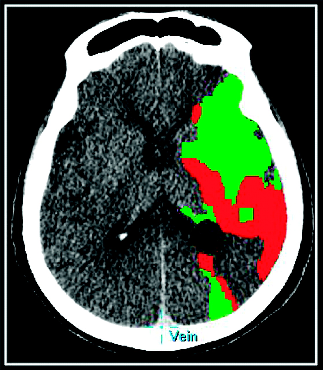
- Fig 1.
A 75-year-old patient with a left MCA ischemic stroke. Prognostic PCT map shows a mixed pattern of infarct core (red) and penumbra (green).
- Fig 2.
Absolute permeability (milliliters per 100 g per minute) maps for the reference standard 90- to 240-second delayed acquisition and for the 0- to 90-second first-pass acquisition with delay correction, the 30- to 90-second first-pass acquisition with delay correction, and the 0- to 90-second first-pass acquisition without delay correction. BBBP values calculated from the delay-corrected 30- to 90-second first-pass acquisition overlapped with the ones calculated from the reference standard 90- to 240-second delayed acquisition in the ischemic parenchymal tissue and were slightly underestimated in the nonischemic parenchymal tissue. The delay-corrected first-pass did not show the overestimation of BBBP values in the penumbra that were seen with the first-pass acquisition without delay correction.


Tables
Patients with Stroke No. of patients 23 No. of men (%) 8 (35%) Age (yr) Median = 74 Interquartile range = 59.5–82.5 Range = 26–92 Time from stroke to PCT (hr) Median = 2.25 Interquartile range = 1.5–5 Range = 1–11.75 Stroke location: ACA and MCA territories 3 MCA territory 20 ACA territory 0 PCA territory 0 90–240 Seconds 30–90 Seconds 0–90 Seconds 0–90 Seconds Reference Standard Delay Correction Delay Correction No Delay Correction Nonischemic 2.07 (1.83–2.34) 1.54 (1.39–1.71) 1.57 (1.24–2.00) 1.95 (1.74–2.18) P < .001 P = .002 P = .125 Infarct 2.48 (2.15–2.87) 2.09 (1.85–2.37) 1.66 (1.30–2.11) 2.49 (2.21–2.79) P = .074 P < .001 P = .874 Tissue at risk 2.43 (2.16–2.74) 2.45 (2.21–2.72) 1.93 (1.52–2.46) 3.49 (3.14–3.88) P = .921 P = .066 P < .001 -
a Mean absolute permeability values and corresponding 95% CIs shown for different regions-of-interest and acquisition-time datasets, as well as P values derived from the GEE models.
-
90–240 Seconds 30–90 Seconds 0–90 Seconds 0–90 Seconds Reference Standard Delay Correction Delay Correction No Delay Correction Nonischemic 1.56 (1.36,1.79) 1.12 (0.97–1.30) 2.38 (1.74–3.26) 4.04 (3.04–5.36) P < .001 P = .002 P < .001 Infarct 1.79 (1.49, 2.15) 1.19 (1.02–1.39) 2.54 (1.85–3.49) 4.05 (3.03–5.38) P < .001 P = .026 P < .001 Tissue at risk 1.65 (1.45, 1.87) 1.14 (1.00–1.29) 2.86 (2.07–3.95) 4.25 (3.24–5.56) P< .001 P< .001 P< .001 -
a Mean √MSE and corresponding 95% CIs are shown for different region-of-interest and acquisition-time datasets, as well as P values derived from the GEE models. √MSE is a measure of variability of data points around a straight line: a value close to zero indicates a smaller spread of data points around the line, corresponding to a better fit.
-


