
Article Figures & Data
Figures
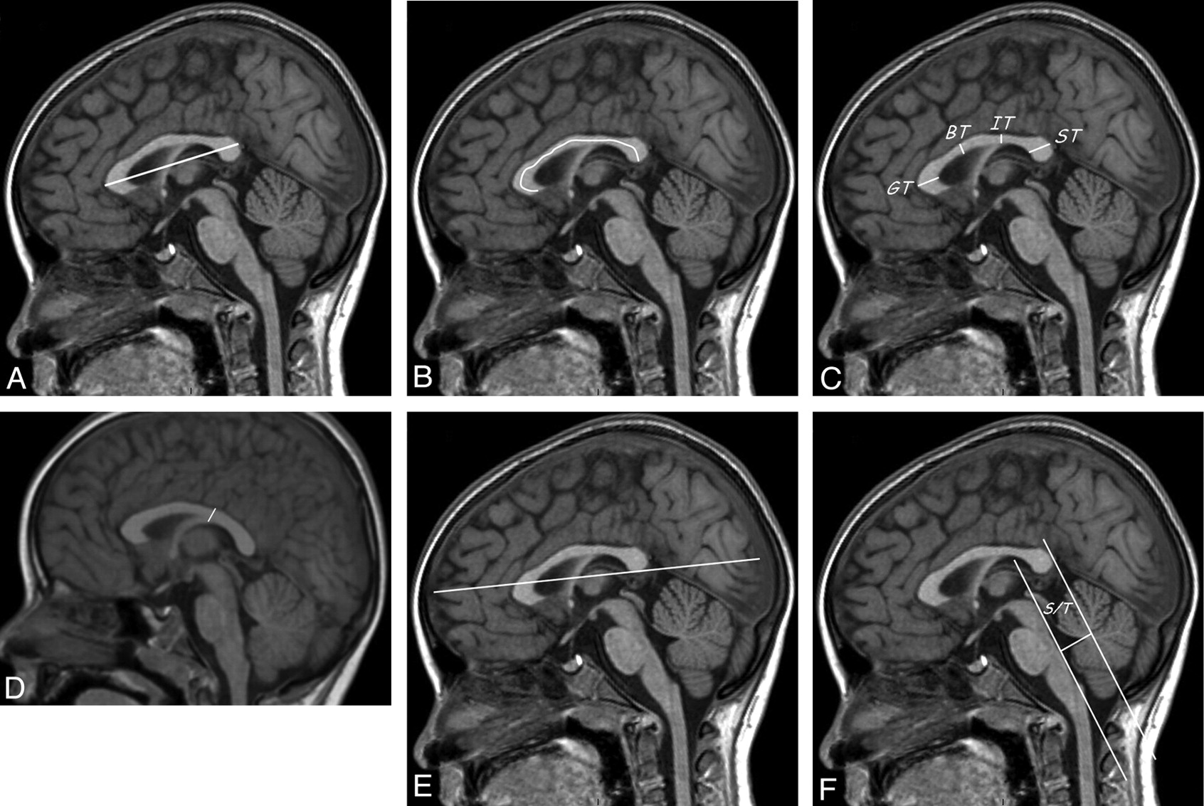
- Fig 1.
Description of the different biometric parameters measured with MR imaging. A, Measurement of the APD of the CC, the distance between the anterior aspect of the genu and the posterior aspect of the splenium. B, Measurement of the true LCC, the curvilinear distance between the rostrum and the splenium at midthickness of the CC. C, Measurement of the thickness of the CC, at the level of the genu (GT), body (BT), isthmus (IT), and splenium (ST). D, measurement of the IT when the isthmus could not be identified because of insufficient CC modeling. IT was measured at the level where the fornix abuts the CC (CC-fornix junction). E, Measurement of the FOD, the distance between the extreme points of the frontal and occipital lobes. F, Evaluation of the position of the splenium. A line was drawn along the dorsal surface of the brain stem. Another line was drawn parallel to the first one and passing at the level of the most posterior point of the splenium. The S/T distance between those lines was measured at the level of the fastigium.
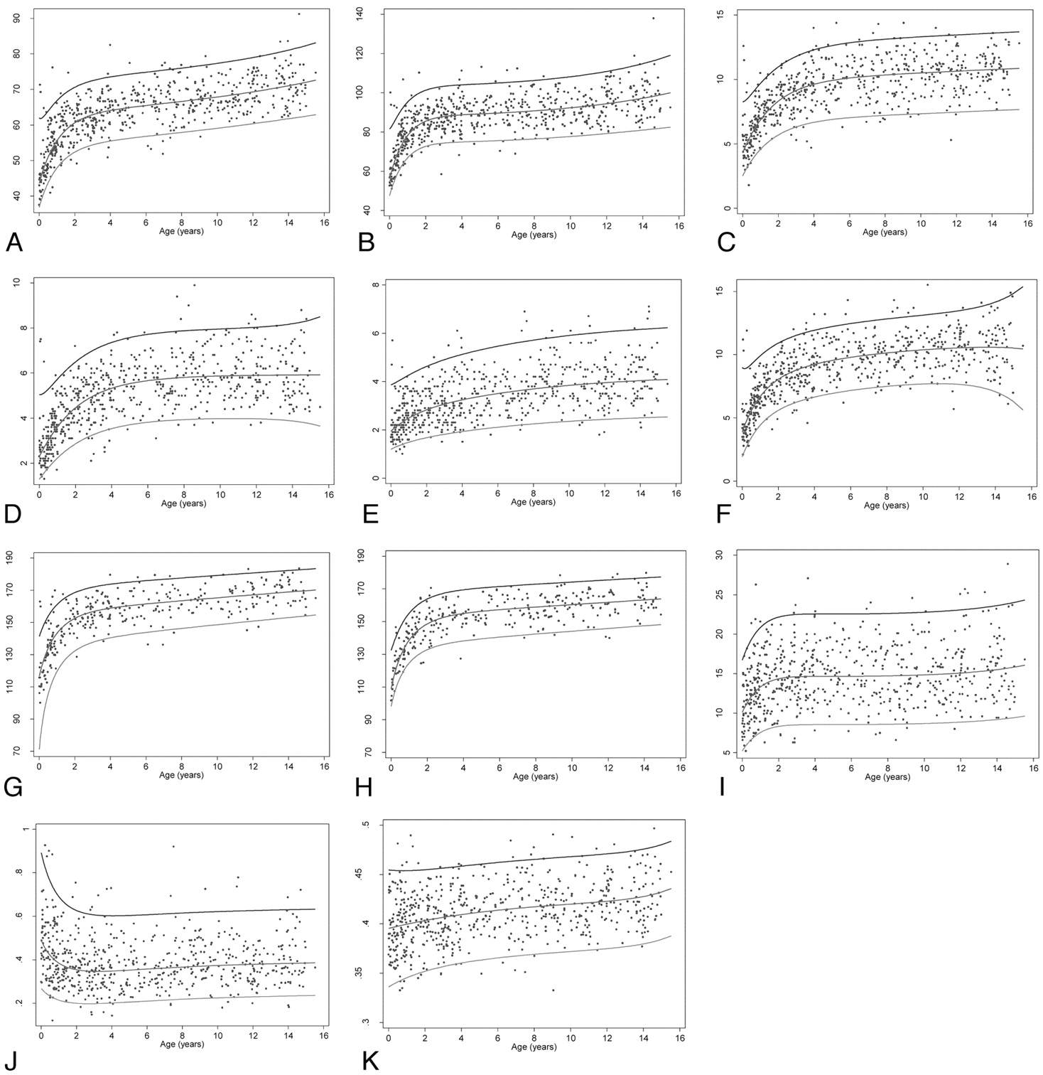
- Fig 2.
Reference intervals (3rd, 50th, 97th) for the different parameters: A = APD; B = LCC; C = GT; D = BT; E = IT; F = ST; G = FOD for males; H = FOD for females; I = S/T; J = IT/ST; and K= ADP/FOD.
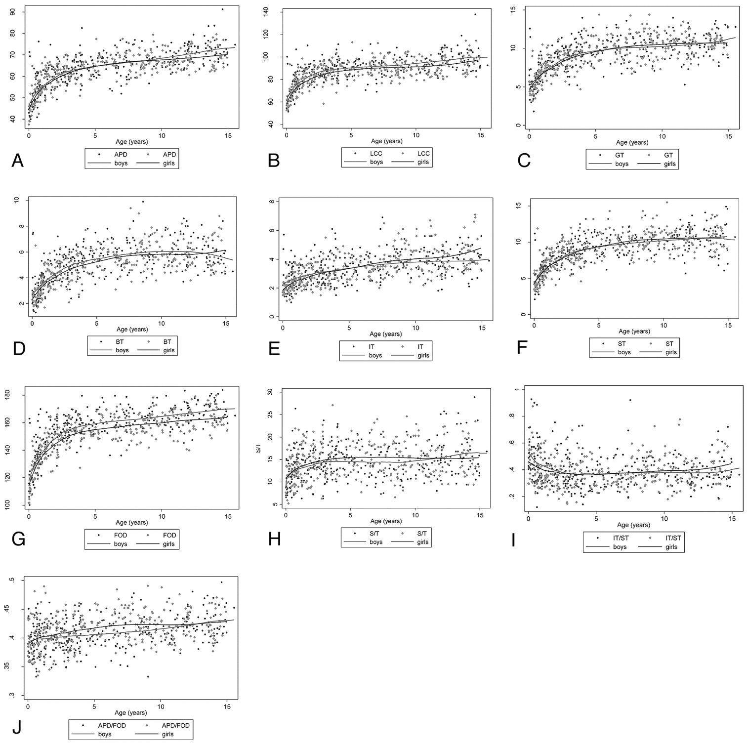
- Fig 3.
Loess smoothing curves of the different parameters depending on age (years) and sex. A = APD; B = LCC; C = GT; D = BT; E = IT; F = ST; G = FOD; H = S/T; I = IT/ST; J = ADP/FOD..


Tables
Abbreviation Parameter Definition Fig APD Anteroposterior diameter of the CC Distance between the anterior aspect of the genu and the posterior aspect of the splenium 1A LCC True length of the CC Curvilinear distance between the rostrum and the splenium at midthickness of the CC 1B GT Thickness of the genu of the CC Thickness of the CC, measured at the level of the genu 1C BT Thickness of the body of the CC Thickness of the CC, measured at the level of the body 1C IT Thickness of the isthmus of the CCa Thickness of the CC, measured at the level of the isthmus 1C ST Thickness of the splenium of the CC Thickness of the CC, measured at the level of the splenium 1C FOD Fronto-occipital diameterb Distance between the extreme points of the frontal and occipital lobes 1E S/T Distance splenium/tegmentum Distance at the level of the fastigium, between a line drawn along the dorsal surface of the brain stem and another line parallel to the first one and passing through the level of the most posterior point of the splenium 1F -
a When the isthmus could not be identified because of insufficient CC modeling, the IT was measured at the level where the fornix abuts the CC (CC-fornix junction) (Fig 1D).
-
b Sometimes the frontal and occipital lobes were not visible on the midsagittal section due to enlargement of the interhemispheric fissure. The FOD was then acquired on a slightly parasagittal section.
-
Age Class in Years Males (n = 320) Females (n = 302) All (N = 622) <1 65 51 116 1–2 26 32 58 2–3 32 27 59 3–4 39 23 62 4–5 15 12 27 5–6 15 13 28 6–7 15 14 29 7–8 15 16 31 8–9 10 22 32 9–10 9 19 28 10–11 12 18 30 11–12 18 14 32 12–13 14 15 29 13–14 16 13 29 14–15 19 13 32 - Table 3:
Values of the median, 3rd, and 97th percentiles for the different parameters, as a function of age
Percentile Age (yr) 0 0.5 1 1.5 2 2.5 3 4 5 6 7 8 9 10 11 12 13 14 15 APD 3rd 36.8 43.7 47.9 50.6 52.4 53.6 54.4 55.5 56.2 56.8 57.4 57.9 58.5 59.1 59.7 60.4 61 61.7 62.5 Median 43.6 50.9 55.6 58.6 60.6 61.9 62.9 64.1 64.8 65.5 66 66.6 67.2 67.9 68.6 69.3 70.1 71 72 97th 62 63.9 66.8 69.1 70.6 71.7 72.5 73.5 74.2 74.8 75.4 76 76.6 77.3 78.1 79 79.9 81.1 82.3 LCC 3rd 47.6 60 66.9 70.7 72.7 73.8 74.4 75 75.3 75.6 76 76.5 77.1 77.7 78.4 79.2 80.1 81 82 Median 56.3 70.2 78.3 82.9 85.5 87 87.9 88.7 89.2 89.7 90.2 90.8 91.5 92.3 93.3 94.4 95.7 97.2 99 97th 81.7 89.1 95.3 99.1 101.3 102.6 103.3 104.1 104.5 105 105.6 106.3 107.1 108.2 109.4 110.9 112.7 114.8 117.5 GT 3rd 2.5 3.7 4.6 5.2 5.7 6 6.3 6.7 6.9 7 7.1 7.2 7.3 7.3 7.4 7.5 7.5 7.6 7.6 Median 4.3 5.8 6.9 7.7 8.3 8.8 9.1 9.6 9.9 10.1 10.2 10.3 10.4 10.5 10.6 10.6 10.7 10.8 10.8 97th 8.3 8.9 9.7 10.4 11 11.4 11.8 12.3 12.6 12.9 13 13.1 13.2 13.3 13.4 13.5 13.5 13.6 13.7 BT 3rd 1.3 1.8 2.2 2.6 2.9 3.1 3.3 3.5 3.7 3.8 3.9 3.9 4 4 4 4 3.9 3.9 3.7 Median 2.3 3 3.6 4.1 4.5 4.8 5 5.3 5.5 5.7 5.8 5.8 5.8 5.9 5.9 5.9 5.9 5.9 5.9 97th 5 5.3 5.7 6.1 6.5 6.8 7 7.4 7.6 7.7 7.8 7.9 7.9 8 8 8 8.1 8.2 8.4 IT 3rd 1.2 1.4 1.5 1.6 1.7 1.7 1.8 1.9 2 2.1 2.2 2.2 2.3 2.4 2.4 2.4 2.5 2.5 2.5 Median 1.9 2.2 2.5 2.7 2.8 3 3.1 3.2 3.4 3.5 3.6 3.7 3.8 3.8 3.9 4 4 4 4.1 97th 3.9 4.1 4.3 4.5 4.6 4.8 4.9 5.1 5.3 5.5 5.6 5.7 5.8 5.9 6 6 6.1 6.2 6.2 ST 3rd 1.9 3.4 4.4 5.1 5.6 6 6.2 6.7 6.9 7.2 7.4 7.5 7.6 7.7 7.7 7.7 7.5 7.1 6.3 Median 3.9 5.6 6.7 7.5 8.1 8.5 8.8 9.2 9.5 9.8 10 10.1 10.3 10.4 10.5 10.5 10.6 10.6 10.5 97th 9 9.2 9.9 10.5 10.9 11.3 11.5 11.9 12.2 12.5 12.7 12.8 13 13.1 13.3 13.5 13.7 14.1 14.8 FOD boys 3rd 69.8 105.2 119.5 127.4 132.3 135.4 137.6 140.5 142.3 143.8 145.1 146.3 147.5 148.6 149.7 150.8 151.9 153 154 Median 116.3 133.9 143.3 148.9 152.5 154.9 156.6 158.8 160.3 161.5 162.5 163.4 164.4 165.3 166.2 167.1 168 168.9 169.7 97th 141 153.7 161.1 165.6 168.5 170.5 171.9 173.7 175 175.9 176.8 177.6 178.4 179.2 180 180.7 181.5 182.2 183 FOD girls 3rd 98.1 115.9 124.7 129.8 132.9 134.9 136.3 138.2 139.5 140.6 141.5 142.4 143.3 144.2 145 145.8 146.7 147.5 148.3 Median 109 128.5 138.7 144.8 148.7 151.2 152.9 155 156.3 157.3 158.1 158.9 159.6 160.4 161.1 161.8 162.5 163.2 163.9 97th 132.5 146.5 155.2 160.5 163.8 166.1 167.6 169.5 170.6 171.5 172.2 172.9 173.5 174.2 174.8 175.5 176.1 176.8 177.4 S/T 3rd 14 22.9 26.6 28.6 29.8 30.4 30.9 31.4 31.8 32.2 32.4 32.6 32.8 32.9 33 33 33 33 33.1 Median 22.6 28.9 32 33.8 34.8 35.4 35.8 36.4 36.8 37.1 37.4 37.6 37.8 37.9 38 38.1 38.1 38.1 38.1 97th 34.7 37.3 39 40 40.7 41.1 41.4 41.8 42.2 42.5 42.8 43.1 43.3 43.4 43.5 43.6 43.7 43.7 43.7 Ratio IT/ST 3rd 0.27 0.23 0.21 0.2 0.2 0.2 0.2 0.2 0.21 0.21 0.21 0.22 0.22 0.22 0.23 0.23 0.23 0.23 0.23 Median 0.49 0.42 0.38 0.36 0.35 0.35 0.35 0.35 0.35 0.36 0.36 0.37 0.37 0.37 0.38 0.38 0.38 0.38 0.38 97th 0.89 0.76 0.69 0.65 0.63 0.61 0.61 0.6 0.6 0.61 0.61 0.61 0.62 0.62 0.62 0.63 0.63 0.63 0.63 Ratio APD/FOD 3rd 0.34 0.34 0.35 0.35 0.35 0.35 0.36 0.36 0.36 0.37 0.37 0.37 0.37 0.37 0.37 0.37 0.38 0.38 0.38 Median 0.4 0.4 0.4 0.4 0.4 0.41 0.41 0.41 0.41 0.41 0.42 0.42 0.42 0.42 0.42 0.42 0.42 0.43 0.43 97th 0.45 0.45 0.45 0.45 0.45 0.46 0.46 0.46 0.46 0.46 0.46 0.47 0.47 0.47 0.47 0.47 0.47 0.48 0.48 Interobserver Agreement Intraobserver Agreement Agreement/Parameter Mean Bias 95% LOA ICC (95% CI) Mean Bias 95% LOA ICC (95% CI) APD −0.08 (−1.70–1.53) 0.995 (0.991–0.997) 0.12 (−1.11; 1.35) 0.997 (0.995–0.998) LCC 4.40 (−1.62–10.32) 0.88 (0.80–0.) −0.22 (−4.49; 4.06) 0.98 (0.96–0.99) GT 0.07 (−1.46–1.61) 0.95 (0.91–0.97) 0.42 (−0.98–1.82) 0.94 (0.90–0.97) BT 0.02 (−1.67–1.71) 0.83 (0.73–0.90) 0.03 (−1.20–1.27) 0.91 (0.85–0.95) IT 0.14 (−1.44–1.72) 0.72 (0.56–0.83) −0.02 (−1.09–1.06) 0.87 (0.78–0.92) ST −0.15 (−1.16–1.99) 0.92 (0.87–0.96) 0.36 (−2.57–3.28) 0.80 (0.67–0.88) FOD 2.90 (−6.23–12.11) 0.93 (0.89–0.96) 0.64 (−6.64–7.92) 0.97 (0.95–0.98) S/T −0.03 (−3.06–3.00) 0.89 (0.82–0.94) 0.16 (−1.98–2.30) 0.93 (0.88–0.96) - Table 5:
Equations for estimating reference values for the different parameters of the CCa–d
A μ = 145.6716 + 1.695221 X−0.5 − 3.059521ln(X) − 55.04029 X3 B μ = 86.22472 + 1.378669 X−0.5 − 31.29894 X3 − 27.07181 X3 ln(X) σ = 14.22583 + 0.0235383 X−1 − 1.237668 X3 μ = 7.679148 + 0.0202749 X−1 − 1.347937 X3 γ = −0.0593074 − 0.381785 X3 γ = −0.0442746 − 0.3954789 X3 θ = 1.26317 C μ = 9.896679 − 0.2111402 ln(X) − 5.590998 X2 D μ = 3.403339 − 0.5878885 X − 1.706435 X2 σ = 1.596243 − 0.346624 X σ = 0.5801882 + 0.0000132 X−2 γ = 0.0632866 − 0.4274138 X3 γ = 0.0081176 − 0.3621435 X3 θ = 0.6632529 E μ = 1.703424 − 0.6519961 X0.5 − 0.3350363 X3 F μ = 11.11401 − 0.0000336 X−2 − 3.188784 X0.5 − 4.048755 X3 σ = 0.3169457 + 0.0246416 X0.5 σ = 1.40096 + 0.0001188 X−2 γ = 0.0197081 − 0.253855 X3 γ = −0.0102384 − 0.4045783 X3 θ = 0.2095135 G μ = 1218829 − 80972.3 ln(X) − 710209.9 X3 H μ = 471589.4 − 23025.27 ln(X) − 288840.5 X3 σ = 211807.3 σ = 73751.38 + 165.8814 X−0.5 − 40108.09 X3 θ = 2.993395 γ = −0.0057655 − 0.430995 X3 θ = 2.802485 I μ = 5.331432 + 0.003554 X−1 − 1.174509 X3 − 1.438436 X3 ln(X) J μ = −0.9268794 − 0.2575655 X0.5 + 0.46828822 X3 σ = 0.8882193 σ = 0.2561955 + 0.0638789 X0.5 θ = 0.4664711 K μ = 0.4266512 + 0.000001123 X−2 − 0.0313357 X0.5 σ = 0.0255449 + 0.0059902 X2 -
a A = APD; B = LCC; C = GT; D = BT; E = IT; F = ST; G = FOD for males; H = FOD for females; I = S/T; J = IT/ST; K = APD/FOD.
-
b With
 where T1 = 0 and Tn = 15.53 denote minimum and maximum ages respectively and ρ, a preselected constant equal to 0.01.
 -
c Six measurements need prior Box-Cox transformation: A, D, E, G, H, I; 1 measurement needs natural logarithmic transformation.
-
d Seven models are EN: A, B, C, D, E, F, H; all others are normal.
-
In this issue


Jump to section
Related Articles
Cited By...
- Missense ABI2 variants linked to a neurodevelopmental disorder with intellectual disability, epilepsy, hypoplasia of the corpus callosum, and white matter abnormalities
- Pituitary Gland Duplication Syndrome: An International Imaging Analysis
- Neuroradiologic, Clinical, and Genetic Characterization of Cerebellar Heterotopia: A Pediatric Multicentric Study
- Imaging Findings and MRI Patterns in a Cohort of 18q Chromosomal Abnormalities
- Biallelic pathogenic variants in TRMT1 disrupt tRNA modification and induce a syndromic neurodevelopmental disorder
- Neuroanatomical Features of NAA10- and NAA15-Related Neurodevelopmental Syndromes
- A transposase-derived gene required for human brain development
- A transposase-derived gene required for human brain development
- Common Neuroimaging Findings in Bosch-Boonstra-Schaaf Optic Atrophy Syndrome
- Distinctive Brain Malformations in Zhu-Tokita-Takenouchi-Kim Syndrome
- Brain Abnormalities in Patients with Germline Variants in H3F3: Novel Imaging Findings and Neurologic Symptoms Beyond Somatic Variants and Brain Tumors
- Feasibility and Added Value of Fetal DTI Tractography in the Evaluation of an Isolated Short Corpus Callosum: Preliminary Results
- Systematic Analysis of Brain MRI Findings in Adaptor Protein Complex 4-Associated Hereditary Spastic Paraplegia
- Neuroradiologic Phenotyping of Galactosemia: From the Neonatal Form to the Chronic Stage
- CDK5RAP2 primary microcephaly is associated with hypothalamic, retinal and cochlear developmental defects
- Biometry of the Cerebellar Vermis and Brain Stem in Children: MR Imaging Reference Data from Measurements in 718 Children
- Persisting Embryonal Infundibular Recess in Morning Glory Syndrome: Clinical Report of a Novel Association
- A variant in TAF1 is associated with a new syndrome with severe intellectual disability and characteristic dysmorphic features