
Article Figures & Data
Figures
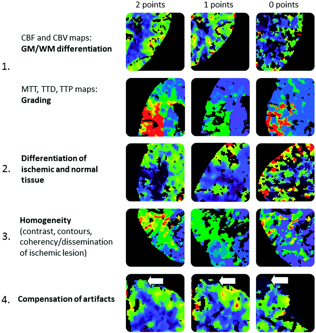
- Fig 1.
Qualitative scoring system.
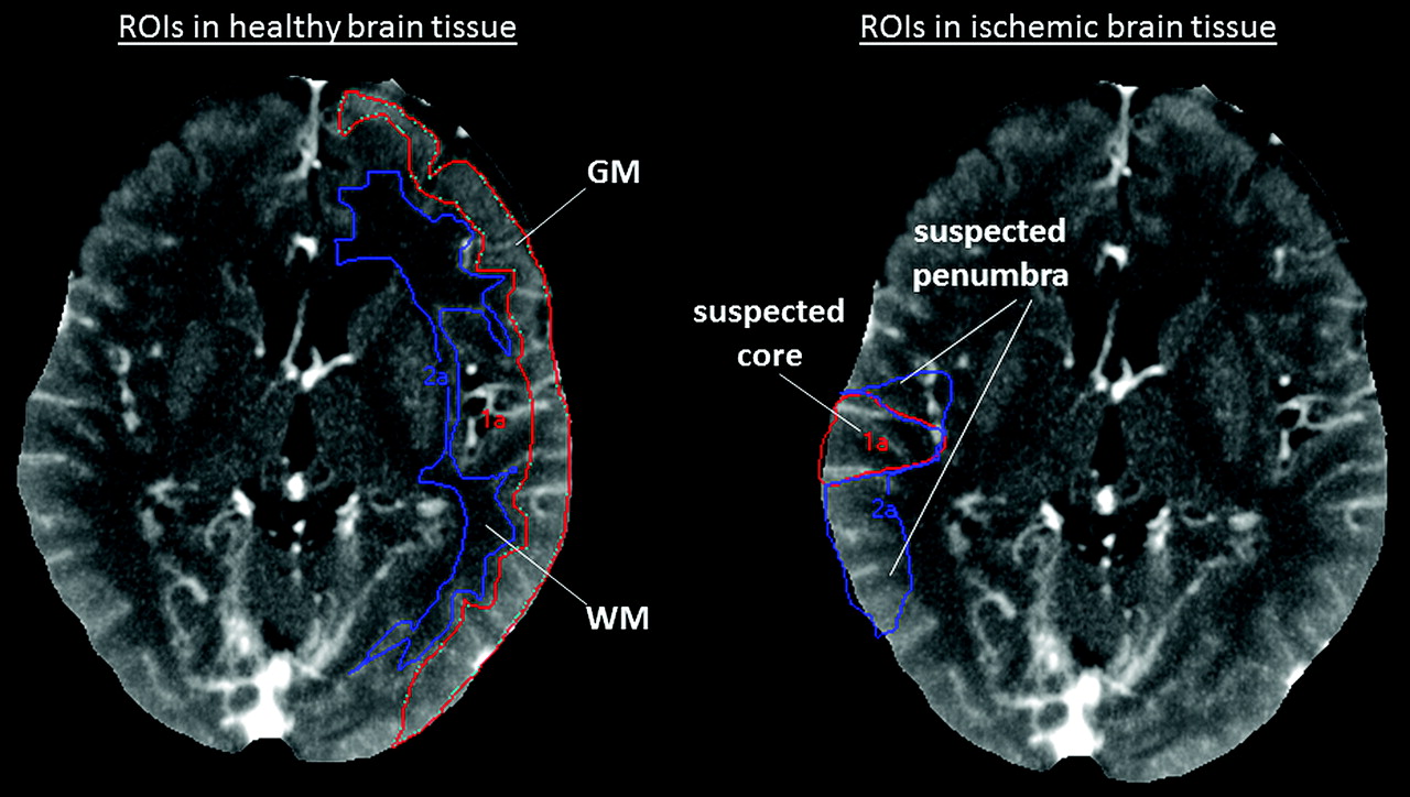
- Fig 2.
In the nonischemic hemisphere, GM and WM regions of interest are placed as shown on the left part of the figure. In the ischemic vascular territory, the GM region with significantly reduced CBF but normal or elevated CBV is outlined as a “suspected penumbra” region of interest. The GM region with significantly reduced CBF and significantly reduced CBV is outlined as a “suspected core” region of interest. All regions of interest were used to determine quantitative perfusion values only; they were not used to predict final tissue fate (final tissue fate was not verified by follow-up imaging).
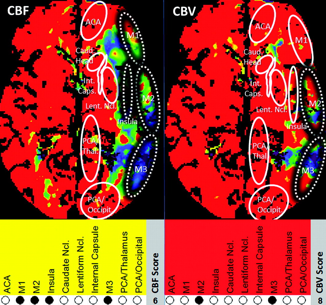
- Fig 3.
CTP scoring system in relationship to ASPECTS. M1, M2, M3, insular ribbon, caudate head, lentiform nucleus, internal capsule, anterior cerebral artery, PCA/occipital lobe, and PCA/thalamus were assigned 1 point each if nonaffected and 0 points if affected. Maximum score (healthy tissue) = 10, minimum score (complete infarction) = 0. For each case, CBF (TAR + NVT) and CBV (NVT) CTP lesion scores were derived from the CBF and CBV color maps, respectively.
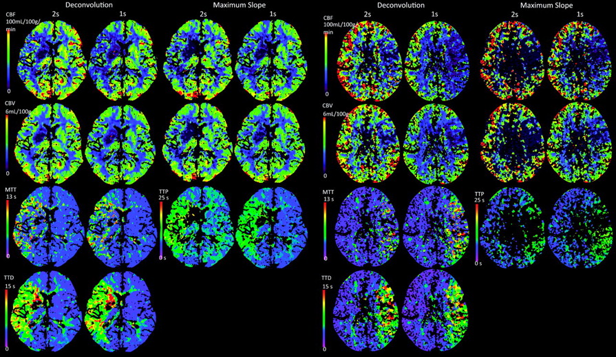
- Fig 4.
Left panel: Example of a case with good source data quality. Note color map quality is almost identical for 2-second TR (column 1) versus 1-second TR (column 2) by using DC and MS (columns 3 and 4) techniques. Right panel: Example of a case with low dataset quality. Note that color map quality at 2-second TR is slightly reduced when using DC (columns 1 and 2) and significantly reduced when using MS (columns 3 and 4) processing techniques.
- Fig 5.
Box-and-whisker plots comparing 2-second and 1-second TR perfusion values.
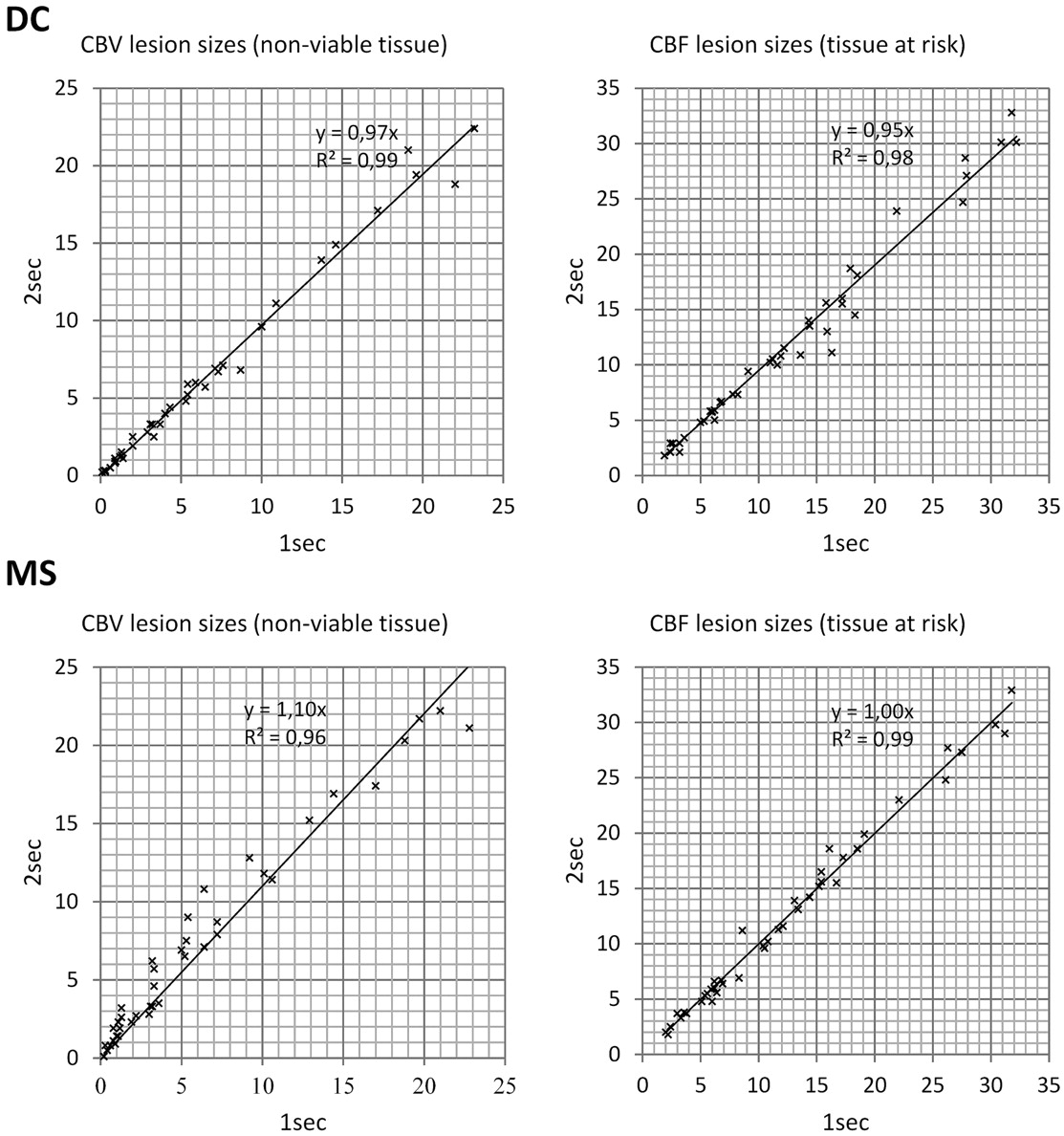
- Fig 6.
Scatterplots comparing CTP lesion sizes: 2-second versus 1-second TR for CBV and CBF perfusion parameters. All units are in cubic centimeters.


Tables
TR CBF CBV TTD MTT TTP 2-second 1-second 2-second 1-second 2-second 1-second 2-second 1-second 2-second 1-second DC Quality score 7.0 (5.6–7.0) 7.0 (5.6–7.0) 7.0 (6.0–7.0) 7.0 (6.0–7.0) 7.5 (6.1–8.0) 7.5 (6.1–8.0) 3.5 (2.0–4.0) 3.5 (2.5–4.0) N/A N/A Level of significance Not significant Not significant Not significant P < .05 N/A Category I 81% 81% 83% 86% 90% 90% 12% 14% N/A N/A II 19% 19% 17% 14% 10% 10% 67% 64% N/A N/A III 0% 0% 0% 0% 0% 0% 21% 21% N/A N/A MS Quality score 5.8 (3.1–6.5) 6.5 (5.1–7.5) 6.5 (4.0–7.5) 6.5 (5.5–7.5) N/A N/A N/A N/A 6.5 (5.0–6.5) 6.5 (5.0–6.5) Level of significance P < .001 P < .05 N/A N/A P < .05 Category I 62% 76% 69% 81% N/A N/A N/A N/A 76% 76% II 26% 19% 24% 14% N/A N/A N/A N/A 21% 24% III 12% 5% 7% 5% N/A N/A N/A N/A 2% 0% -
↵a All values are median (interquartile range). Quality scores are on a scale from 0 (worst) to 8 (best) points.
-
CTP Algorithm CBF score (TAR) CBV score (NVT) DC 2-second TR 6.4 ± 2.0, 7.0 (5.0–8.0) 7.5 ± 1.9, 8.0 (7.0–9.0) 1-second TR 6.1 ± 2.1, 7.0 (5.0–8.0) 7.5 ± 1.9, 8.0 (6.3–9.0) Significance P < .05 Not significant MS 2-second TR 5.9 ± 2.0, 6.0 (4.0–7.8) 6.8 ± 2.1, 7.0 (5.0–8.0) 1-second TR 5.9 ± 2.0, 6.0 (5.0–7.0) 7.5 ± 1.9, 8.0 (6.3–9.0) Significance not significant P < .001 -
↵a All values are given as mean ± SD and median (interquartile range). Scores for 1 brain section in orientation to ASPECTS: healthy = 10, completely infarcted = 0.
-
In this issue


Jump to section
Related Articles
Cited By...
- Clinical Applications of Conebeam CTP Imaging in Cerebral Disease: A Systematic Review
- Optimal Computed Tomographic Perfusion Scan Duration for Assessment of Acute Stroke Lesion Volumes
- Exposing Hidden Truncation-Related Errors in Acute Stroke Perfusion Imaging
- Whole-Brain Adaptive 70-kVp Perfusion Imaging with Variable and Extended Sampling Improves Quality and Consistency While Reducing Dose
- Radiation Doses of Cerebral Blood Volume Measurements Using C-Arm CT: A Phantom Study
- Vertebral Artery Hypoplasia: Frequency and Effect on Cerebellar Blood Flow Characteristics
- Can Iterative Reconstruction Improve Imaging Quality for Lower Radiation CT Perfusion? Initial Experience
- Stroke and CT Perfusion