Advertisement

Main menu

  • Home
  • Content
    • Current Issue
    • Accepted Manuscripts
    • Article Preview
    • Past Issue Archive
    • AJNR Case Collection
    • Case of the Week Archive
    • Classic Case Archive
    • Case of the Month Archive
  • Special Collections
    • Spinal CSF Leak Articles (Jan 2020-June 2024)
    • 2024 AJNR Journal Awards
    • Most Impactful AJNR Articles
  • Multimedia
    • AJNR Podcast
    • AJNR Scantastics
    • Video Articles
  • For Authors
    • Submit a Manuscript
    • Author Policies
    • Fast publishing of Accepted Manuscripts
    • Graphical Abstract Preparation
    • Manuscript Submission Guidelines
    • Imaging Protocol Submission
    • Submit a Case for the Case Collection
  • About Us
    • About AJNR
    • Editorial Board
  • More
    • Become a Reviewer/Academy of Reviewers
    • Subscribers
    • Permissions
    • Alerts
    • Feedback
    • Advertisers
    • ASNR Home
  • Other Publications
    • ajnr

User menu

  • Alerts
  • Log in

Search

  • Advanced search
American Journal of Neuroradiology

American Journal of Neuroradiology

ASHNR American Society of Functional Neuroradiology ASHNR American Society of Pediatric Neuroradiology ASSR
  • Alerts
  • Log in

Advanced Search

  • Home
  • Content
    • Current Issue
    • Accepted Manuscripts
    • Article Preview
    • Past Issue Archive
    • AJNR Case Collection
    • Case of the Week Archive
    • Classic Case Archive
    • Case of the Month Archive
  • Special Collections
    • Spinal CSF Leak Articles (Jan 2020-June 2024)
    • 2024 AJNR Journal Awards
    • Most Impactful AJNR Articles
  • Multimedia
    • AJNR Podcast
    • AJNR Scantastics
    • Video Articles
  • For Authors
    • Submit a Manuscript
    • Author Policies
    • Fast publishing of Accepted Manuscripts
    • Graphical Abstract Preparation
    • Manuscript Submission Guidelines
    • Imaging Protocol Submission
    • Submit a Case for the Case Collection
  • About Us
    • About AJNR
    • Editorial Board
  • More
    • Become a Reviewer/Academy of Reviewers
    • Subscribers
    • Permissions
    • Alerts
    • Feedback
    • Advertisers
    • ASNR Home
  • Follow AJNR on Twitter
  • Visit AJNR on Facebook
  • Follow AJNR on Instagram
  • Join AJNR on LinkedIn
  • RSS Feeds

Welcome to the new AJNR, Updated Hall of Fame, and more. Read the full announcements.


AJNR is seeking candidates for the position of Associate Section Editor, AJNR Case Collection. Read the full announcement.


Case ReportCase Report

Anomalous Extraocular Muscles with Strabismus

M.D. Dobbs, L.A. Mawn and S.P. Donahue
American Journal of Neuroradiology October 2011, 32 (9) E167-E168; DOI: https://doi.org/10.3174/ajnr.A2291
M.D. Dobbs
  • Find this author on Google Scholar
  • Find this author on PubMed
  • Search for this author on this site
L.A. Mawn
  • Find this author on Google Scholar
  • Find this author on PubMed
  • Search for this author on this site
S.P. Donahue
  • Find this author on Google Scholar
  • Find this author on PubMed
  • Search for this author on this site
  • Article
  • Figures & Data
  • Info & Metrics
  • Responses
  • References
  • PDF
Loading

Abstract

SUMMARY: An 8-month-old boy with Gorlin syndrome presented with a large right-face turn and constant exotropia of the left eye. Eight-millimeter recession of the left lateral rectus muscle was performed at 23 months of age without complete postoperative improvement. Orbital imaging revealed bilateral anomalous extraocular muscles inferolateral to the optic nerves. Surgical resection of the tissue confirmed the accessory musculature with postoperative correction of the strabismus. To our knowledge, this appears to be the first reported case in the radiologic literature.

An 8-month-old boy with a history of Gorlin syndrome (basal cell nevus syndrome) presented to pediatric ophthalmology with a large right-face turn (to compensate for left eye deviation). He was observed for nearly a year without change in his ocular mobility and was thought to have type II Duane syndrome. He underwent a large left lateral rectus recession at 23 months of age, which produced a moderate improvement in adduction and the face turn. The movements of the right eye were normal. However, as he became older, a limitation of elevation of the left eye with marked globe retraction became clearly evident, which prompted imaging to assess for any congenital accessory orbital structures. An orbital CT was performed.

Case Report

Orbital unenhanced CT revealed bilateral anomalous tissue within the intraconal regions inferolateral to the optic nerves, medial to the lateral rectus, and superior to the inferior rectus muscles, larger on the left side (Figs 1 and 2). This tissue had CT attenuation similar to that of muscle and had the visual appearance of a taut muscular structure. The tissue extended from the posterior orbital apex near the annulus of Zinn and inserted directly on the posterior globe. This tissue did not insert or originate from any of the surrounding normal extraocular muscles. All of the remaining extraocular muscles were present with normal size and location, with no other intraconal or extraconal abnormalities. The findings were thought to represent bilateral accessory extraocular muscles. The patient underwent surgery the following month. During surgery, the ophthalmologists placed a radiopaque string around what was thought to be the anomalous muscle tissue and performed an intraoperative CT scan to confirm that the correct tissue was resected.

Fig 1.
  • Download figure
  • Open in new tab
  • Download powerpoint
Fig 1.

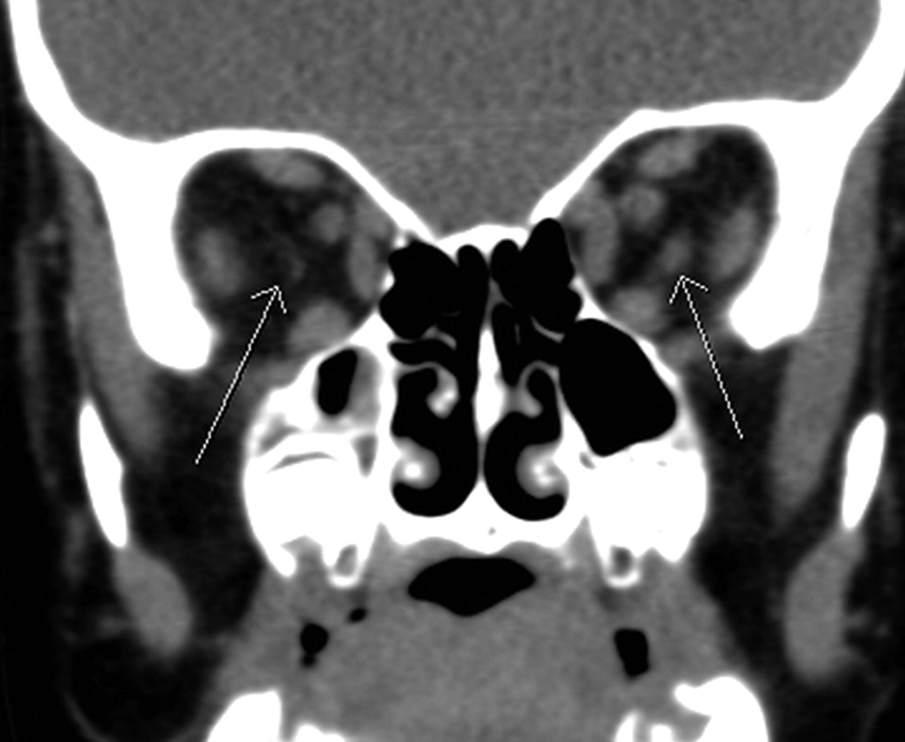
Coronal unenhanced CT scan of the orbits at the level of the ethmoid sinuses demonstrates bilateral accessory extraocular muscles (arrows) medial to the lateral rectus muscles, superior to the inferior rectus muscles, and inferolateral to the optic nerves. Attenuation is similar to that of surrounding extraocular muscles. The structure is much larger on the left side.

Fig 2.
  • Download figure
  • Open in new tab
  • Download powerpoint
Fig 2.

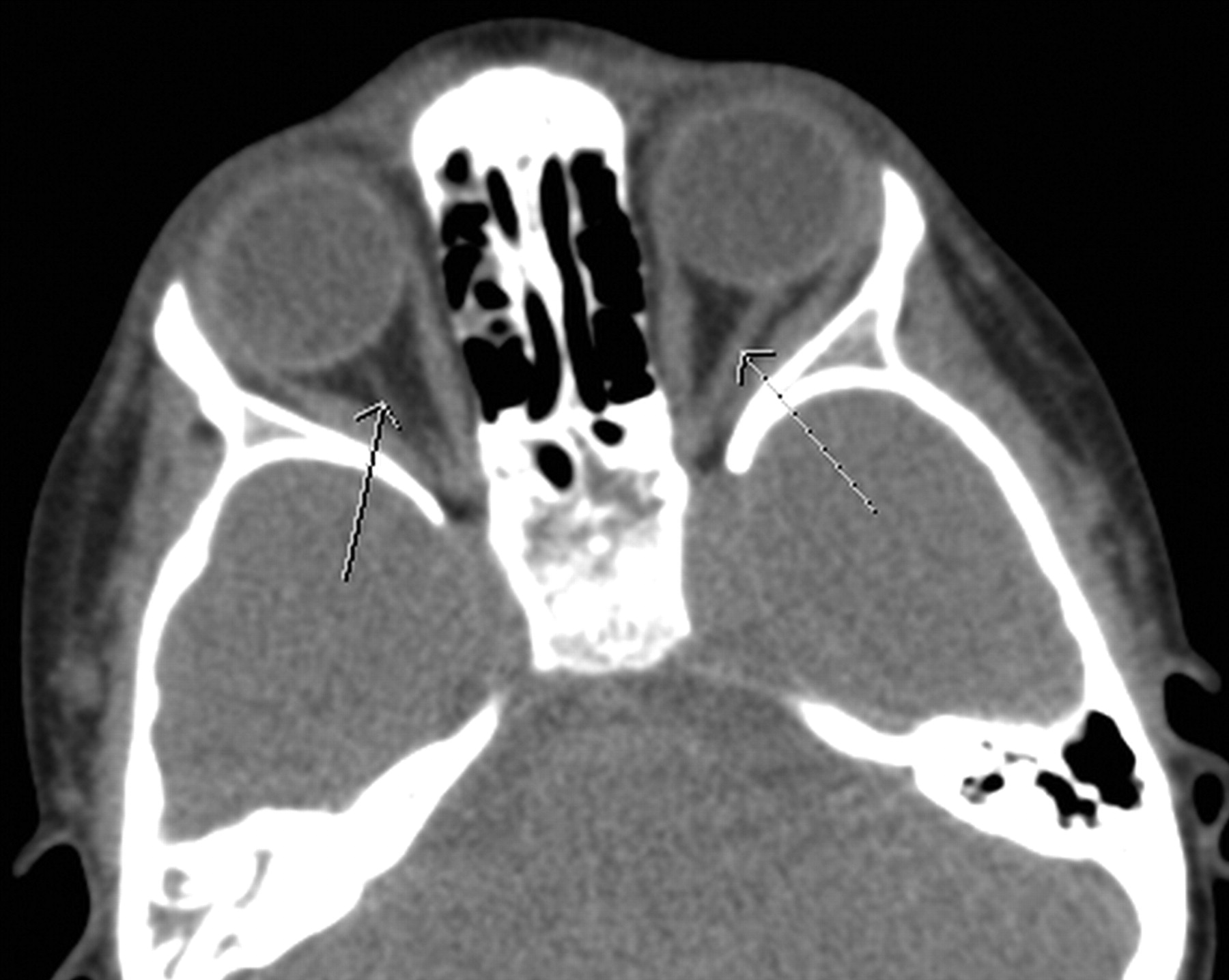
Axial unenhanced CT scan of the orbits at the level of the greater sphenoid wing demonstrates accessory extraocular muscles (arrows) medial to the lateral rectus muscles and lateral to the optic nerves (not shown on this image) that arise from the orbital apex at the annulus of Zinn and insert directly on the posterior globe, with no bridging connection or assimilation into the surrounding muscles.

Frozen and formalin-fixed sections of skeletal muscle stained with hematoxylin-eosin, Gomori trichrome, adenosine triphosphatase (pH 9.4 and 4.2), and nicotinamide adenine dinucleotide (diphosphopyridine nucleotide) showed preservation of fascicular architecture with no increase in connective tissue and no evidence of inflammation or vasculitis. There was mild variation in fiber sizes and a slight increase in the internal nuclei; both features are expected for extraocular muscle.

Following surgery, the patient's ocular deviation improved markedly. The thin sliver of tissue within the contralateral right orbit was not resected because it did not cause any clinical abnormality.

Discussion

Accessory extraocular muscles are very rare, having only been described in scattered case reports. The true prevalence is unknown due to the rare presentation. A few similar cases have been reported in the ophthalmic and pathologic literature.1,2 To our knowledge, there are no reports in the radiologic literature.

Three broad types of accessory extraocular tissue have been reported including anomalous bands of muscle bridging 2 muscles; fibrous tissues adjacent to the muscles, which may attach to the globe; and muscles arising from the posterior orbit and inserting on the globe or extraocular muscles.1

An interesting hypothesis of accessory musculature was proposed by Whitnall in 1911,3 asserting that these fibromuscular bands of tissue may represent an atavistic retractor bulbi muscle. This is a muscle found normally in reptiles, amphibians, and some ruminants but not in Homo sapiens, allowing retraction of the globe into the orbital cavity for protection.

Our patient did show retraction of the globe with attempted extraocular movements. Rather than an atavistic retractor bulbi muscle, our patient's findings could be considered as an accessory lateral rectus muscle. One argument against an accessory lateral rectus muscle is that this anomalous muscle did not appear radiographically to originate on the lateral rectus as described by Narasimhan et al.4 The anomalous muscle seen in our patient originated at the orbital apex at the optic foramen. Surgically, the muscle was extremely taut and inserted directly into the posterior globe, slightly inferolateral to the optic nerve.

An argument against this muscle being a vestigial retractor bulbi muscle is that it was an isolated distinct muscle and did not have pathologic features suggestive of skeletal muscle but rather had features typical of extraocular muscle.5 The described atavistic retractor bulbi muscles have either a cone shape or multiple tendon slips that surround the optic nerve region.6 Our patient lacked these findings.

We believe that this accessory muscle resulted from a disturbance of the mesodermal development of the extraocular muscles. Releasing this anomalous muscle from the globe relieved the globe retraction and significantly improved the exotropia.

Our patient coincidentally has Gorlin syndrome, which is due to a mutation at chromosome location 9q22·3-q31.7 This mutation causes loss of function of the PTCH1 receptor, of which sonic hedgehog protein is the ligand.8,9 Gorlin syndrome is associated with subsequent excessive hedgehog signaling, one of the pathways that directs development during embryogenesis, and is associated with the increased risk of basal cell carcinoma and medulloblastoma seen in patients with this syndrome.9 No association has been made between Gorlin syndrome and accessory extraocular muscles, but animal research shows induction and promotion of muscle development due to hedgehog signaling.10–13 One recent report found a patient presenting with exotropia similar to that in our patient, but the underlying cause was found to be an odontogenic keratocyst (a classic finding in Gorlin syndrome) of the alveolar ridge causing outward deviation of the globe.14

This case provides an example of a rare anatomic anomaly that has significant clinical pertinence. Awareness of such an entity can improve the search pattern of radiologists evaluating patients with atypical strabismus because this anomaly is amenable to surgical resection.

Footnotes

  • L.A. Mawn is funded by a grant from Research to Prevent Blindness, New York, New York.

References

  1. 1.↵
    1. Lueder GT
    . Anomalous orbital structures resulting in unusual strabismus. Surv Ophthalmol 2002; 47: 27–35
    CrossRefPubMed
  2. 2.↵
    1. Kirkham TH
    . Accessory extraocular muscle. Br J Ophthalmol 1970; 54: 276–77
    FREE Full Text
  3. 3.↵
    1. Whitnall SE
    . An instance of the retractor bulbi muscle in man. J Anat Physiol 1911; 46: 36–40
    PubMed
  4. 4.↵
    1. Narasimhan A,
    2. Tychsen L,
    3. Poukens V,
    4. et al
    . Horizontal rectus muscle anatomy in naturally and artificially strabismic monkeys. Invest Ophthalmol Vis Sci 2007; 48: 2576–88
    Abstract/FREE Full Text
  5. 5.↵
    1. Porter JD,
    2. Baker RS,
    3. Ragusa RJ,
    4. et al
    . Extraocular muscles: basic and clinical aspects of structure and function. Surv Ophthalmol 1995; 39: 451–84
    CrossRefPubMed
  6. 6.↵
    1. von Ludinghausen M
    . Bilateral supernumerary rectus muscles of the orbit. Clin Anat 1998; 11: 271–77
    CrossRefPubMed
  7. 7.↵
    1. Farndon PA,
    2. Del Mastro RG,
    3. Evans DG,
    4. et al
    . Location of gene for Gorlin syndrome. Lancet 1992; 339: 581–82
    CrossRefPubMed
  8. 8.↵
    1. Johnson RL,
    2. Rothman AL,
    3. Xie J,
    4. et al
    . Human homolog of patched, a candidate gene for the basal cell nevus syndrome. Science 1996; 272: 1668–71
    Abstract
  9. 9.↵
    1. Epstein EH
    . Basal cell carcinomas: attack of the hedgehog. Nat Rev Cancer 2008; 8: 743–54
    CrossRefPubMed
  10. 10.↵
    1. Duprez D,
    2. Fournier-Thibault C,
    3. Le Douarin N
    . Sonic hedgehog induces proliferation of committed skeletal muscle cells in the chick limb. Development 1998; 125: 495–505
    Abstract
  11. 11.↵
    1. Duprez D,
    2. Lapointe F,
    3. Edom-Vovard F,
    4. et al
    . Sonic hedgehog (SHH) specifies muscle pattern at tissue and cellular chick level, in the chick limb bud. Mech Dev 1999; 82: 151–63
    CrossRefPubMed
  12. 12.↵
    1. Li X,
    2. Blagden CS,
    3. Bildsoe H,
    4. et al
    . Hedgehog can drive terminal differentiation of amniote slow skeletal muscle. BMC Dev Biol 2004; 4: 9
    CrossRefPubMed
  13. 13.↵
    1. Feng X,
    2. Adiarte EG,
    3. Devoto SH
    . Hedgehog acts directly on the zebrafish dermomyotome to promote myogenic differentiation. Dev Biol 2006; 300: 736–46. Epub 2006 Aug 30
    CrossRefPubMed
  14. 14.↵
    1. Hall J,
    2. Johnston KA,
    3. McPhillips JP,
    4. et al
    . Nevoid basal cell carcinoma syndrome in a black child. J Am Acad Dermatol 1998; 38: 363–65
    CrossRefPubMed
  • Received July 6, 2011.
  • Accepted after revision July 15, 2011.
  • © 2011 by American Journal of Neuroradiology
View Abstract
PreviousNext
Back to top

In this issue

American Journal of Neuroradiology: 32 (9)
American Journal of Neuroradiology
Vol. 32, Issue 9
1 Oct 2011
  • Table of Contents
  • Index by author
Advertisement
Print
Download PDF
Email Article

Thank you for your interest in spreading the word on American Journal of Neuroradiology.

NOTE: We only request your email address so that the person you are recommending the page to knows that you wanted them to see it, and that it is not junk mail. We do not capture any email address.

Enter multiple addresses on separate lines or separate them with commas.
Anomalous Extraocular Muscles with Strabismus
(Your Name) has sent you a message from American Journal of Neuroradiology
(Your Name) thought you would like to see the American Journal of Neuroradiology web site.
CAPTCHA
This question is for testing whether or not you are a human visitor and to prevent automated spam submissions.
Cite this article
M.D. Dobbs, L.A. Mawn, S.P. Donahue
Anomalous Extraocular Muscles with Strabismus
American Journal of Neuroradiology Oct 2011, 32 (9) E167-E168; DOI: 10.3174/ajnr.A2291

Citation Manager Formats

  • BibTeX
  • Bookends
  • EasyBib
  • EndNote (tagged)
  • EndNote 8 (xml)
  • Medlars
  • Mendeley
  • Papers
  • RefWorks Tagged
  • Ref Manager
  • RIS
  • Zotero
0 Responses
Respond to this article
Share
Bookmark this article
Anomalous Extraocular Muscles with Strabismus
M.D. Dobbs, L.A. Mawn, S.P. Donahue
American Journal of Neuroradiology Oct 2011, 32 (9) E167-E168; DOI: 10.3174/ajnr.A2291
del.icio.us logo Twitter logo Facebook logo Mendeley logo
  • Tweet Widget
  • Facebook Like
  • Google Plus One
Purchase

Jump to section

  • Article
    • Abstract
    • Case Report
    • Discussion
    • Footnotes
    • References
  • Figures & Data
  • Info & Metrics
  • Responses
  • References
  • PDF

Related Articles

  • No related articles found.
  • PubMed
  • Google Scholar

Cited By...

  • No citing articles found.
  • Crossref (18)
  • Google Scholar

This article has been cited by the following articles in journals that are participating in Crossref Cited-by Linking.

  • Mutations inPOMGNT1cause non-syndromic retinitis pigmentosa
    Mingchu Xu, Takeyuki Yamada, Zixi Sun, Aiden Eblimit, Irma Lopez, Feng Wang, Hiroshi Manya, Shan Xu, Li Zhao, Yumei Li, Adva Kimchi, Dror Sharon, Ruifang Sui, Tamao Endo, Robert K. Koenekoop, Rui Chen
    Human Molecular Genetics 2016 25 8
  • Accessory Extraocular Muscle as a Cause of Restrictive Strabismus
    A. Molinari, D. Plager, P. Merino, M. M. Galan, M. Swaminathan, S. Ramasuramanian, J. T. H. N. de Faber
    Strabismus 2016 24 4
  • Double-bellied superior rectus muscle
    Satheesha B. Nayak, Surekha D. Shetty, Naveen Kumar, Ashwini P. Aithal
    Surgical and Radiologic Anatomy 2019 41 6
  • Accessory Lateral Rectus in a Patient With Normal Ocular Motor Control
    Yaping Joyce Liao, Jaclyn J. Hwang
    Journal of Neuro-Ophthalmology 2014 34 2
  • Strabismus resulting from an anomalous extraocular muscle in Gorlin syndrome
    Peter B. Knowlton, Louise A. Mawn, James B. Atkinson, Sean P. Donahue
    Journal of American Association for Pediatric Ophthalmology and Strabismus 2014 18 5
  • Pathologic Study of Supernumerary Orbital Band in Type I Duane Syndrome
    Muhammad Hassaan Ali, Stacy L. Pineles, Federico G. Velez, Anika K. Tandon, Ben J. Glasgow
    Ocular Oncology and Pathology 2019 5 5
  • The presence of anomalous extraocular bands in Duane retraction syndrome
    E. Cumhur Sener, Pinar Topcu Yilmaz, Arzu Sağlam
    Journal of American Association for Pediatric Ophthalmology and Strabismus 2021 25 1
  • Retracting Globe
    Cristy A. Ku, Christopher W. Bailey, Thomas D. Roberts, Jeffery P. Hogg, Jennifer A. Sivak-Callcott
    Journal of Neuro-Ophthalmology 2015 35 2
  • Supernumerary Extraocular Muscle: A Rare Cause of Atypical Restrictive Strabismus
    Xiangjun Wang, Tao Shen, Mengya Han, Jianhua Yan
    Medicina 2022 58 11
  • Accessory Lateral Rectus Muscle in Graves’ Orbitopathy: A Case Report
    N. Fichter, G. von Arx, E. C. Kirsch
    Clinical Neuroradiology 2013

More in this TOC Section

  • Atypical Diffusion-Restricted Lesion in 5-Fluorouracil Encephalopathy
  • Multimodal Imaging of Spike Propagation: A Technical Case Report
  • Dural Infantile Hemangioma Masquerading as a Skull Vault Lesion
Show more Case Reports

Similar Articles

Advertisement

Indexed Content

  • Current Issue
  • Accepted Manuscripts
  • Article Preview
  • Past Issues
  • Editorials
  • Editors Choice
  • Fellow Journal Club
  • Letters to the Editor

Cases

  • Case Collection
  • Archive - Case of the Week
  • Archive - Case of the Month
  • Archive - Classic Case

Special Collections

  • Special Collections

Resources

  • News and Updates
  • Turn around Times
  • Submit a Manuscript
  • Author Policies
  • Manuscript Submission Guidelines
  • Evidence-Based Medicine Level Guide
  • Publishing Checklists
  • Graphical Abstract Preparation
  • Imaging Protocol Submission
  • Submit a Case
  • Become a Reviewer/Academy of Reviewers
  • Get Peer Review Credit from Publons

Multimedia

  • AJNR Podcast
  • AJNR SCANtastic
  • Video Articles

About Us

  • About AJNR
  • Editorial Board
  • Not an AJNR Subscriber? Join Now
  • Alerts
  • Feedback
  • Advertise with us
  • Librarian Resources
  • Permissions
  • Terms and Conditions

American Society of Neuroradiology

  • Not an ASNR Member? Join Now

© 2025 by the American Society of Neuroradiology All rights, including for text and data mining, AI training, and similar technologies, are reserved.
Print ISSN: 0195-6108 Online ISSN: 1936-959X

Powered by HighWire