
Article Figures & Data
Figures
- Fig 1.
Visibility of identified structures. The percentage of each assigned grade on 3D and 2D FLAIR images and 2D T2WI. A, Bar graphs show the distribution of the assigned visibility grades for identified structures at the midbrain on 3D and 2D FLAIR images and 2D T2WI in all subjects. Grades 2 and 3 are more frequently assigned to the CST, SCP, and DSCP on 3D FLAIR than on 2D FLAIR images and 2D T2WI. B, Bar graphs show the distribution of the assigned visibility grades for identified structures at the pons on 3D and 2D FLAIR images and 2D T2WI in all subjects. Grades 2 and 3 are more frequently assigned to the CTT, CST, SCP, and MCP on 3D FLAIR than on 2D FLAIR images and 2D T2WI.
- Fig 2.
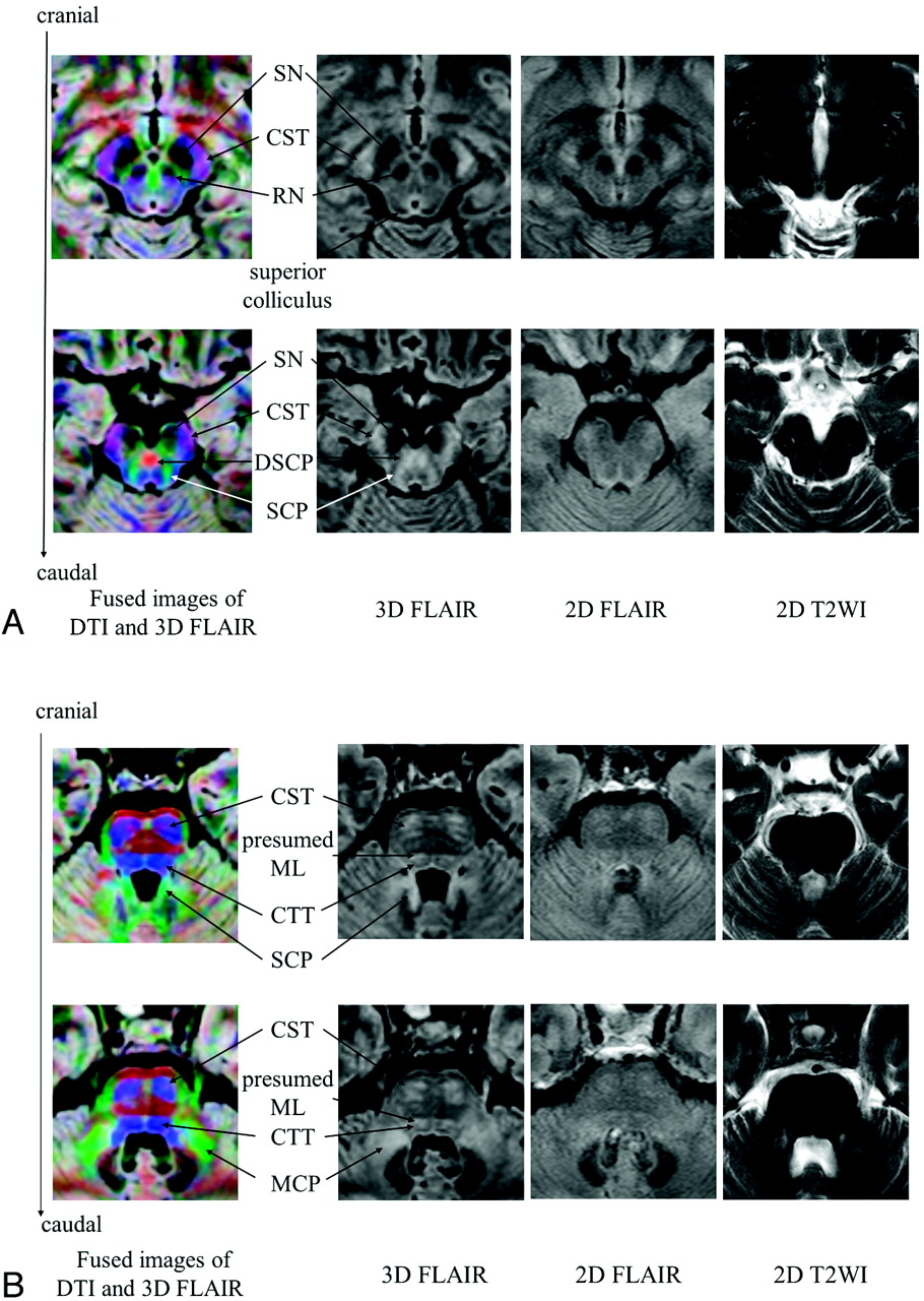
Midbrain and pontine anatomy. A, Midbrain: left column, fused images of 3D FLAIR and color maps; 2nd column from left, 3D FLAIR images; 3rd column, corresponding sections on 2D FLAIR images; right column, corresponding sections on 2D T2WI. Blue indicates superior-inferior orientation; green, anteroposterior orientation; red, laterolateral orientation. B, Pons: Left column, fused images of 3D FLAIR images and color maps; 2nd column from left, 3D FLAIR; 3rd column, corresponding sections on 2D FLAIR images; right column, corresponding sections on 2D T2WI. For color codes, see A.
- Fig 3.
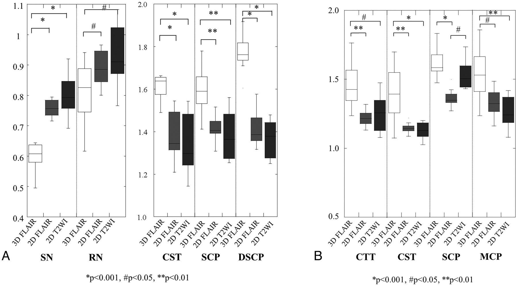
Statistical results for the contrast ratio of anatomic structures on 3D and 2D FLAIR images and 2D T2WI. A, Statistical results for the contrast ratio of anatomic structures on 3D and 2D FLAIR images and 2D T2WI at the midbrain. B, Statistical results for the contrast ratio of anatomic structures on 3D and 2D FLAIR images and 2D T2WI at the pons.


Tables
- Table 1:
Mean contrast ratio of the anatomic structures on 3D and 2D FLAIR imaging and 2D T2WI in the midbrain
SN Mean (SD) RN Mean (SD) CST Mean (SD) SCP Mean (SD) DSCP Mean (SD) 3D FLAIR 0.60 (0.07) 0.82 (0.10) 1.63 (0.09) 1.60 (0.10) 1.77 (0.08) 2D FLAIR 0.76 (0.03) 0.88 (0.06) 1.37 (0.12) 1.41 (0.07) 1.41 (0.08) 2D T2WI 0.80 (0.07) 0.93 (0.10) 1.34 (0.14) 1.38 (0.11) 1.37 (0.09) - Table 2:
Mean contrast ratio of the anatomic structures on 3D and 2D FLAIR imaging and 2D T2WI in the pons
CTT Mean (SD) CST Mean (SD) SCP Mean (SD) MCP Mean (SD) 3D FLAIR 1.45 (0.16) 1.40 (0.18) 1.61 (0.11) 1.55 (0.20) 2D FLAIR 1.22 (0.05) 1.15 (0.04) 1.36 (0.07) 1.32 (0.10) 2D T2WI 1.21 (0.26) 1.13 (0.06) 1.50 (0.16) 1.24 (0.29)
In this issue


Jump to section
Related Articles
Cited By...
- Effectiveness of 3D T2-Weighted FLAIR FSE Sequences with Fat Suppression for Detection of Brain MR Imaging Signal Changes in Children
- Reduction of Oxygen-Induced CSF Hyperintensity on FLAIR MR Images in Sedated Children: Usefulness of Magnetization-Prepared FLAIR Imaging
- Transition into Driven Equilibrium of the Balanced Steady-State Free Precession as an Ultrafast Multisection T2-Weighted Imaging of the Brain