
Article Figures & Data
Figures
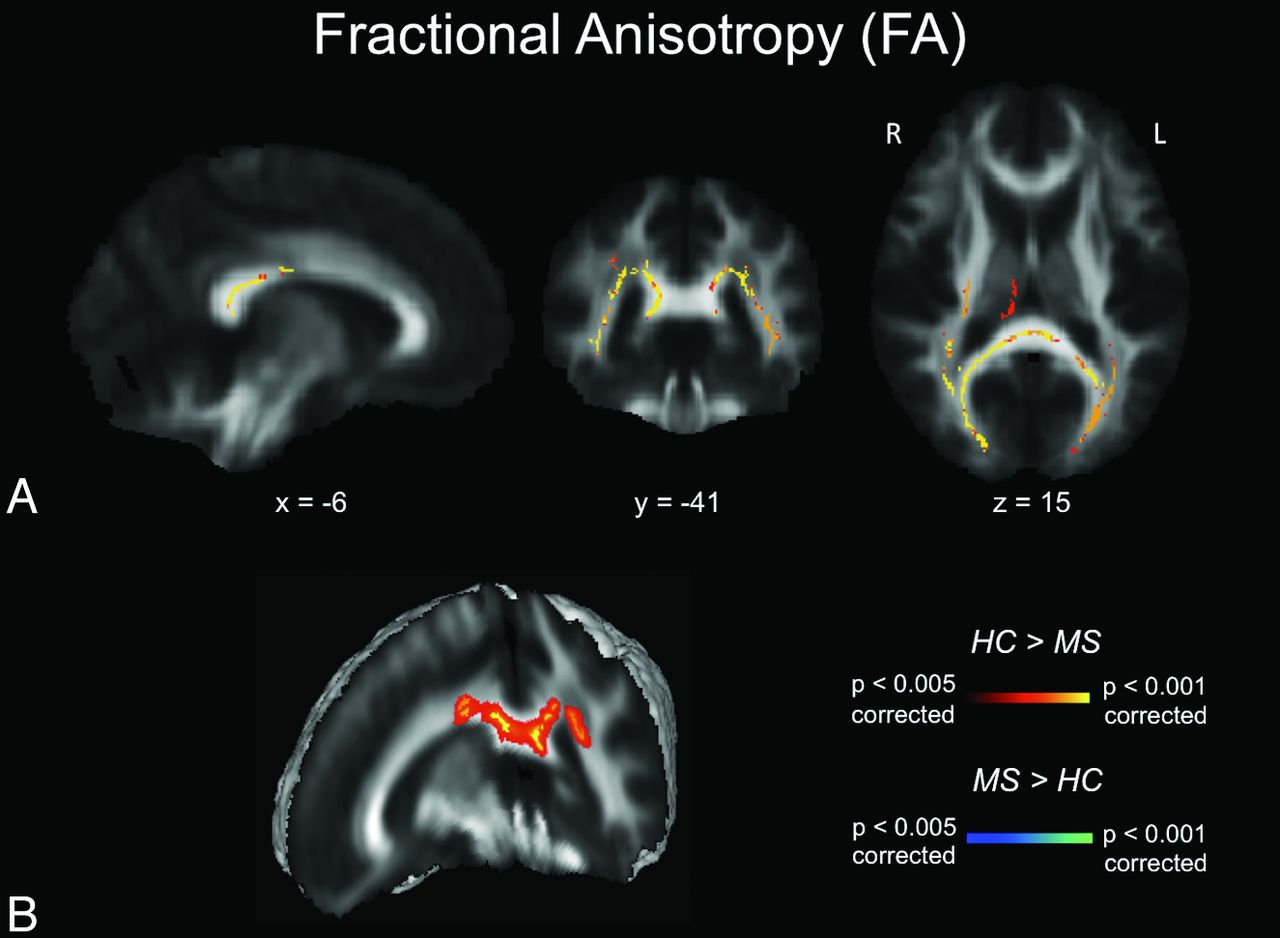
- Fig 1.
FA maps. A, TBSS FA results for healthy subjects compared with patients with MS. Significant clusters of decreased FA values in patients with MS compared with healthy controls are shown as corresponding P values in red-orange (scale ranging from red to yellow for the comparison HC > MS and scale ranging from blue to light blue for the comparison MS > HC) and have been thresholded at P < .005 for between-group comparisons (corrected for multiple comparisons). B, 3D visualization of significantly (P < .01, corrected for multiple comparisons) different white matter clusters between the 2 groups. Note that the results are thickened for visual purposes only. L indicates left; R, right (radiologic convention); HC, healthy controls.
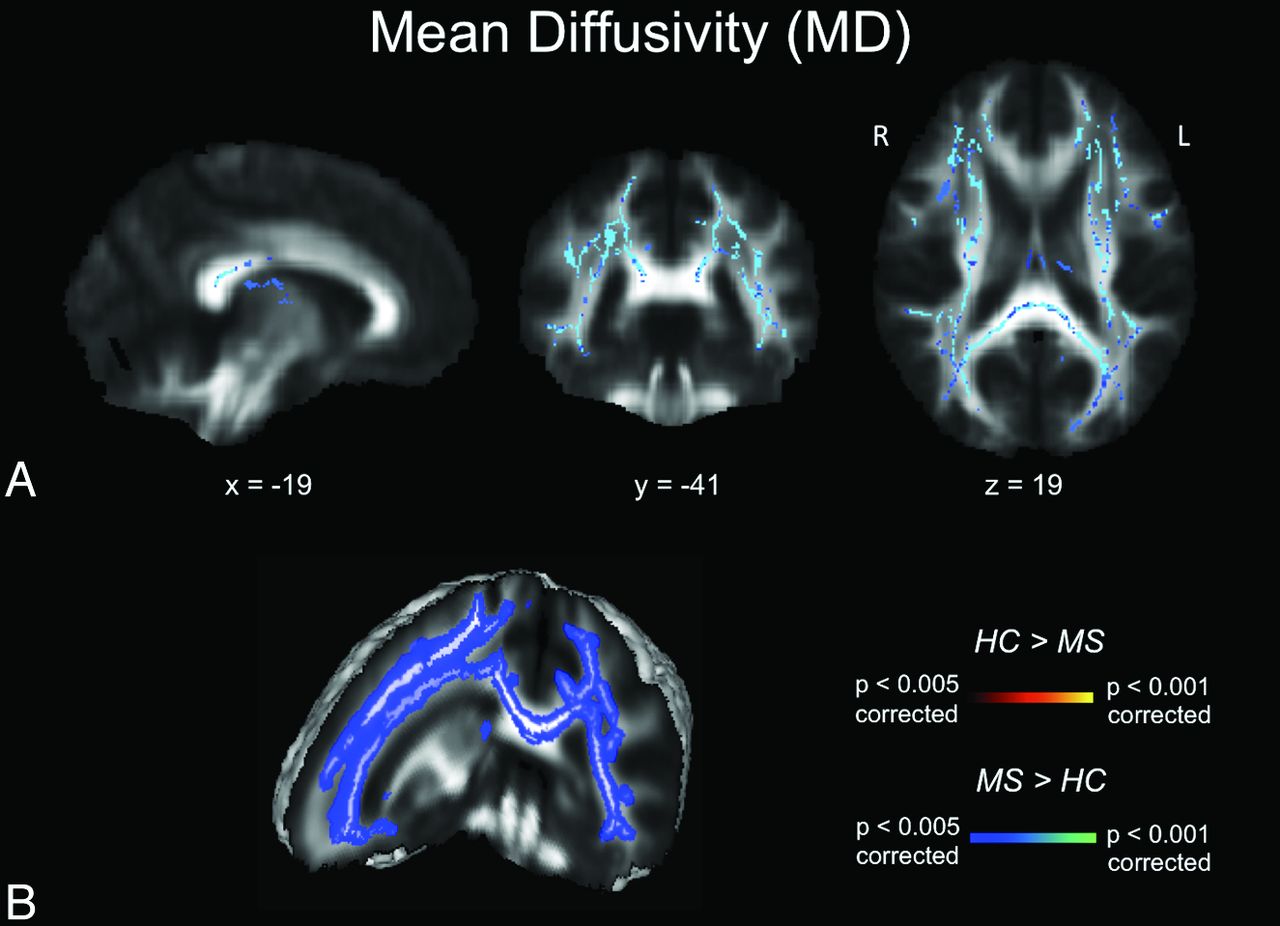
- Fig 2.
MD maps. A, TBSS MD results for healthy subjects compared with patients with MS. Significant clusters of increased MD values in patients with MS compared with healthy controls are shown as corresponding P values in red-orange (scale ranging from red to yellow for the comparison HC > MS and scale ranging from blue to light blue for the comparison MS > HC) and have been thresholded at P < .005 for between-group comparisons (corrected for multiple comparisons). B, 3D visualization of significantly (P < .01, corrected for multiple comparisons) different white matter clusters between the 2 groups. Note that the results are thickened for visual purposes only. L indicates left; R, right (radiologic convention); HC, healthy controls.
- Fig 3.
AD maps. A, TBSS AD results for healthy subjects compared with patients with MS. Significant clusters of increased AD values in patients with MS compared with healthy controls are shown as corresponding P values in red-orange (scale ranging from red to yellow for the comparison HC > MS and scale ranging from blue to light blue for the comparison MS > HC) and have been thresholded at P < .05 for between-group comparisons (corrected for multiple comparisons). B, 3D visualization of significantly (P < .01, corrected for multiple comparisons) different white matter clusters between the 2 groups. Note that the results are thickened for visual purposes only. L indicates left; R, right (radiologic convention); HC, healthy controls.
- Fig 4.
RD maps. A, TBSS RD results for healthy subjects compared with patients with MS. Significant clusters of increased RD values in patients with MS compared with healthy controls are shown as corresponding P values in red-orange (scale ranging from red to yellow for the comparison HC > MS and scale ranging from blue to light blue for the comparison MS > HC) and have been thresholded at P < .005 for between-group comparisons (corrected for multiple comparisons). B, 3D visualization of significantly (P < .01, corrected for multiple comparisons) different white matter clusters between the 2 groups. Note that the results are thickened for visual purposes only. L indicates left; R, right (radiologic convention); HC, healthy controls.


Jump to section
Related Articles
Cited By...
- No citing articles found.