Advertisement

Main menu

  • Home
  • Content
    • Current Issue
    • Accepted Manuscripts
    • Article Preview
    • Past Issue Archive
    • AJNR Case Collection
    • Case of the Week Archive
    • Classic Case Archive
    • Case of the Month Archive
  • Special Collections
    • Spinal CSF Leak Articles (Jan 2020-June 2024)
    • 2024 AJNR Journal Awards
    • Most Impactful AJNR Articles
  • Multimedia
    • AJNR Podcast
    • AJNR Scantastics
    • Video Articles
  • For Authors
    • Submit a Manuscript
    • Author Policies
    • Fast publishing of Accepted Manuscripts
    • Graphical Abstract Preparation
    • Manuscript Submission Guidelines
    • Imaging Protocol Submission
    • Submit a Case for the Case Collection
  • About Us
    • About AJNR
    • Editorial Board
  • More
    • Become a Reviewer/Academy of Reviewers
    • Subscribers
    • Permissions
    • Alerts
    • Feedback
    • Advertisers
    • ASNR Home
  • Other Publications
    • ajnr

User menu

  • Alerts
  • Log in

Search

  • Advanced search
American Journal of Neuroradiology

American Journal of Neuroradiology

ASHNR American Society of Functional Neuroradiology ASHNR American Society of Pediatric Neuroradiology ASSR
  • Alerts
  • Log in

Advanced Search

  • Home
  • Content
    • Current Issue
    • Accepted Manuscripts
    • Article Preview
    • Past Issue Archive
    • AJNR Case Collection
    • Case of the Week Archive
    • Classic Case Archive
    • Case of the Month Archive
  • Special Collections
    • Spinal CSF Leak Articles (Jan 2020-June 2024)
    • 2024 AJNR Journal Awards
    • Most Impactful AJNR Articles
  • Multimedia
    • AJNR Podcast
    • AJNR Scantastics
    • Video Articles
  • For Authors
    • Submit a Manuscript
    • Author Policies
    • Fast publishing of Accepted Manuscripts
    • Graphical Abstract Preparation
    • Manuscript Submission Guidelines
    • Imaging Protocol Submission
    • Submit a Case for the Case Collection
  • About Us
    • About AJNR
    • Editorial Board
  • More
    • Become a Reviewer/Academy of Reviewers
    • Subscribers
    • Permissions
    • Alerts
    • Feedback
    • Advertisers
    • ASNR Home
  • Follow AJNR on Twitter
  • Visit AJNR on Facebook
  • Follow AJNR on Instagram
  • Join AJNR on LinkedIn
  • RSS Feeds

Welcome to the new AJNR, Updated Hall of Fame, and more. Read the full announcements.


AJNR is seeking candidates for the position of Associate Section Editor, AJNR Case Collection. Read the full announcement.


Getting new auth cookie, if you see this message a lot, tell someone!
Review ArticleReview Articles
Open Access

A Decade of DTI in Traumatic Brain Injury: 10 Years and 100 Articles Later

M.B. Hulkower, D.B. Poliak, S.B. Rosenbaum, M.E. Zimmerman and M.L. Lipton
American Journal of Neuroradiology November 2013, 34 (11) 2064-2074; DOI: https://doi.org/10.3174/ajnr.A3395
M.B. Hulkower
cAlbert Einstein College of Medicine (M.B.H., D.B.P., S.B.R.), Bronx, New York.
  • Find this author on Google Scholar
  • Find this author on PubMed
  • Search for this author on this site
D.B. Poliak
cAlbert Einstein College of Medicine (M.B.H., D.B.P., S.B.R.), Bronx, New York.
  • Find this author on Google Scholar
  • Find this author on PubMed
  • Search for this author on this site
S.B. Rosenbaum
cAlbert Einstein College of Medicine (M.B.H., D.B.P., S.B.R.), Bronx, New York.
  • Find this author on Google Scholar
  • Find this author on PubMed
  • Search for this author on this site
M.E. Zimmerman
bSaul R. Korey Department of Neurology (M.E.Z.)
  • Find this author on Google Scholar
  • Find this author on PubMed
  • Search for this author on this site
M.L. Lipton
aFrom the Gruss Magnetic Resonance Research Center (M.L.L.)
  • Find this author on Google Scholar
  • Find this author on PubMed
  • Search for this author on this site
  • Article
  • Figures & Data
  • Supplemental
  • Info & Metrics
  • Responses
  • References
  • PDF
Loading

Article Figures & Data

Figures

  • Tables
  • Fig 1.
    • Download figure
    • Open in new tab
    • Download powerpoint
    Fig 1.

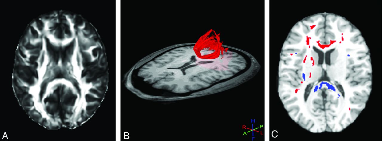
    FA image (A) reveals no abnormality in a patient with TBI. Tractography (B) can be used to delineate a region of interest for analysis. In this case, the forceps major (red) appears normal, but quantitative analysis of FA within this tract showed lower FA in the TBI group compared with controls. Whole-brain voxelwise analysis (C) reveals areas of low (blue) and high (red) FA. Low FA, consistent with TAI, is present within the forceps major at the splenium of the corpus callosum, as well as elsewhere.

  • Fig 2.
    • Download figure
    • Open in new tab
    • Download powerpoint
    Fig 2.

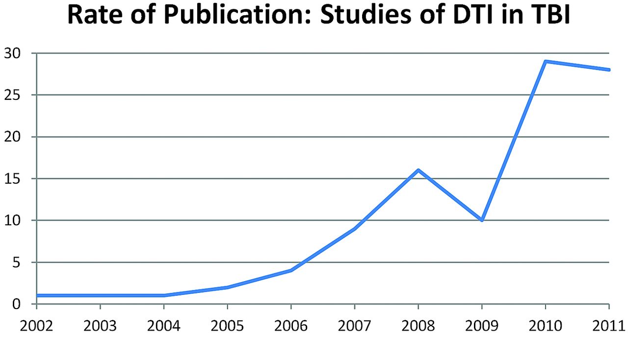
    The number of publications per year reporting DTI in TBI.

  • Fig 3.
    • Download figure
    • Open in new tab
    • Download powerpoint
    Fig 3.

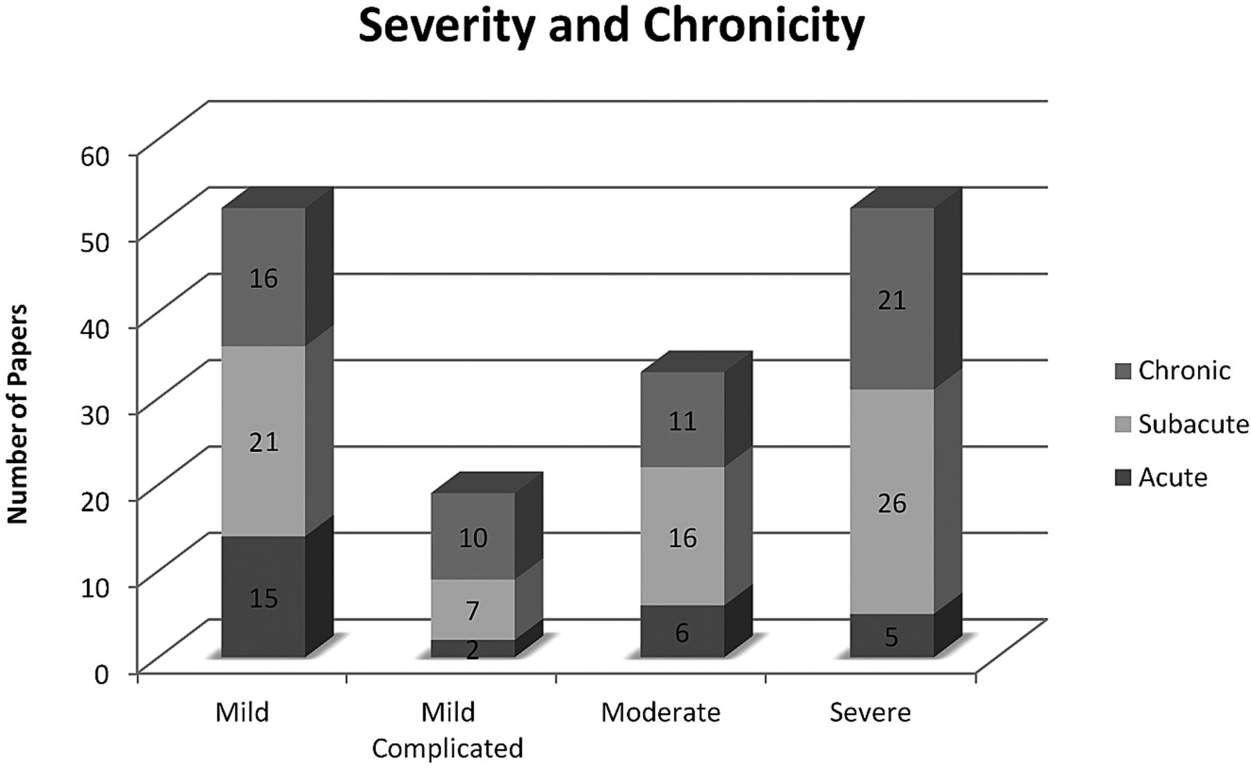
    The number of articles that studied patients at each timeframe and level of injury severity. Articles were only included if there was sufficient information to determine both the severity and the chronicity of individual patient injuries. Articles may be included multiple times if they studied subjects with multiple severities and/or multiple chronicities. A fully referenced version of this figure is available in On-line Table I.

  • Fig 4.
    • Download figure
    • Open in new tab
    • Download powerpoint
    Fig 4.

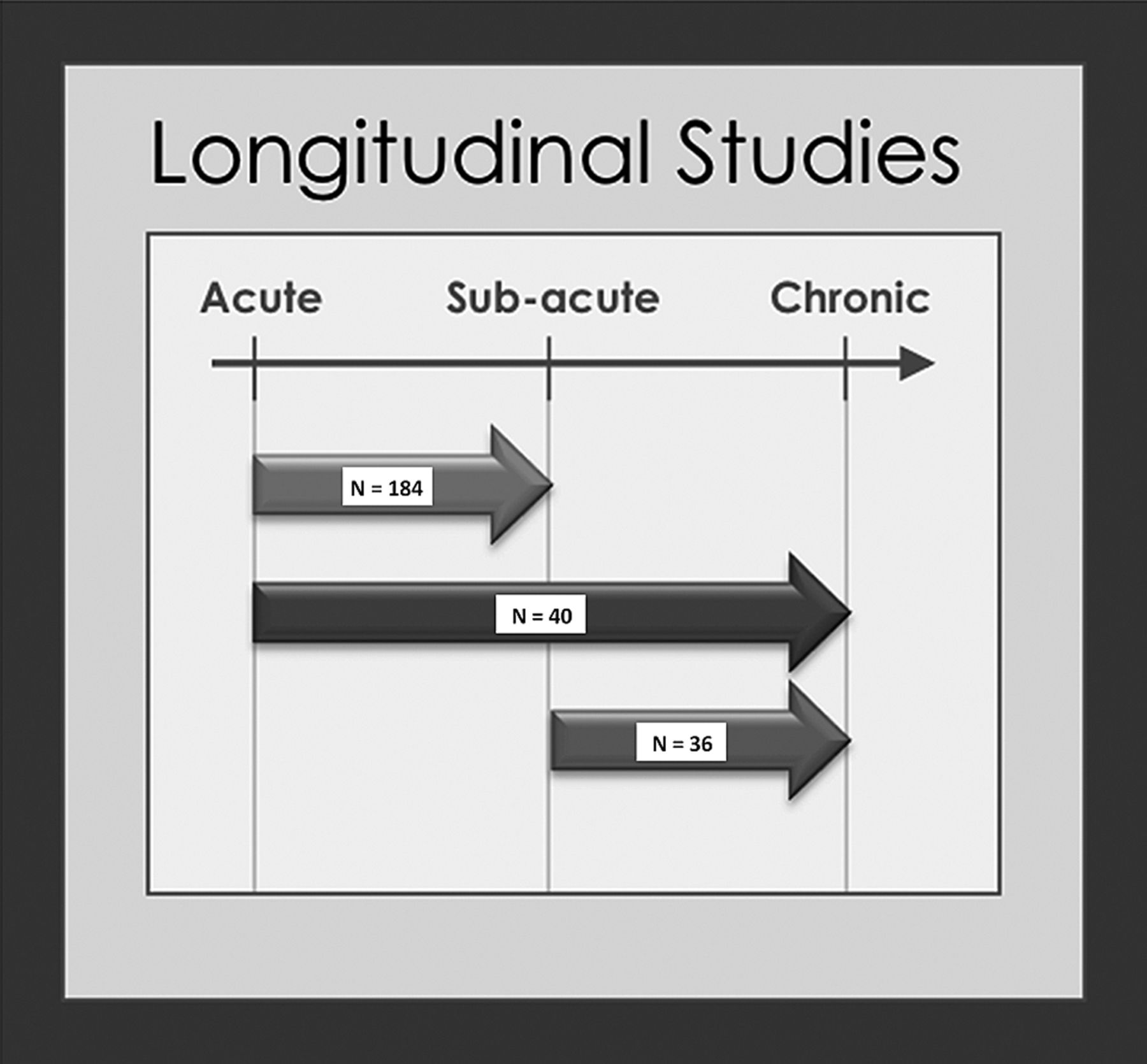
    Thirteen studies used a longitudinal design. Numbers represent patients from all studies imaged at 2 time points. Nine studies assessed patients at both acute and subacute time points.3,29,39,47,54,56,67,83,85 One study assessed patients at both acute and chronic time points.102 Two studies assessed patients at both subacute and chronic time points.24,93 One study (n = 47) assessed patients twice during the subacute period and, therefore, was omitted from the figure.49

Tables

  • Figures
    • View popup
    Table 1:

    Most common locations of abnormal FA by ROI analysisa

    LocationsFindings
    Corpus callosum, anterior/genu22b/30
    Corpus callosum, posterior/splenium21b/32
    Posterior limb of the internal capsule11/22
    Corpus callosum, body10/18
    Frontal lobe7/10
    Corona radiata6b/10
    Cingulum bundle7/8
    Centrum semiovale6/11
    Brain stem5/8
    Cerebral peduncle5/7
    • ↵a Values indicate the number of articles reporting abnormally low FA. Denominators represent the number of studies that assessed FA at these locations, including those that did not find abnormal FA.

    • ↵b Includes articles reporting abnormally high FA. A fully referenced version of this Table is available in On-line Table 2.

    • View popup
    Table 2:

    Most common locations of abnormal FA by tractographya

    LocationsFindings
    Corpus callosum, total10b/11
    Corpus callosum, anterior/genu8/8
    Corpus callosum, posterior/splenium7/8
    Cingulum bundle6/10
    Fornix5/7
    Corpus callosum, body4/6
    Fronto-occipital fasciculus4/5
    Inferior longitudinal fasciculus4/5
    Uncinate fasciculus4/5
    Hippocampus3/3
    • ↵a Values indicate the number of articles reporting abnormally low FA. Denominators represent the number of studies that assessed FA at these locations, including those that did not find abnormal FA.

    • ↵b Includes articles reporting abnormally high FA. A fully referenced version of this Table is available in On-line Table 3.

    • View popup
    Table 3:

    Most common locations of abnormal FA by whole-brain analysisa

    LocationsFindings
    Superior longitudinal fasciculus7/25
    Corpus callosum, anterior/genu7/25
    Inferior longitudinal fasciculus7/25
    Posterior limb of the internal capsule6/25
    Fronto-occipital fasciculus6/25
    Cingulum bundle5/25
    Corona radiata5/25
    Corpus callosum, overall5/25
    Corpus callosum, body5/25
    Fornix5/25
    Frontal lobe5/25
    Temporal lobe5/25
    • ↵a Values indicate the number of articles reporting low FA in these locations. Twenty-five articles used voxelwise analysis to assess FA throughout the entire brain. Because whole-brain analyses examine all brain regions, denominators are identical for all brain regions. A fully referenced version of this Table is available in On-line Table 4.

    • View popup
    Table 4:

    Most common locations of abnormal mean diffusivity by ROI analysisa

    LocationsFindings
    Corpus callosum, posterior/splenium10b/20
    Corpus callosum, anterior/genu10/16
    Frontal lobe9/10
    White matter7/7
    Thalamus4/6
    • ↵a Values indicate the number of articles reporting abnormally low MD. Denominators represent the number of studies that assessed MD at these locations, including those that did not find abnormal MD.

    • ↵b Includes articles reporting abnormally high MD. A fully referenced version of this Table is available in On-line Table 5.

    • View popup
    Table 5:

    Most common locations of abnormal mean diffusivity by tractography analysisa

    LocationsFindings
    Corpus callosum, anterior/genu4/4
    Fronto-occipital fasciculus4/5
    Inferior longitudinal fasciculus4/5
    Uncinate fasciculus4/4
    Cingulum bundle3b/7
    • ↵a Values indicate the number of articles reporting abnormally low MD. Denominators represent the number of studies that assessed MD at these locations, including those that did not find abnormal MD.

    • ↵b Includes articles reporting abnormally high MD. A fully referenced version of this Table is available in On-line Table 6.

    • View popup
    Table 6:

    Most common locations of abnormal mean diffusivity by whole-brain analysisa

    LocationsFindings
    Cingulum bundle6/13
    Corpus callosum, total5/13
    Superior longitudinal fasciculus4/13
    Posterior limb of the internal capsule4/13
    Fronto-occipital fasciculus4/13
    Frontal lobe4/13
    • ↵a Values indicate the number of articles reporting abnormally increased MD in these locations. Thirteen articles used whole-brain analysis to assess MD throughout the entire brain. Because whole-brain analyses examine all brain regions, denominators are identical for all brain regions. A fully referenced version of this Table is available in On-line Table 7.

    • View popup
    Table 7:

    Relationship of DTI metrics to cognitive outcome measuresa

    DTI MeasureCorrelationAttentionExecutive FunctionMemoryMotorPsychomotor/Processing SpeedVisuospatialIQ
    FAPositive correlation119144542
    Negative correlation6520200
    No correlation2661106
    MDPositive correlation3220010
    Negative correlation4670130
    No correlation1431200
    • Note:—IQ indicates intelligence quotient.

    • ↵a Total number of articles assessing relationships between DTI measures and cognitive outcomes. Cognitive-outcome measures have been categorized as 7 domains (top row). Articles are classified as reporting positive correlation, negative correlation, or no correlation. Positive correlation indicates a correlation coefficient greater than zero. Negative correlation indicates a correlation coefficient less than zero. No correlation includes articles that reported analyzing relationships between the DTI measures and cognitive outcomes within a domain but either reported finding no correlation (correlation coefficient equal to zero) or a correlation with a P value > .05. A fully referenced version of this Table is available in On-line Table 8.

    • View popup
    Table 8:

    Relationship of DTI metrics to general clinical assessmentsa

    DTI MeasureCorrelationGlobal Outcome MeasuresGCSPostconcussion Symptoms
    FAPositive correlation1153
    Negative correlation413
    No correlation386
    MDPositive correlation111
    Negative correlation542
    No correlation001
    • ↵a Total number of articles assessing relationships between DTI measures and global outcome measures (see “Functional Outcomes after TBI”), GCS, or postconcussive symptoms. Articles are classified as reporting positive correlation, negative correlation, or no correlation. Positive correlation indicates a correlation coefficient greater than zero. Negative correlation indicates a correlation coefficient less than zero. No correlation includes articles that reported analyzing relationships between the DTI measure and cognitive outcomes within a domain but either reported finding no correlation (correlation coefficient equal to zero) or a correlation with a P value >.05. A fully referenced version of this Table is available in On-line Table 9.

PreviousNext
Back to top

In this issue

American Journal of Neuroradiology: 34 (11)
American Journal of Neuroradiology
Vol. 34, Issue 11
1 Nov 2013
  • Table of Contents
  • Index by author
  • Complete Issue (PDF)
Advertisement
Print
Download PDF
Email Article

Thank you for your interest in spreading the word on American Journal of Neuroradiology.

NOTE: We only request your email address so that the person you are recommending the page to knows that you wanted them to see it, and that it is not junk mail. We do not capture any email address.

Enter multiple addresses on separate lines or separate them with commas.
A Decade of DTI in Traumatic Brain Injury: 10 Years and 100 Articles Later
(Your Name) has sent you a message from American Journal of Neuroradiology
(Your Name) thought you would like to see the American Journal of Neuroradiology web site.
CAPTCHA
This question is for testing whether or not you are a human visitor and to prevent automated spam submissions.
Cite this article
M.B. Hulkower, D.B. Poliak, S.B. Rosenbaum, M.E. Zimmerman, M.L. Lipton
A Decade of DTI in Traumatic Brain Injury: 10 Years and 100 Articles Later
American Journal of Neuroradiology Nov 2013, 34 (11) 2064-2074; DOI: 10.3174/ajnr.A3395

Citation Manager Formats

  • BibTeX
  • Bookends
  • EasyBib
  • EndNote (tagged)
  • EndNote 8 (xml)
  • Medlars
  • Mendeley
  • Papers
  • RefWorks Tagged
  • Ref Manager
  • RIS
  • Zotero
0 Responses
Respond to this article
Share
Bookmark this article
A Decade of DTI in Traumatic Brain Injury: 10 Years and 100 Articles Later
M.B. Hulkower, D.B. Poliak, S.B. Rosenbaum, M.E. Zimmerman, M.L. Lipton
American Journal of Neuroradiology Nov 2013, 34 (11) 2064-2074; DOI: 10.3174/ajnr.A3395
del.icio.us logo Twitter logo Facebook logo Mendeley logo
  • Tweet Widget
  • Facebook Like
  • Google Plus One
Purchase

Jump to section

  • Article
    • Abstract
    • ABBREVIATIONS:
    • Subjects with TBI
    • Severity, Chronicity, and Study Design
    • Data Acquisition Parameters
    • Data Analysis Methods
    • Specific Diffusion Measures Studied
    • Brain Regions
    • Functional Outcomes after TBI
    • Assessment of Individual Patients with TBI
    • Implications, Limitations, and Possibilities
    • Acknowledgments
    • Footnotes
    • REFERENCES
  • Figures & Data
  • Supplemental
  • Info & Metrics
  • Responses
  • References
  • PDF

Related Articles

  • PubMed
  • Google Scholar

Cited By...

  • Probabilistic Mapping and Automated Segmentation of Human Brainstem White Matter Bundles
  • Imaging traumatic brain injuries in mice with potassium channel PET tracer [18F]3F4AP
  • Longitudinal multimodal neuroimaging after traumatic brain injury
  • Dense Longitudinal Precision Neuroimaging of Recovery from Traumatic Brain Injury
  • Regionally specific resting-state beta neural power predicts brain injury and symptom recovery in adolescents with concussion: a longitudinal study
  • Evidence Suggesting Prolonged Neuroinflammation in a Subset of Children after Moderate/Severe TBI: A UCLA RAPBI Study
  • Volumetric and Diffusion Tensor Imaging biomarkers indicating long lasting post-concussion abnormalities in a youth pig model of mild Traumatic Brain Injury
  • Brain volume changes following blast-related mild TBI in service members and veterans: a LIMBIC-CENC study
  • Disrupted maturation of white matter microstructure after concussion contributes to internalizing behavior problems in female children
  • Automated detection of axonal damage along white matter tracts in acute severe traumatic brain injury
  • Towards Understanding Comprehensive Morphometric Changes and Its Correlation with Cognition and Exposure to Fighting in Active Professional Boxers
  • Abnormal Neurite Density and Orientation Dispersion in Frontal Lobe Link to Elevated Hyperactive/Impulsive Behaviors in Young Adults with Traumatic Brain Injury
  • White Matter Disruption in Pediatric Traumatic Brain Injury: Results From ENIGMA Pediatric Moderate to Severe Traumatic Brain Injury
  • Multi-tract multi-symptom relationships in pediatric concussion
  • Traumatic Cerebral Microbleeds in the Subacute Phase Are Practical and Early Predictors of Abnormality of the Normal-Appearing White Matter in the Chronic Phase
  • Connectomic Assessment of Injury Burden and Longitudinal Structural Network Alterations in Moderate-to-severe Traumatic Brain Injury
  • White matter and concussion: Are we on the right tract?
  • Neurofilament light as a biomarker in traumatic brain injury
  • Time course and diagnostic utility of NfL, tau, GFAP, and UCH-L1 in subacute and chronic TBI
  • Tractography-Pathology Correlations in Traumatic Brain Injury: A TRACK-TBI Study
  • Neuropsychological outcomes following traumatic brain injury
  • Randomised controlled clinical trial of a structured cognitive rehabilitation in patients with attention deficit following mild traumatic brain injury: study protocol
  • Structural abnormalities in thalamo-prefrontal tracks revealed by high angular resolution diffusion imaging predict working memory scores in concussed children
  • Relationship between white matter integrity and post-traumatic cognitive deficits: a systematic review and meta-analysis
  • Defining an Analytic Framework to Evaluate Quantitative MRI Markers of Traumatic Axonal Injury: Preliminary Results in a Mouse Closed Head Injury Model
  • Diverging white matter trajectories in children after traumatic brain injury: The RAPBI study
  • Trauma Imaging: A Literature Review
  • Analysis of head impact exposure and brain microstructure response in a season-long application of a jugular vein compression collar: a prospective, neuroimaging investigation in American football
  • Principal Component Analysis of Diffusion Tensor Images to Determine White Matter Injury Patterns Underlying Postconcussive Headache
  • Imaging assessment of traumatic brain injury
  • Callosal Function in Pediatric Traumatic Brain Injury Linked to Disrupted White Matter Integrity
  • Imaging Evidence and Recommendations for Traumatic Brain Injury: Advanced Neuro- and Neurovascular Imaging Techniques
  • Classification algorithms using multiple MRI features in mild traumatic brain injury
  • Single-Neuron NMDA Receptor Phenotype Influences Neuronal Rewiring and Reintegration following Traumatic Injury
  • Crossref (354)
  • Google Scholar

This article has been cited by the following articles in journals that are participating in Crossref Cited-by Linking.

  • Traumatic brain injuries
    Kaj Blennow, David L. Brody, Patrick M. Kochanek, Harvey Levin, Ann McKee, Gerard M. Ribbers, Kristine Yaffe, Henrik Zetterberg
    Nature Reviews Disease Primers 2016 2 1
  • Aging of cerebral white matter
    Huan Liu, Yuanyuan Yang, Yuguo Xia, Wen Zhu, Rehana K. Leak, Zhishuo Wei, Jianyi Wang, Xiaoming Hu
    Ageing Research Reviews 2017 34
  • White matter involvement after TBI: Clues to axon and myelin repair capacity
    Regina C. Armstrong, Amanda J. Mierzwa, Christina M. Marion, Genevieve M. Sullivan
    Experimental Neurology 2016 275
  • Traumatic Brain Injury as a Disorder of Brain Connectivity
    Jasmeet P. Hayes, Erin D. Bigler, Mieke Verfaellie
    Journal of the International Neuropsychological Society 2016 22 2
  • Fixel-based Analysis of Diffusion MRI: Methods, Applications, Challenges and Opportunities
    Thijs Dhollander, Adam Clemente, Mervyn Singh, Frederique Boonstra, Oren Civier, Juan Dominguez Duque, Natalia Egorova, Peter Enticott, Ian Fuelscher, Sanuji Gajamange, Sila Genc, Elie Gottlieb, Christian Hyde, Phoebe Imms, Claire Kelly, Melissa Kirkovski, Scott Kolbe, Xiaoyun Liang, Atul Malhotra, Remika Mito, Govinda Poudel, Tim J. Silk, David N. Vaughan, Julien Zanin, David Raffelt, Karen Caeyenberghs
    NeuroImage 2021 241
  • Time course and diagnostic utility of NfL, tau, GFAP, and UCH-L1 in subacute and chronic TBI
    Pashtun Shahim, Adam Politis, Andre van der Merwe, Brian Moore, Vindhya Ekanayake, Sara M. Lippa, Yi-Yu Chou, Dzung L. Pham, John A. Butman, Ramon Diaz-Arrastia, Henrik Zetterberg, Kaj Blennow, Jessica M. Gill, David L. Brody, Leighton Chan
    Neurology 2020 95 6
  • A Review of the Effectiveness of Neuroimaging Modalities for the Detection of Traumatic Brain Injury
    Franck Amyot, David B. Arciniegas, Michael P. Brazaitis, Kenneth C. Curley, Ramon Diaz-Arrastia, Amir Gandjbakhche, Peter Herscovitch, Sidney R. Hinds, Geoffrey T. Manley, Anthony Pacifico, Alexander Razumovsky, Jason Riley, Wanda Salzer, Robert Shih, James G. Smirniotopoulos, Derek Stocker
    Journal of Neurotrauma 2015 32 22
  • Neurofilament light as a biomarker in traumatic brain injury
    Pashtun Shahim, Adam Politis, Andre van der Merwe, Brian Moore, Yi-Yu Chou, Dzung L. Pham, John A. Butman, Ramon Diaz-Arrastia, Jessica M. Gill, David L. Brody, Henrik Zetterberg, Kaj Blennow, Leighton Chan
    Neurology 2020 95 6
  • Neuropsychology of traumatic brain injury: An expert overview
    P. Azouvi, A. Arnould, E. Dromer, C. Vallat-Azouvi
    Revue Neurologique 2017 173 7-8
  • Spatial patterns of progressive brain volume loss after moderate-severe traumatic brain injury
    James H Cole, Amy Jolly, Sara de Simoni, Niall Bourke, Maneesh C Patel, Gregory Scott, David J Sharp
    Brain 2018 141 3

More in this TOC Section

  • Ultra-High-Field MR Neuroimaging
  • Mechanisms of Healing in Coiled Intracranial Aneurysms: A Review of the Literature
  • Armies of Pestilence: CNS Infections as Potential Weapons of Mass Destruction
Show more Review Articles

Similar Articles

Advertisement

Indexed Content

  • Current Issue
  • Accepted Manuscripts
  • Article Preview
  • Past Issues
  • Editorials
  • Editors Choice
  • Fellow Journal Club
  • Letters to the Editor

Cases

  • Case Collection
  • Archive - Case of the Week
  • Archive - Case of the Month
  • Archive - Classic Case

Special Collections

  • Special Collections

Resources

  • News and Updates
  • Turn around Times
  • Submit a Manuscript
  • Author Policies
  • Manuscript Submission Guidelines
  • Evidence-Based Medicine Level Guide
  • Publishing Checklists
  • Graphical Abstract Preparation
  • Imaging Protocol Submission
  • Submit a Case
  • Become a Reviewer/Academy of Reviewers
  • Get Peer Review Credit from Publons

Multimedia

  • AJNR Podcast
  • AJNR SCANtastic
  • Video Articles

About Us

  • About AJNR
  • Editorial Board
  • Not an AJNR Subscriber? Join Now
  • Alerts
  • Feedback
  • Advertise with us
  • Librarian Resources
  • Permissions
  • Terms and Conditions

American Society of Neuroradiology

  • Not an ASNR Member? Join Now

© 2025 by the American Society of Neuroradiology All rights, including for text and data mining, AI training, and similar technologies, are reserved.
Print ISSN: 0195-6108 Online ISSN: 1936-959X

Powered by HighWire