
Article Figures & Data
Figures
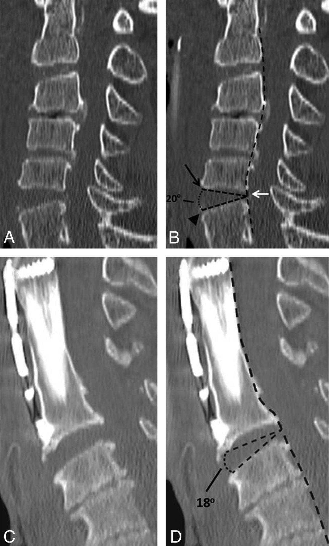
- Fig 1.
A, Sagittal CT reconstruction of the cervical spine in a 65-year-old man with blunt trauma demonstrating (B) measurement of the IDA in a level with ADL disruption, measured between the anterior superior endplate (black arrow) and anterior inferior endplate (arrowhead), with the apex of the angle at the midpoint of the posterior disk (white arrow) at the posterior vertebral body margin (broken black line). C, Sagittal CT reconstruction of the cervical spine in an 82-year-old man with a history of fall demonstrating measurement of an IDA in the presence of anterior osteophytes and vertebral body distraction, resulting in parallel endplates, at a level with ADL disruption. D, The IDA measurement excludes the osteophytes and is still measured at the midpoint of the distracted posterior disk.
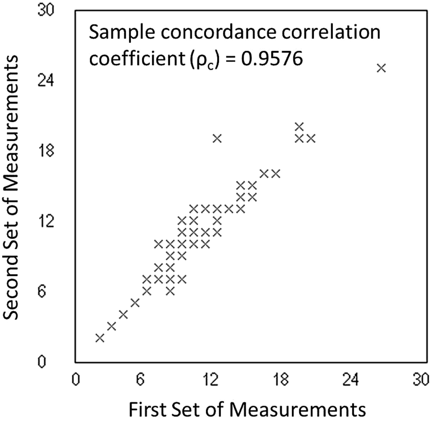
- Fig 2.
Concordance correlation coefficient plot measuring both precision and accuracy to determine how far the measured IDAs from the 2 different observers deviate from the line of perfect concordance (the line at 45° on a square scatterplot). The Lin coefficient increases in value as a function of the nearness of the data's reduced major axis to the line of perfect concordance (the accuracy of the data) and of the tightness of the data about its reduced major axis (the precision of the data).
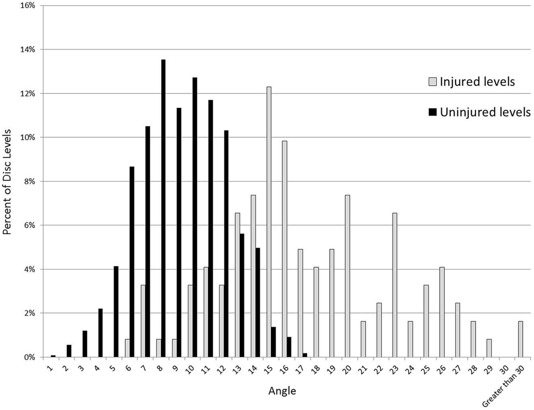
- Fig 3.
Distribution of disk angles among injured and uninjured disks, showing a Gaussian-like distribution of angles among uninjured disks, somewhat overlapping with angles in injured disks predominantly at 14° and below. Above an angle of 14°, there is minimal overlap.
- Fig 4.
ROC curves for IDA measurements. A, ROC curve for progressively smaller IDAs, demonstrating marked increase in sensitivity with only minor loss in specificity as the angle is decreased from 20° (white diamond) to 13° (black diamond). B, ROC curve for deviation of the IDA from normal values, demonstrating an increase in sensitivity with only mild loss in specificity as the range is changed from 2 SDs from normal values (white triangle) to 1 SD (black triangle). C, ROC curve for deviation of IDA from the average IDA of the remaining disks also demonstrating an increase in sensitivity with only mild loss in specificity as the range is changed from 2 SDs from the average (white open circle) to 1 SD (black open circle).


Tables
Trauma Patients Control Patients Total No. of patients 103 104 207 No. of male patients (%) 77 (76) 73 (70) 150 (72) Mean age, y (range) 56 (17–93) 52 (17–93) 54 (17–93) Disk Level Average IDA 95% CI C2–3 9.9 5.3–14.5 C3–4 9.4 3.8–15.1 C4–5 9.2 3.1–15.3 C5–6 8.6 2.2–14.9 C6–7 8.9 3.0–14.8 C7–T1 9.6 4.9–14.4 - Table 3.
Comparison of diagnostic performance among subjective disk angle measurements and IDA measurements
Subjective Disk Widening IDA of 13 IDA > 2 SD from Other Levels IDA > 1 SD from Normal Values Sensitivity (%) 16.4 82.0 72.1 86.1 Specificity (%) 99.4 89.1 100 84.7 PPV (%) 87.0 45.7 100 38.6 NPV (%) 82.3 97.8 97.0 98.2 AUC 0.580 0.854 0.860 0.853 Interpretation Poor discrimination Excellent discrimination Excellent discrimination Excellent discrimination Note:— NPV indicates negative predictive value; PPV, positive predictive value; SD, standard deviation.


