Advertisement

Main menu

  • Home
  • Content
    • Current Issue
    • Accepted Manuscripts
    • Article Preview
    • Past Issue Archive
    • AJNR Case Collection
    • Case of the Week Archive
    • Classic Case Archive
    • Case of the Month Archive
  • Special Collections
    • Spinal CSF Leak Articles (Jan 2020-June 2024)
    • 2024 AJNR Journal Awards
    • Most Impactful AJNR Articles
  • Multimedia
    • AJNR Podcast
    • AJNR Scantastics
    • Video Articles
  • For Authors
    • Submit a Manuscript
    • Author Policies
    • Fast publishing of Accepted Manuscripts
    • Graphical Abstract Preparation
    • Manuscript Submission Guidelines
    • Imaging Protocol Submission
    • Submit a Case for the Case Collection
  • About Us
    • About AJNR
    • Editorial Board
  • More
    • Become a Reviewer/Academy of Reviewers
    • Subscribers
    • Permissions
    • Alerts
    • Feedback
    • Advertisers
    • ASNR Home
  • Other Publications
    • ajnr

User menu

  • Alerts
  • Log in

Search

  • Advanced search
American Journal of Neuroradiology

American Journal of Neuroradiology

ASHNR American Society of Functional Neuroradiology ASHNR American Society of Pediatric Neuroradiology ASSR
  • Alerts
  • Log in

Advanced Search

  • Home
  • Content
    • Current Issue
    • Accepted Manuscripts
    • Article Preview
    • Past Issue Archive
    • AJNR Case Collection
    • Case of the Week Archive
    • Classic Case Archive
    • Case of the Month Archive
  • Special Collections
    • Spinal CSF Leak Articles (Jan 2020-June 2024)
    • 2024 AJNR Journal Awards
    • Most Impactful AJNR Articles
  • Multimedia
    • AJNR Podcast
    • AJNR Scantastics
    • Video Articles
  • For Authors
    • Submit a Manuscript
    • Author Policies
    • Fast publishing of Accepted Manuscripts
    • Graphical Abstract Preparation
    • Manuscript Submission Guidelines
    • Imaging Protocol Submission
    • Submit a Case for the Case Collection
  • About Us
    • About AJNR
    • Editorial Board
  • More
    • Become a Reviewer/Academy of Reviewers
    • Subscribers
    • Permissions
    • Alerts
    • Feedback
    • Advertisers
    • ASNR Home
  • Follow AJNR on Twitter
  • Visit AJNR on Facebook
  • Follow AJNR on Instagram
  • Join AJNR on LinkedIn
  • RSS Feeds

Welcome to the new AJNR, Updated Hall of Fame, and more. Read the full announcements.


AJNR is seeking candidates for the position of Associate Section Editor, AJNR Case Collection. Read the full announcement.


Getting new auth cookie, if you see this message a lot, tell someone!
Review ArticleReview Articles
Open Access

Facial Reanimation Procedures Depicted on Radiologic Imaging

D.T. Ginat, P. Bhama, M.E. Cunnane and T.A. Hadlock
American Journal of Neuroradiology September 2014, 35 (9) 1662-1666; DOI: https://doi.org/10.3174/ajnr.A3684
D.T. Ginat
aFrom the Department of Radiology (D.T.G.), Division of Neuroradiology, Massachusetts General Hospital, Boston, Massachusetts
  • Find this author on Google Scholar
  • Find this author on PubMed
  • Search for this author on this site
P. Bhama
bDepartment of Otolaryngology (P.B., T.A.H.), Division of Facial Plastic and Reconstructive Surgery
  • Find this author on Google Scholar
  • Find this author on PubMed
  • Search for this author on this site
M.E. Cunnane
cDepartment of Radiology (M.E.C.), Massachusetts Eye and Ear Infirmary, Boston, Massachusetts.
  • Find this author on Google Scholar
  • Find this author on PubMed
  • Search for this author on this site
T.A. Hadlock
bDepartment of Otolaryngology (P.B., T.A.H.), Division of Facial Plastic and Reconstructive Surgery
  • Find this author on Google Scholar
  • Find this author on PubMed
  • Search for this author on this site
  • Article
  • Figures & Data
  • Info & Metrics
  • Responses
  • References
  • PDF
Loading

Abstract

SUMMARY: Various facial reanimation procedures can be performed for treating patients with chronic facial nerve paralysis. The radiologic imaging features of static and dynamic techniques are reviewed in this article with clinical correlation, including brow lift, eyelid weights and springs, gracilis free flaps, fascia lata grafts, temporalis flaps, and Gore-Tex suspension slings. Although the anatomic alterations resulting from facial reanimation surgery may not necessarily be the focus of the imaging examination, it is important to recognize such changes and be familiar with MR imaging compatibility of the associated implanted materials. Furthermore, imaging is sometimes used to specifically evaluate the postoperative results, such as vessel patency following free gracilis transfer.

Chronic facial nerve paralysis can result in significant morbidity, including brow ptosis, lagophthalmos, ectropion, exposure keratopathy, nasal alar collapse, effacement of the nasolabial fold, ptosis of the oral commissure, and drooling. Static and dynamic facial reanimation surgical techniques are available to prevent and treat these complications. Static facial nerve rehabilitation procedures include brow lift, eyelid weight implantation, lower eyelid canthoplasty and tightening, fascia lata and alloplastic slings, and cheiloplasty. Dynamic facial reanimation procedures include nerve transfer and grafting, eyelid springs, free muscle transfer, regional muscle transfer, and lengthening myoplasty. The anatomic changes brought about by these procedures can be delineated by using conventional radiologic imaging modalities, including radiographs, CT, sonography, and MR imaging. Furthermore, in certain instances, imaging may be requested specifically to evaluate the results of facial reanimation surgery, such as interrogating the patency of the vascular pedicle following gracilis muscle transfer. The imaging findings after selected static and dynamic facial reanimation surgeries are described and depicted in the following sections.

Brow Lift

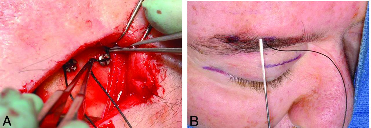
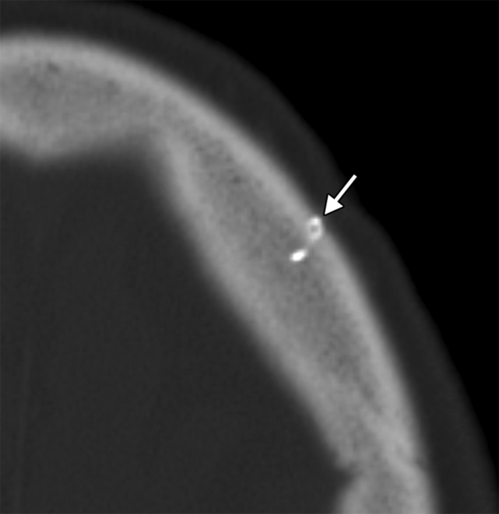
Brow ptosis can be addressed by performing a brow lift. Several methods exist to elevate the brow, one of which consists of implanting a fixation device into the frontal calvaria to which a permanent suture is secured (Fig 1).1 Brow lift can be performed in conjunction with ablation of the brow depressor muscles and blepharoplasty. Numerous fixation devices can be used, including pins, screws, tacks, K-wires, and tissue adhesives. Metallic fixation devices can be depicted on CT and should not be mistaken for unintended foreign bodies (Fig 2).

Fig 1.
  • Download figure
  • Open in new tab
  • Download powerpoint
Fig 1.

Brow lift. Intraoperative photographs show screws in the frontal bone used to secure sutures tunneled under the subcutaneous tissues toward the brow.

Fig 2.
  • Download figure
  • Open in new tab
  • Download powerpoint
Fig 2.

Brow lift. Axial CT image shows a metallic left frontal bone pin (arrow) used for suture fixation.

Eyelid Weights

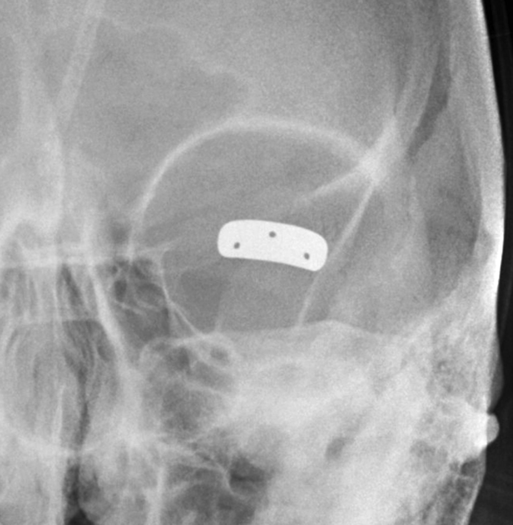
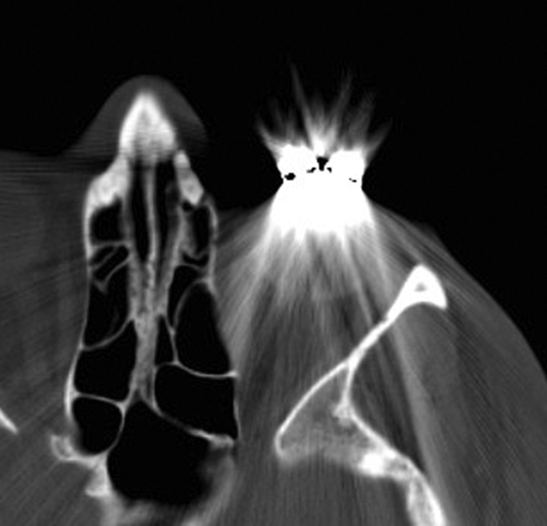
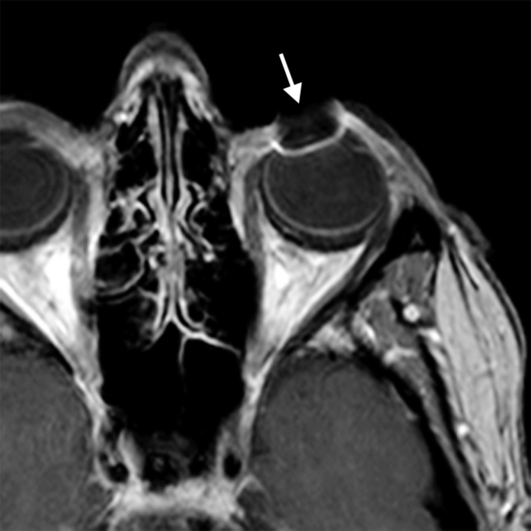
Implantation of gold and platinum eyelid weights into the upper eyelid is a static reanimation procedure for treating lagophthalmos.2 Although gold weights are more commonly used, platinum weights have a thinner profile and therefore offer an increased aesthetic benefit.3 Available eyelid implant designs include curved metal sheets with holes to permit anchoring with a suture (Fig 3) and flexible metal chains. In-growth of fibrous tissue through the holes also helps secure the weight in position. Eyelid weights are secured to the superficial aspect of the upper eyelid tarsal plate. The eyelid weights generally produce considerable streak artifacts on CT, which can obscure surrounding structures (Fig 4). Platinum and gold eyelid weights are considered MR imaging–compatible up to 7T4,5 but may cause local field inhomogeneity (Fig 5). Complications related to eyelid weight implantation include suboptimal eyelid contour, infection, allergic reaction, migration, and extrusion.6

Fig 3.
  • Download figure
  • Open in new tab
  • Download powerpoint
Fig 3.

Eyelid weight. Frontal radiograph shows a gold implant containing 3 drill holes at the level of the left upper eyelid.

Fig 4.
  • Download figure
  • Open in new tab
  • Download powerpoint
Fig 4.

Eyelid weight. Axial CT image shows extensive streak artifacts related to the left eyelid weight, which obscures surrounding structures.

Fig 5.
  • Download figure
  • Open in new tab
  • Download powerpoint
Fig 5.

Eyelid weight. Axial post-contrast fat-suppressed T1-weighted image shows field inhomogeneity associated with the left eyelid weight (arrow).

Eyelid Springs

Eyelid springs are used to augment eyelid closure in patients with eyelid paralysis.7,8 The spring has the ability to achieve complete eye closure in the supine position.9 The device is implanted via orbitotomy and consists of a palpebral branch and an orbital branch connected by a spring mechanism at the fulcrum. The springs are generally composed of stainless steel and are MR imaging–compatible up to at least 1.5T.10 The positioning and function of the device can be evaluated on radiographs obtained in the open and closed lid positions, whereby the palpebral branch is expected to descend with lid closure (Fig 6). Potential complications of eyelid springs include dislocation, metal fatigue resulting in failure, and exposure.

Fig 6.
  • Download figure
  • Open in new tab
  • Download powerpoint
Fig 6.

Eyelid spring. Frontal (A) and lateral (B) radiographs show that the inferior limb of the spring is positioned in the superior eyelid (arrows), while the superior limb is positioned along the orbital rim.

Gracilis Free Flap

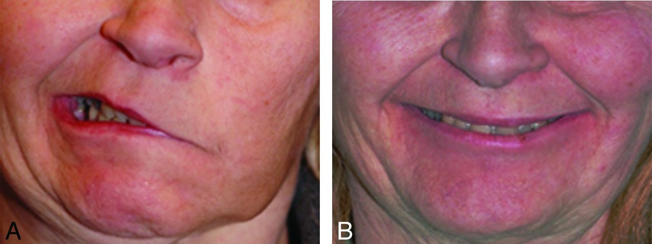
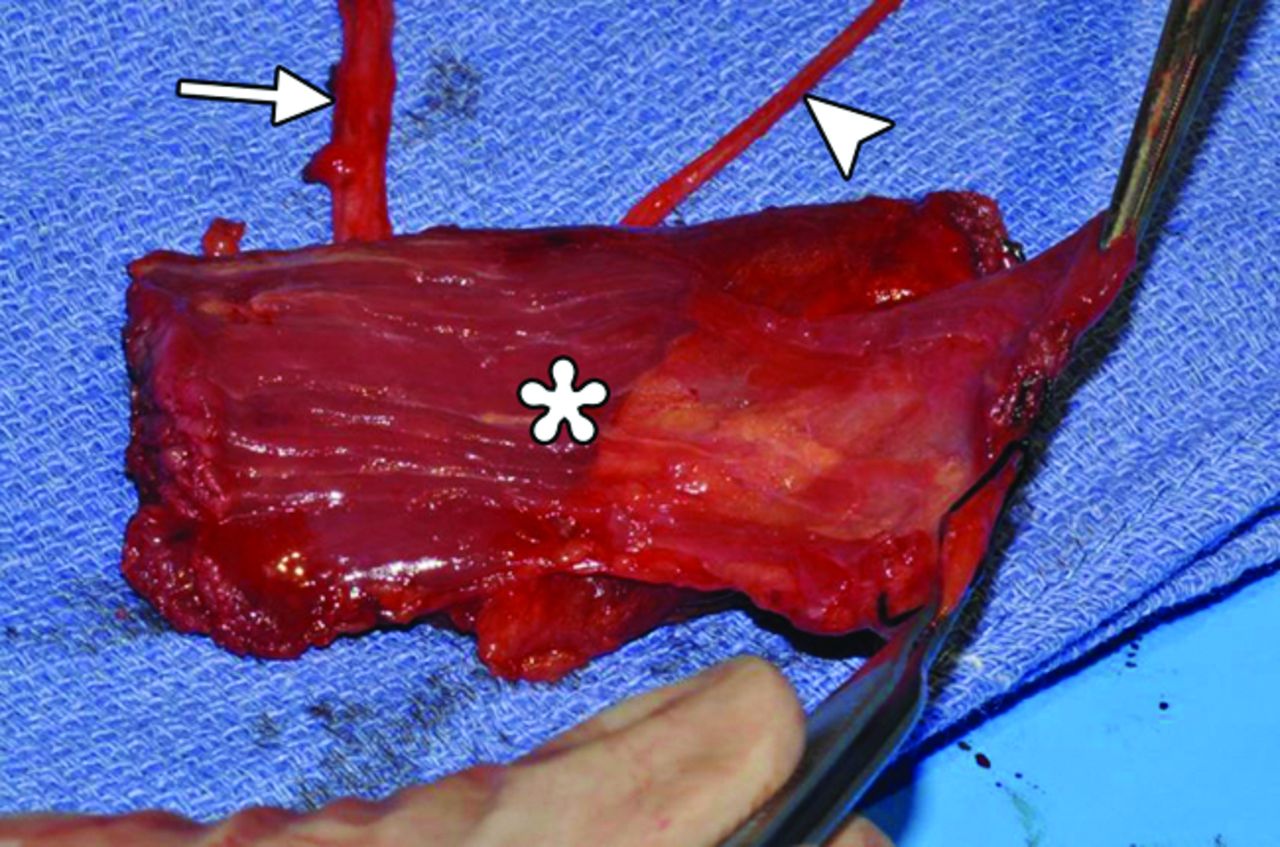
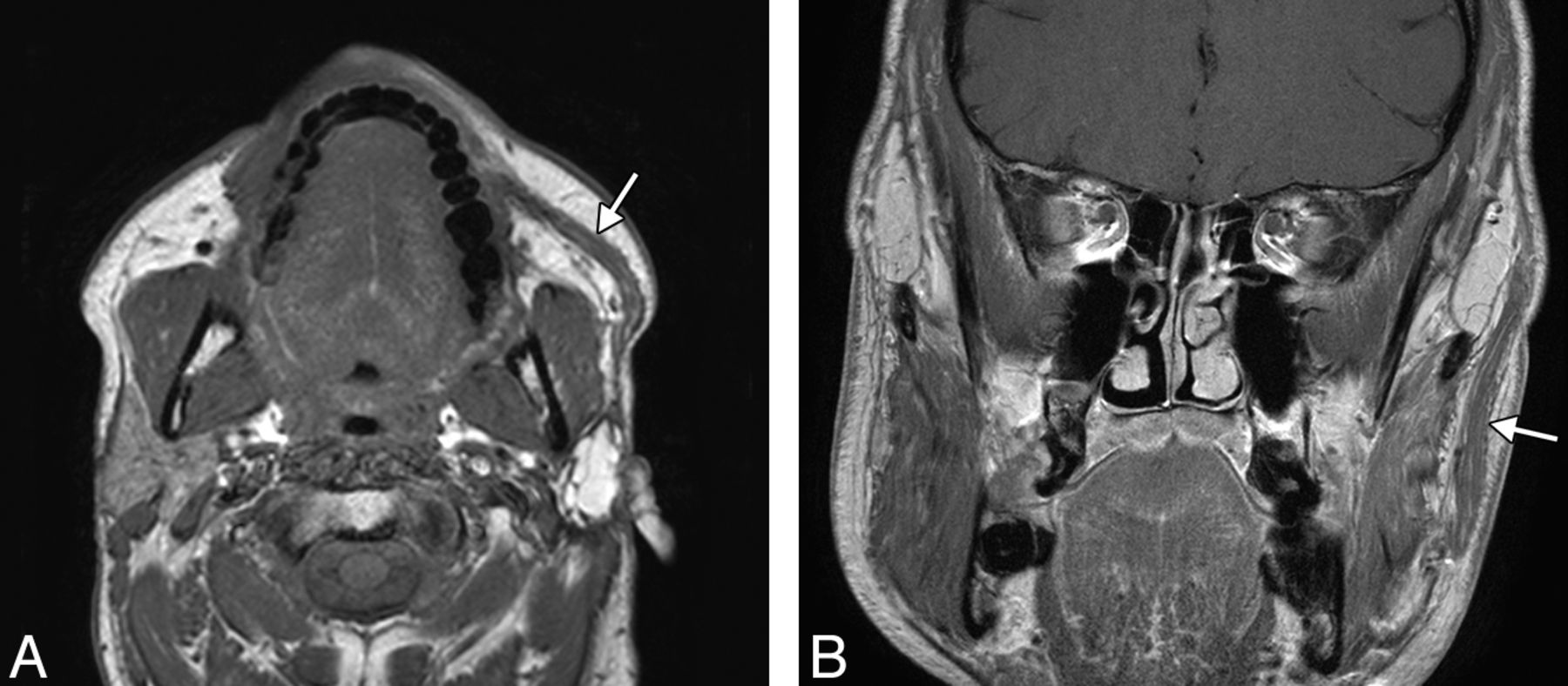
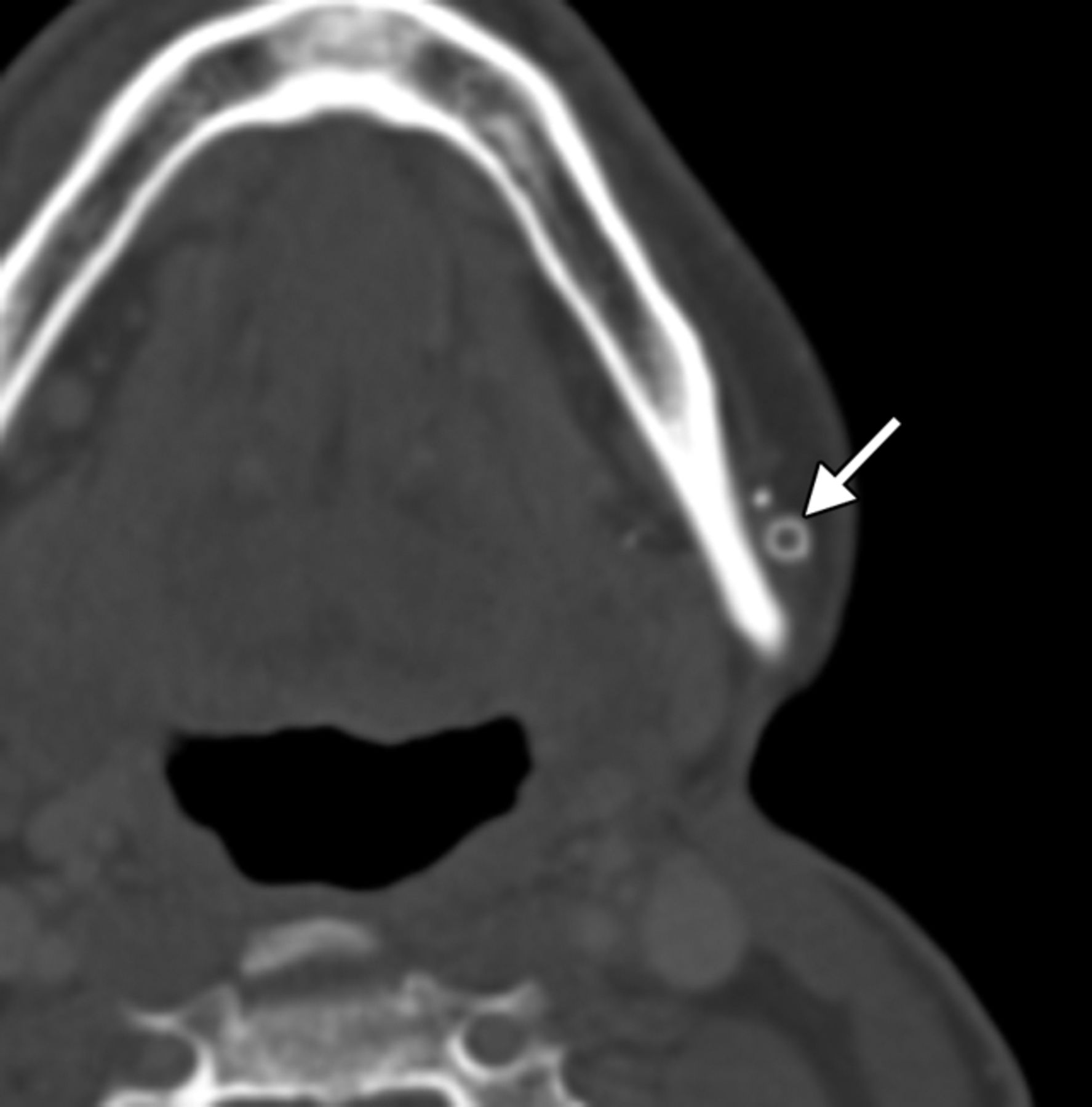
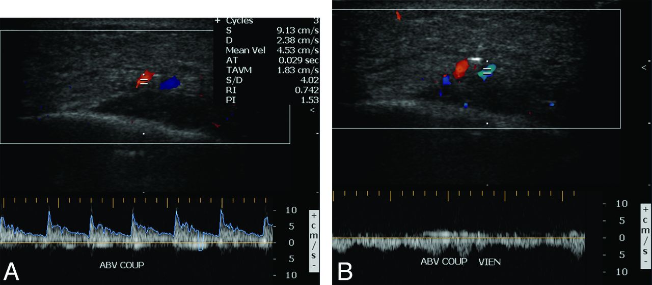
Free gracilis transfer is an effective method of smile rehabilitation for facial paralysis in selected patients (Fig 7).11⇓⇓–14 The gracilis muscle is harvested from the thigh with its neurovascular supply (Fig 8). The graft is then inset in the plane deep to the superficial musculoaponeurotic system and extends from the zygomatic arch to the modiolus of the oral commissure (Fig 9). The vascular pedicle of the graft is typically anastomosed to the facial artery and vein, and the obturator nerve can be anastomosed to a cross–face nerve graft and/or the masseteric branch of the trigeminal nerve.15,16 Vascular ring coupler devices are often used for the venous anastomosis.17 Depending on the particular type, the ring coupler may appear as a hyperattenuated circular structure overlying the angle of the mandible (Fig 10). Postoperative flap monitoring by physical examination alone is challenging and is often supplemented with use of a hand-held Doppler probe. Color Doppler sonography is an effective and noninvasive tool for evaluating arterial and venous flow through the pedicle of the buried free flap, whereby a sharp systolic upstroke should be evident in the artery and continuous flow should be observed in the vein (Fig 11). The examination can potentially avoid wound exploration to verify appropriate muscle perfusion and is typically performed on the first postoperative day.18 The arterial waveform of the graft should normally demonstrate a sharp systolic upstroke, while the vein may normally exhibit a continuous waveform and should be compressible, except at the site of the ring connector device. A good functional outcome correlates with normal muscle structure of the free flap depicted on MR imaging.19 Imaging may also be useful for measuring the graft thickness,20 which potentially relates to function.

Fig 7.
  • Download figure
  • Open in new tab
  • Download powerpoint
Fig 7.

Gracilis free flap. Clinical photographs before (A) and after (B) gracilis free flap reanimation show marked restoration of the patient's smile.

Fig 8.
  • Download figure
  • Open in new tab
  • Download powerpoint
Fig 8.

Gracilis free flap. Intraoperative photograph shows the harvested flap (asterisk) with an attached vascular pedicle (arrow) and nerve (arrowhead).

Fig 9.
  • Download figure
  • Open in new tab
  • Download powerpoint
Fig 9.

Gracilis free flap. Axial (A) and coronal (B) postcontrast T1-weighted MR images show a healthy gracilis flap (arrows), which extends from the zygomatic arch to the oral commissure following total left parotidectomy, with facial nerve sacrifice for resection of mucoepidermoid carcinoma.

Fig 10.
  • Download figure
  • Open in new tab
  • Download powerpoint
Fig 10.

Gracilis free flap. Axial CT image shows a hyperattenuated circular ring connector (arrow) used to facilitate the vascular anastomosis.

Fig 11.
  • Download figure
  • Open in new tab
  • Download powerpoint
Fig 11.

Gracilis free flap. Color Doppler sonographic images of the vascular pedicle show normal arterial (A) and venous (B) waveforms.

Fascia Lata Graft

Autogenous fascia lata grafts can be used as the primary therapeutic option in static facial rebalancing or in conjunction with dynamic muscle reanimation.21 In particular, fascia lata slings can be used to improve oral competence and external nasal valve patency (Fig 12). The fascia lata graft is similarly inset into the subsuperficial musculoaponeurotic system plane and appears as a thin band of soft tissue that courses through the subcutaneous tissues of the face on cross-sectional imaging (Fig 13).

Fig 12.
  • Download figure
  • Open in new tab
  • Download powerpoint
Fig 12.

Fascia lata graft. Intraoperative photograph shows the prepared fascia lata graft (arrow) over its planned course toward the left nasal ala before implantation.

Fig 13.
  • Download figure
  • Open in new tab
  • Download powerpoint
Fig 13.

Fascia lata graft. The patient did not obtain optimal muscular function after the gracilis free flap procedure. Axial CT shows a tenuous right gracilis flap (arrowheads) and a linear band of soft tissue that extends from the gracilis flap to the right alar base, which corresponds to the fascia lata sling resuspension for external nasal valve correction (arrow).
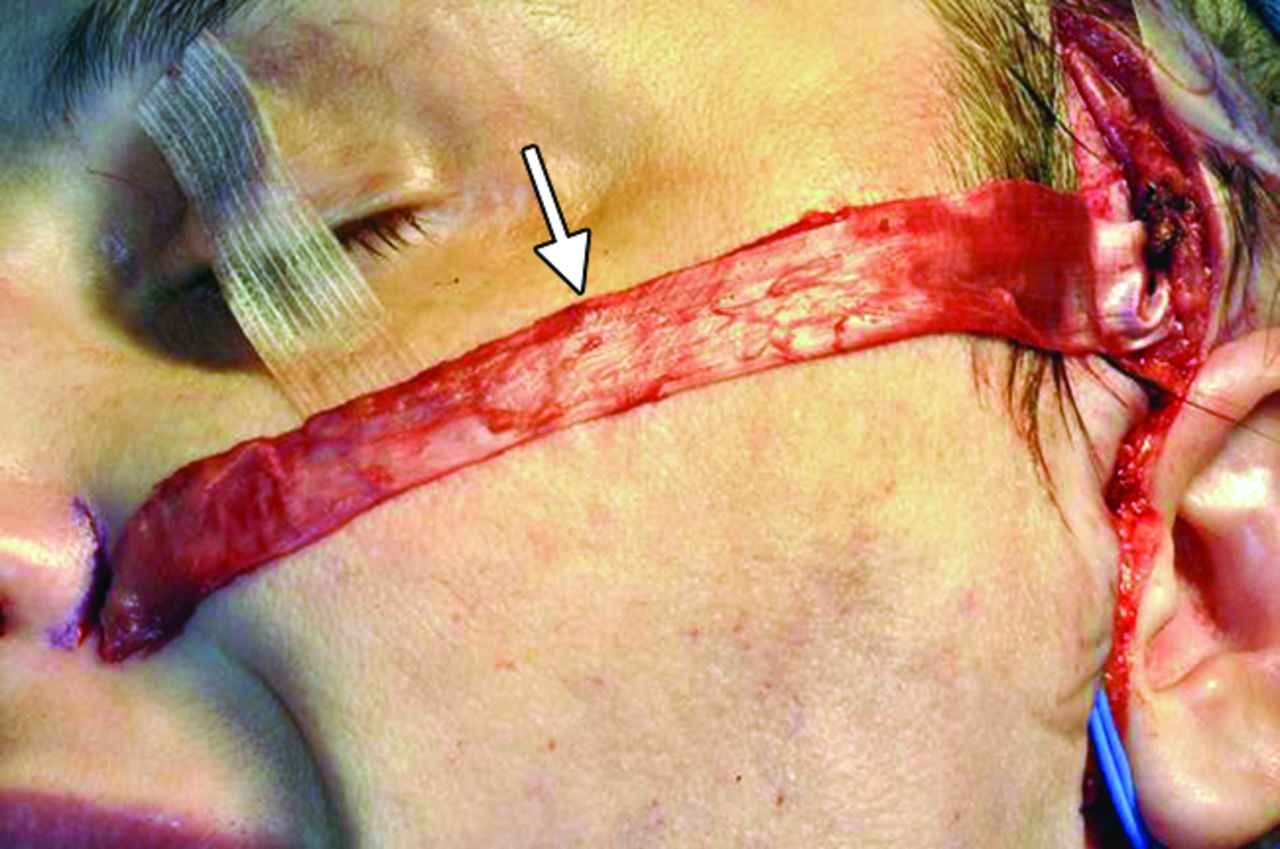
Temporalis Flap

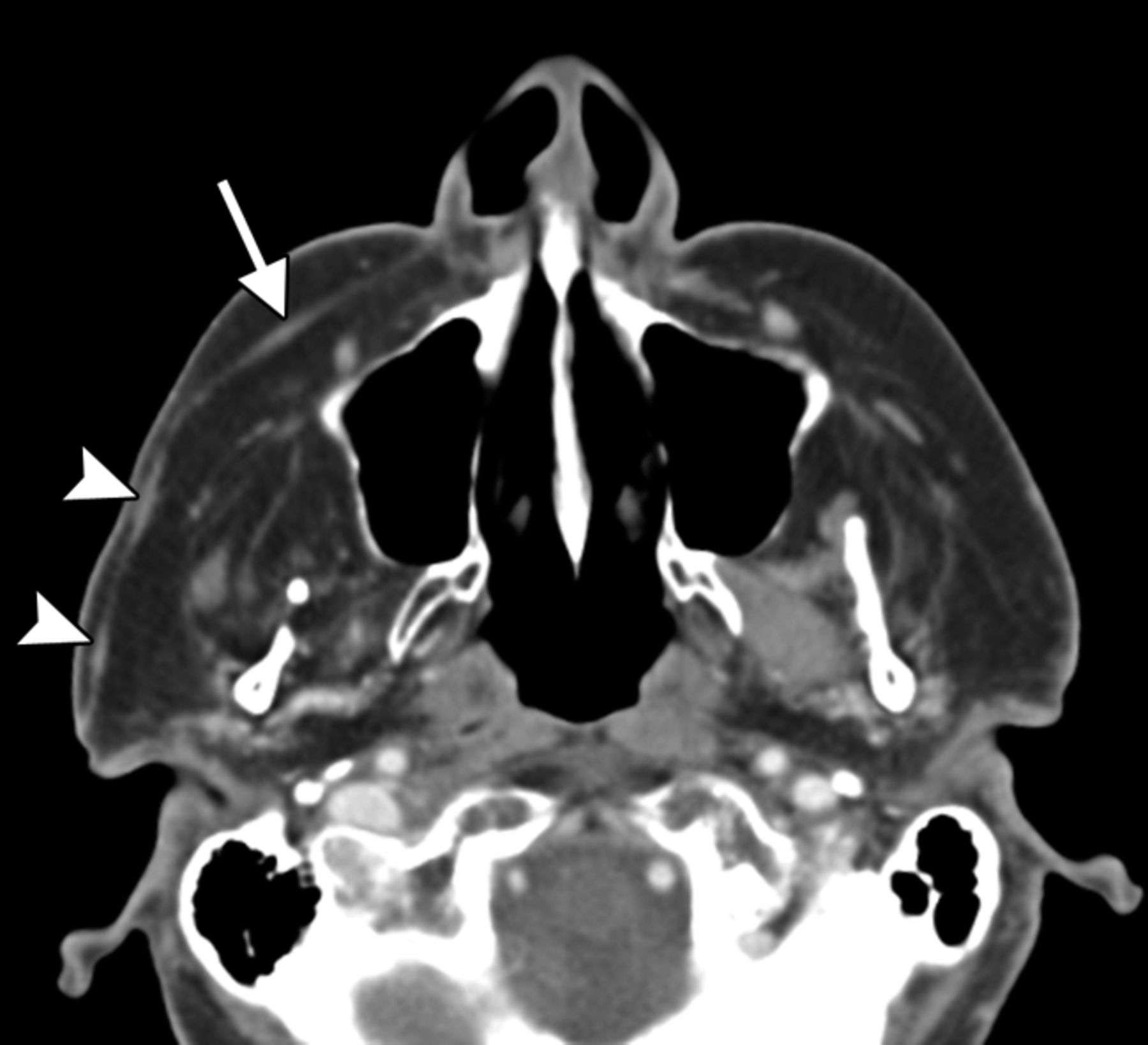
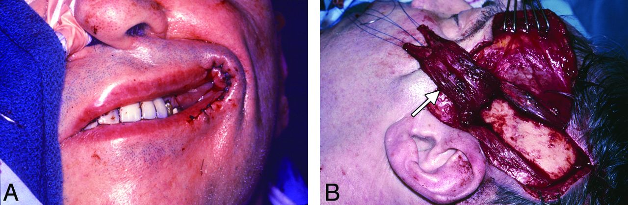
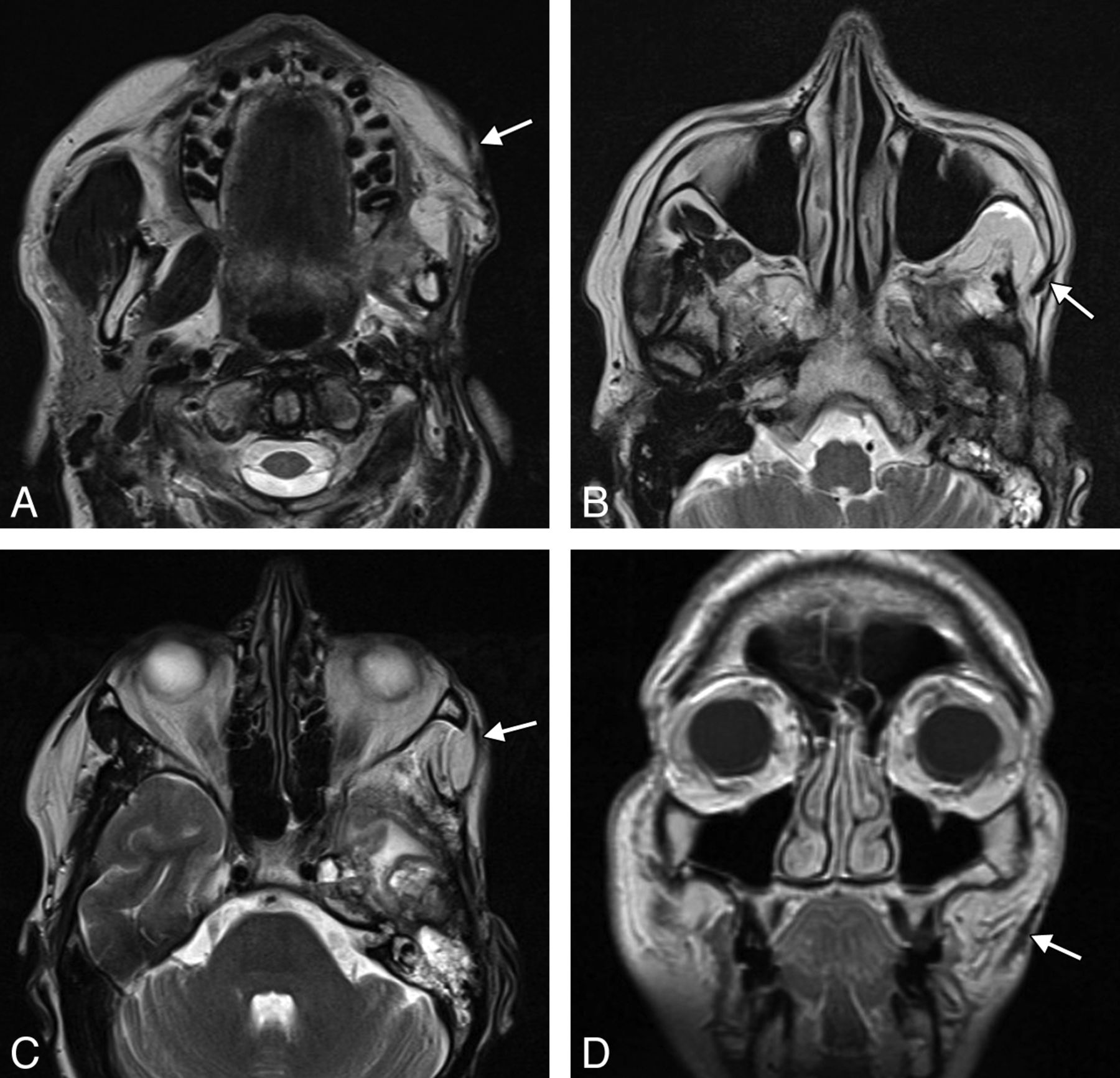
The temporalis flap (temporalis muscle transposition) procedure is an effective option for reanimation of the smile.22 An approximately 1.5-cm-wide strip of the midportion of the temporalis muscle is dissected off the calvaria and reflected in the subdermal plane from the zygomatic arch to the modiolus of the oral commissure (Fig 14). The course of the transposed temporalis muscle can be delineated on cross-sectional imaging as a thin band of soft tissue (Fig 15).

Fig 14.
  • Download figure
  • Open in new tab
  • Download powerpoint
Fig 14.

Temporalis flap. Intraoperative photographs show that the middle portion of the temporalis muscle (arrow) has been dissected free and transposed toward the oral commissure. The flap was subsequently tunneled beneath the subcutaneous tissues.

Fig 15.
  • Download figure
  • Open in new tab
  • Download powerpoint
Fig 15.

Temporalis flap. Axial T2 MR imaging (A–C) and coronal T1 MR imaging (D) show that the left temporalis muscle with the overlying fascia (arrows) is directed retrograde from the temporal fossa to the orbicularis oris.

Gore-Tex Sling Suspension

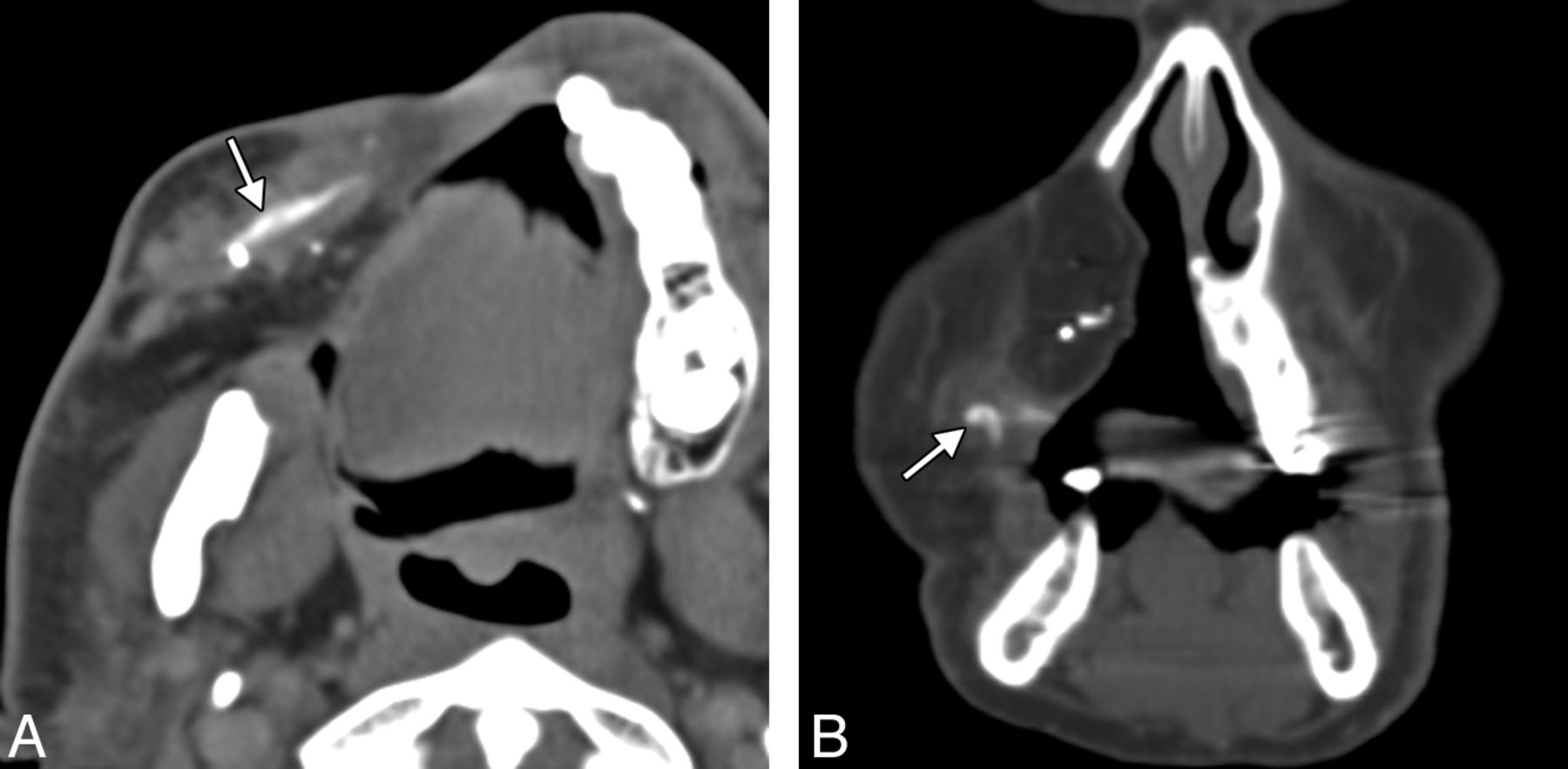
Gore-Tex (expanded polytetrafluoroethylene) (W.L. Gore and Associates, Reisterstown, Maryland) can be used for static suspension in facial paralysis.23,24 It is manufactured in thin 1- to 2-mm sheets, which can be cut into strips and implanted through small incisions. On CT, Gore-Tex slings appear as linear hyperattenuations (Fig 16).25 Although the use of Gore-Tex in facial reanimation eliminates donor site morbidity associated with the harvest of autologous grafts, the allograft is prone to complications, such as delayed wound infection.24

Fig 16.
  • Download figure
  • Open in new tab
  • Download powerpoint
Fig 16.

Gore-Tex sling. Axial (A) and coronal (B) CT images show the linear hyperattenuated strip of Gore-Tex that supports the right oral commissure (arrows). The patient is status post right complete maxillectomy with myocutaneous flap reconstruction.

Footnotes

  • Disclosures: Mary E. Cunnane—UNRELATED: Other: WorldCare Clinical, Comments: I was a radiologist for a clinical trial of a chemotherapy drug for head and neck cancer.

Indicates open access to non-subscribers at www.ajnr.org

REFERENCES

  1. 1.↵
    1. Foustanos A
    . Suture fixation technique for endoscopic brow lift. Semin Plast Surg 2008;22:43–49
    CrossRefPubMed
  2. 2.↵
    1. Bladen JC,
    2. Norris JH,
    3. Malhotra R
    . Cosmetic comparison of gold weight and platinum chain insertion in primary upper eyelid loading for lagophthalmos. Ophthal Plast Reconstr Surg 2012;28:171–75
    CrossRefPubMed
  3. 3.↵
    1. Silver AL,
    2. Lindsay RW,
    3. Cheney ML,
    4. et al
    . Thin-profile platinum eyelid weighting: a superior option in the paralyzed eye. Plast Reconstr Surg 2009;123:1697–703
    CrossRefPubMed
  4. 4.↵
    1. Schrom T,
    2. Thelen A,
    3. Asbach P,
    4. et al
    . Effect of 7.0 Tesla MRI on upper eyelid implants. Ophthal Plast Reconstr Surg 2006;22:480–82
    CrossRefPubMed
  5. 5.↵
    1. Marra S,
    2. Leonetti JP,
    3. Konior RJ,
    4. et al
    . Effect of magnetic resonance imaging on implantable eyelid weights. Ann Otol Rhinol Laryngol 1995;104:448–52
    Abstract/FREE Full Text
  6. 6.↵
    1. Dinces EA,
    2. Mauriello JA Jr.,
    3. Kwartler JA,
    4. et al
    . Complications of gold weight eyelid implants for treatment of fifth and seventh nerve paralysis. Laryngoscope 1997;107:1617–22
    CrossRefPubMed
  7. 7.↵
    1. Fay A,
    2. Santiago YM
    . A modified Levine palpebral spring for the treatment of myogenic ptosis. Ophthal Plast Reconstr Surg 2012;28:372–75
    CrossRefPubMed
  8. 8.↵
    1. Demirci H,
    2. Frueh BR
    . Palpebral spring in the management of lagophthalmos and exposure keratopathy secondary to facial nerve palsy. Ophthal Plast Reconstr Surg 2009;25:270–75
    CrossRefPubMed
  9. 9.↵
    1. Levine RE,
    2. Shapiro JP
    . Reanimation of the paralyzed eyelid with the enhanced palpebral spring or the gold weight: modern replacements for tarsorrhaphy. Facial Plast Surg 2000;16:325–36
    CrossRefPubMed
  10. 10.↵
    1. Gardner TA,
    2. Rak KM
    . Magnetic resonance imaging of eyelid springs and gold weights. Arch Ophthalmol 1991;109:1498
    CrossRefPubMed
  11. 11.↵
    1. Chuang DC
    . Free tissue transfer for the treatment of facial paralysis. Facial Plast Surg 2008;24:194–203
    CrossRefPubMed
  12. 12.↵
    1. Hadlock TA,
    2. Malo JS,
    3. Cheney ML,
    4. et al
    . Free gracilis transfer for smile in children: the Massachusetts Eye and Ear Infirmary Experience in excursion and quality-of-life changes. Arch Facial Plast Surg 2011;13:190–94
    CrossRefPubMed
  13. 13.↵
    1. Boahene KD
    . Dynamic muscle transfer in facial reanimation. Facial Plast Surg 2008;24:204–10
    CrossRefPubMed
  14. 14.↵
    1. Vakharia KT,
    2. Henstrom D,
    3. Plotkin SR,
    4. et al
    . Facial reanimation of patients with neurofibromatosis type 2. Neurosurgery 2012;70:237–43
    PubMed
  15. 15.↵
    1. Bianchi B,
    2. Copelli C,
    3. Ferrari S,
    4. et al
    . Facial animation with free-muscle transfer innervated by the masseter motor nerve in unilateral facial paralysis. J Oral Maxillofac Surg 2010;68:1524–29
    CrossRefPubMed
  16. 16.↵
    1. Faria JC,
    2. Scopel GP,
    3. Busnardo FF,
    4. et al
    . Nerve sources for facial reanimation with muscle transplant in patients with unilateral facial palsy: clinical analysis of 3 techniques. Ann Plast Surg 2007;59:87–91
    CrossRefPubMed
  17. 17.↵
    1. Barker EV,
    2. Enepekides DJ
    . The utility of microvascular anastomotic devices in head and neck reconstruction. Curr Opin Otolaryngol Head Neck Surg 2008;16:331–34
    CrossRefPubMed
  18. 18.↵
    1. Vakharia KT,
    2. Henstrom D,
    3. Lindsay R,
    4. et al
    . Color Doppler ultrasound: effective monitoring of the buried free flap in facial reanimation. Otolaryngol Head Neck Surg 2012;146:372–76
    Abstract/FREE Full Text
  19. 19.↵
    1. Ylä-Kotola TM,
    2. Kauhanen MS,
    3. Koskinen SK,
    4. et al
    . Magnetic resonance imaging of microneurovascular free muscle flaps in facial reanimation. Br J Plast Surg 2005;58:222–27
  20. 20.↵
    1. Salmi A,
    2. Ahovuo J,
    3. Tukiainen E,
    4. et al
    . Use of ultrasonography to evaluate muscle thickness and blood flow in free flaps. Microsurgery 1995;16:601–05
    CrossRefPubMed
  21. 21.↵
    1. Rose EH
    . Autogenous fascia lata grafts: clinical applications in reanimation of the totally or partially paralyzed face. Plast Reconstr Surg 2005;116:20–32, discussion 33–35
    PubMed
  22. 22.↵
    1. White TP,
    2. Faulkner JA,
    3. Markley JM Jr.,
    4. et al
    . Translocation of the temporalis muscle for treatment of facial paralysis. Muscle Nerve 1982;5:500–04
    CrossRefPubMed
  23. 23.↵
    1. Liu YM,
    2. Sherris DA
    . Static procedures for the management of the midface and lower face. Facial Plast Surg 2008;24:211–15
    CrossRefPubMed
  24. 24.↵
    1. Constantinides M,
    2. Galli SK,
    3. Miller PJ
    . Complications of static facial suspensions with expanded polytetrafluoroethylene (ePTFE). Laryngoscope 2001;111:2114–21
    CrossRefPubMed
  25. 25.↵
    1. Schatz CJ,
    2. Ginat DT
    . Imaging of cosmetic facial implants and grafts. AJNR Am J Neuroradiol 2013;34:1674–81
    Abstract/FREE Full Text
  • © 2014 by American Journal of Neuroradiology
View Abstract
PreviousNext
Back to top

In this issue

American Journal of Neuroradiology: 35 (9)
American Journal of Neuroradiology
Vol. 35, Issue 9
1 Sep 2014
  • Table of Contents
  • Index by author
  • Complete Issue (PDF)
Advertisement
Print
Download PDF
Email Article

Thank you for your interest in spreading the word on American Journal of Neuroradiology.

NOTE: We only request your email address so that the person you are recommending the page to knows that you wanted them to see it, and that it is not junk mail. We do not capture any email address.

Enter multiple addresses on separate lines or separate them with commas.
Facial Reanimation Procedures Depicted on Radiologic Imaging
(Your Name) has sent you a message from American Journal of Neuroradiology
(Your Name) thought you would like to see the American Journal of Neuroradiology web site.
CAPTCHA
This question is for testing whether or not you are a human visitor and to prevent automated spam submissions.
Cite this article
D.T. Ginat, P. Bhama, M.E. Cunnane, T.A. Hadlock
Facial Reanimation Procedures Depicted on Radiologic Imaging
American Journal of Neuroradiology Sep 2014, 35 (9) 1662-1666; DOI: 10.3174/ajnr.A3684

Citation Manager Formats

  • BibTeX
  • Bookends
  • EasyBib
  • EndNote (tagged)
  • EndNote 8 (xml)
  • Medlars
  • Mendeley
  • Papers
  • RefWorks Tagged
  • Ref Manager
  • RIS
  • Zotero
0 Responses
Respond to this article
Share
Bookmark this article
Facial Reanimation Procedures Depicted on Radiologic Imaging
D.T. Ginat, P. Bhama, M.E. Cunnane, T.A. Hadlock
American Journal of Neuroradiology Sep 2014, 35 (9) 1662-1666; DOI: 10.3174/ajnr.A3684
del.icio.us logo Twitter logo Facebook logo Mendeley logo
  • Tweet Widget
  • Facebook Like
  • Google Plus One
Purchase

Jump to section

  • Article
    • Abstract
    • Footnotes
    • REFERENCES
  • Figures & Data
  • Info & Metrics
  • Responses
  • References
  • PDF

Related Articles

  • No related articles found.
  • PubMed
  • Google Scholar

Cited By...

  • No citing articles found.
  • Crossref (6)
  • Google Scholar

This article has been cited by the following articles in journals that are participating in Crossref Cited-by Linking.

  • Imaging of the Postoperative Eye and Orbit
    Daniel T. Ginat
    Neuroimaging Clinics of North America 2022 32 1
  • Surgical Implants in the Head and Neck: Normal and Abnormal Imaging Appearances
    Daniel Ginat
    Seminars in Roentgenology 2023 58 3
  • Atlas of Postsurgical Neuroradiology
    Daniel Thomas Ginat, Elizabeth Blair, Hugh D. Curtin
    2017
  • Implants and Foreign Bodies on Head and Neck Imaging
    Daniel T. Ginat
    Neuroimaging Clinics of North America 2022 32 2
  • Post-treatment Imaging of the Orbit
    Daniel Thomas Ginat, Gul Moonis, Suzanne K. Freitag
    2015
  • Postsurgical and Postradiation Findings in the Head and Neck Imaging
    Charles Pierce, Daniel T. Ginat
    Neuroimaging Clinics of North America 2022 32 2

More in this TOC Section

  • Ultra-High-Field MR Neuroimaging
  • Mechanisms of Healing in Coiled Intracranial Aneurysms: A Review of the Literature
  • Armies of Pestilence: CNS Infections as Potential Weapons of Mass Destruction
Show more Review Articles

Similar Articles

Advertisement

Indexed Content

  • Current Issue
  • Accepted Manuscripts
  • Article Preview
  • Past Issues
  • Editorials
  • Editors Choice
  • Fellow Journal Club
  • Letters to the Editor

Cases

  • Case Collection
  • Archive - Case of the Week
  • Archive - Case of the Month
  • Archive - Classic Case

Special Collections

  • Special Collections

Resources

  • News and Updates
  • Turn around Times
  • Submit a Manuscript
  • Author Policies
  • Manuscript Submission Guidelines
  • Evidence-Based Medicine Level Guide
  • Publishing Checklists
  • Graphical Abstract Preparation
  • Imaging Protocol Submission
  • Submit a Case
  • Become a Reviewer/Academy of Reviewers
  • Get Peer Review Credit from Publons

Multimedia

  • AJNR Podcast
  • AJNR SCANtastic
  • Video Articles

About Us

  • About AJNR
  • Editorial Board
  • Not an AJNR Subscriber? Join Now
  • Alerts
  • Feedback
  • Advertise with us
  • Librarian Resources
  • Permissions
  • Terms and Conditions

American Society of Neuroradiology

  • Not an ASNR Member? Join Now

© 2025 by the American Society of Neuroradiology All rights, including for text and data mining, AI training, and similar technologies, are reserved.
Print ISSN: 0195-6108 Online ISSN: 1936-959X

Powered by HighWire