
Article Figures & Data
Figures
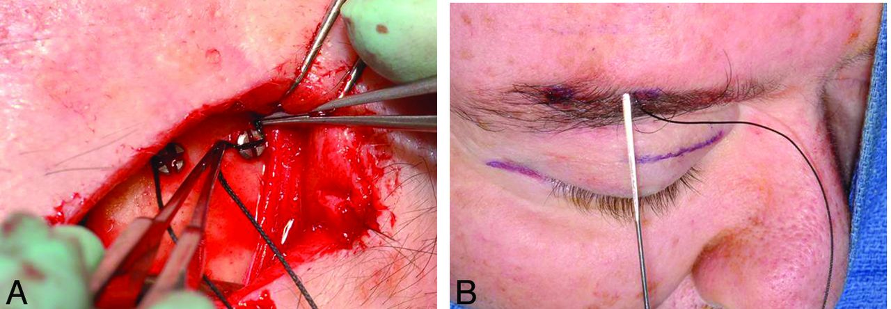
- Fig 1.
Brow lift. Intraoperative photographs show screws in the frontal bone used to secure sutures tunneled under the subcutaneous tissues toward the brow.
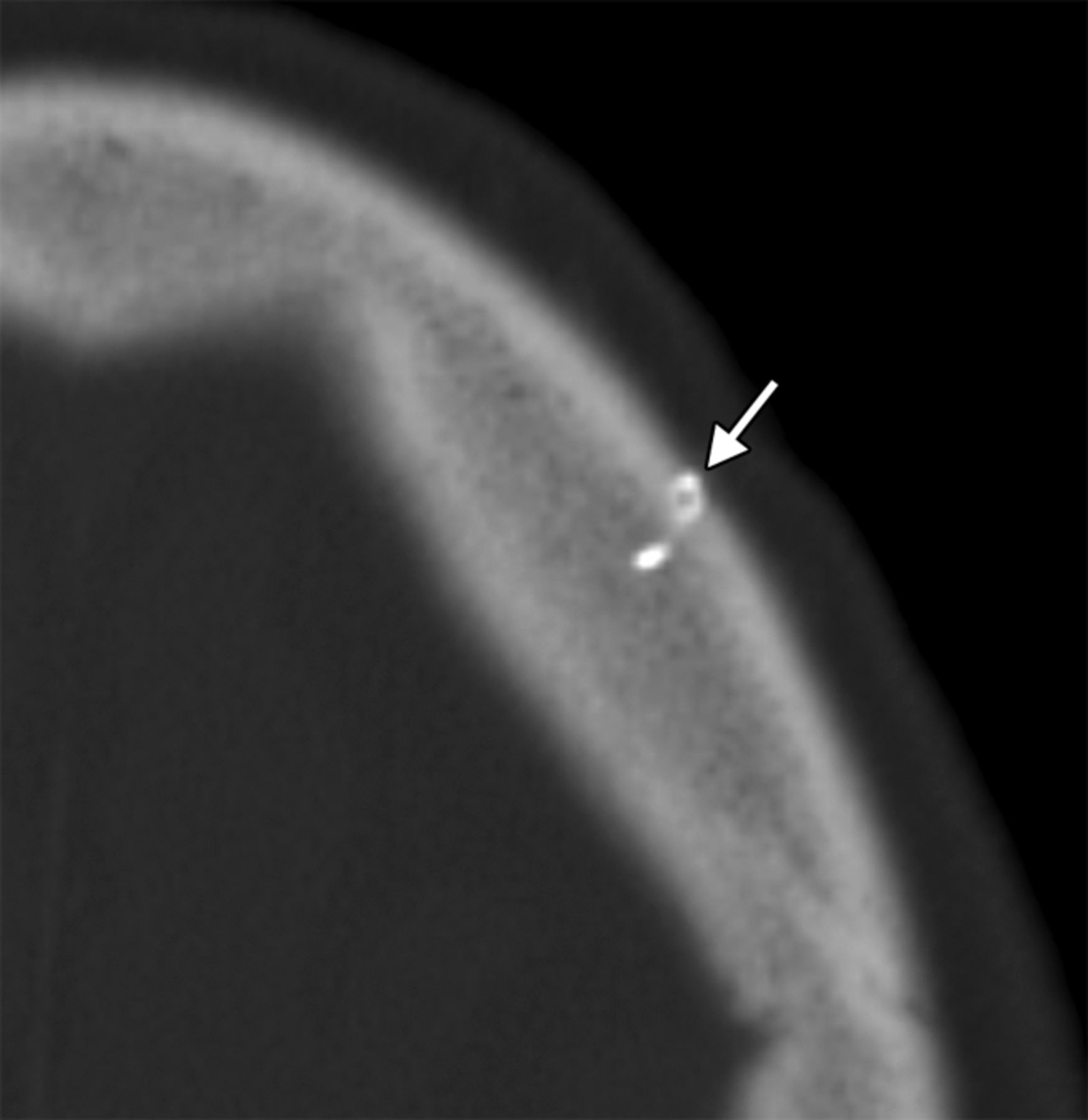
- Fig 2.
Brow lift. Axial CT image shows a metallic left frontal bone pin (arrow) used for suture fixation.
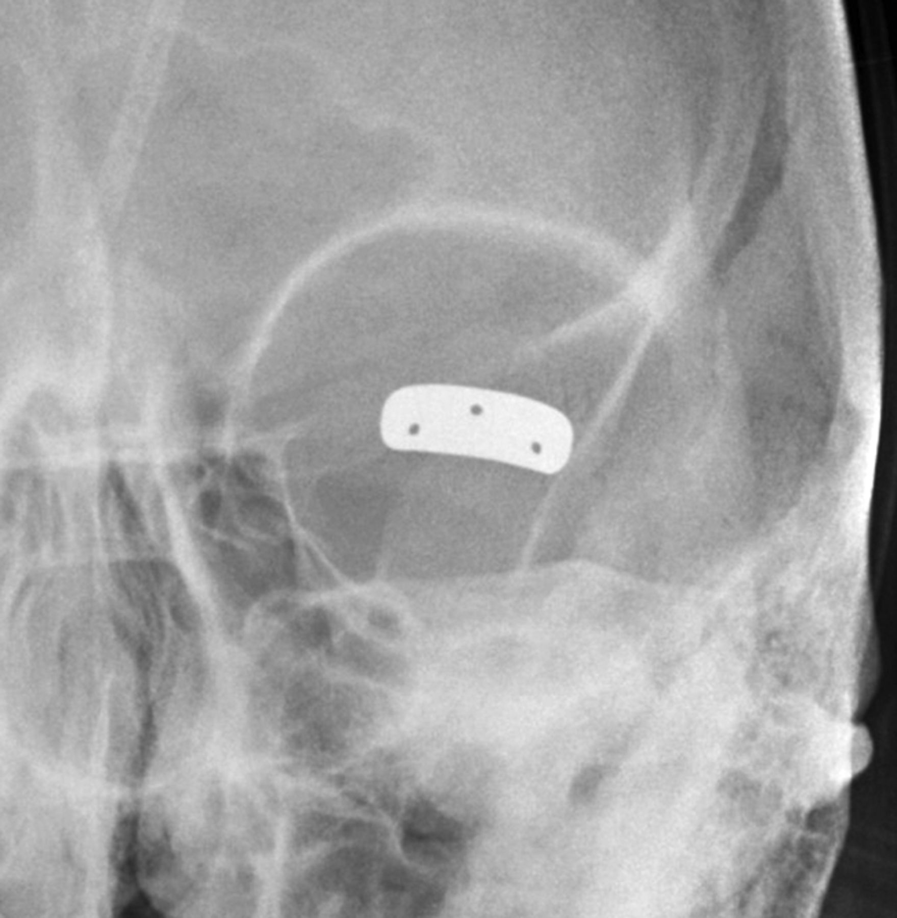
- Fig 3.
Eyelid weight. Frontal radiograph shows a gold implant containing 3 drill holes at the level of the left upper eyelid.
- Fig 4.
Eyelid weight. Axial CT image shows extensive streak artifacts related to the left eyelid weight, which obscures surrounding structures.
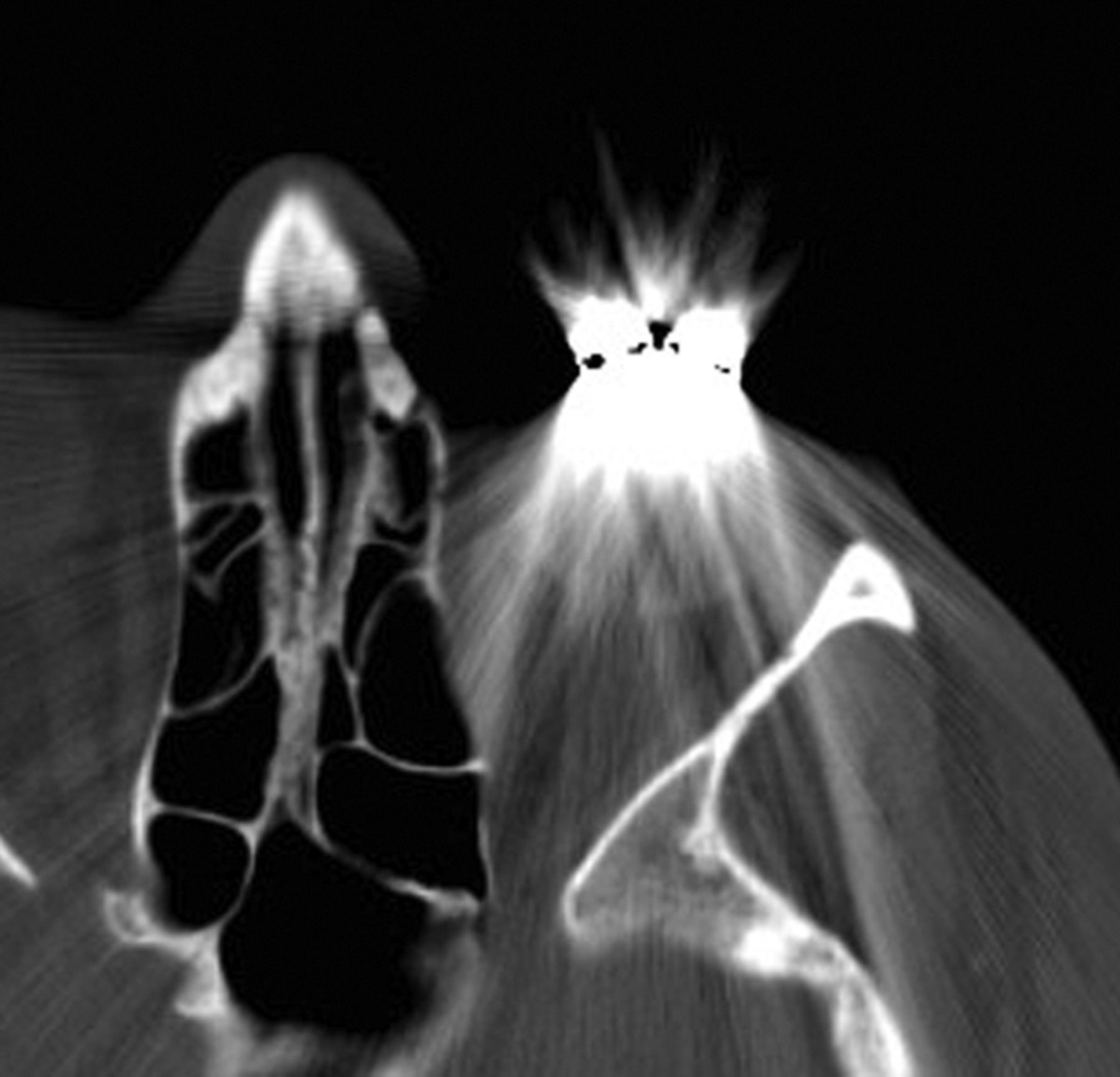
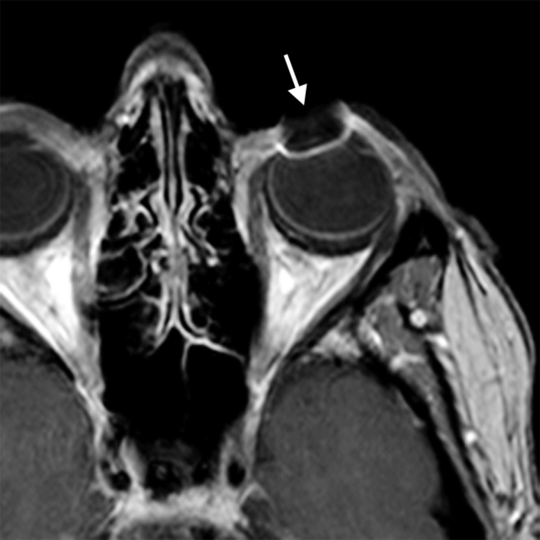
- Fig 5.
Eyelid weight. Axial post-contrast fat-suppressed T1-weighted image shows field inhomogeneity associated with the left eyelid weight (arrow).
- Fig 6.
Eyelid spring. Frontal (A) and lateral (B) radiographs show that the inferior limb of the spring is positioned in the superior eyelid (arrows), while the superior limb is positioned along the orbital rim.
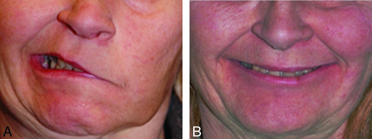
- Fig 7.
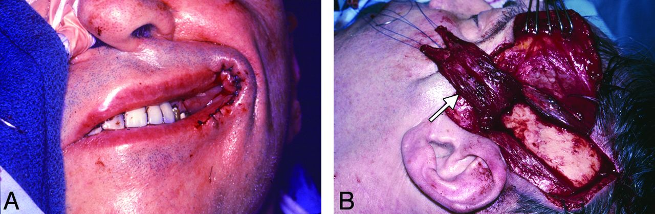
Gracilis free flap. Clinical photographs before (A) and after (B) gracilis free flap reanimation show marked restoration of the patient's smile.
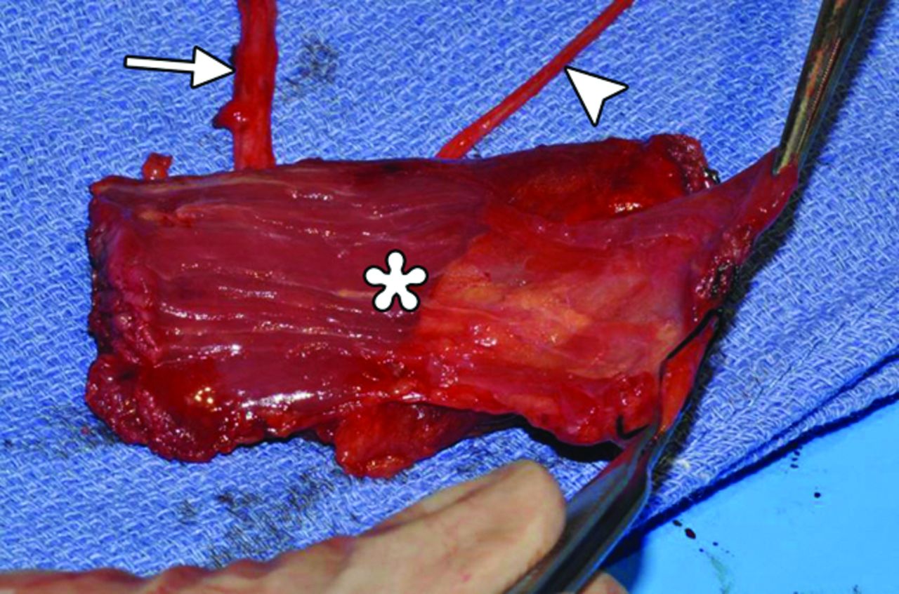
- Fig 8.
Gracilis free flap. Intraoperative photograph shows the harvested flap (asterisk) with an attached vascular pedicle (arrow) and nerve (arrowhead).
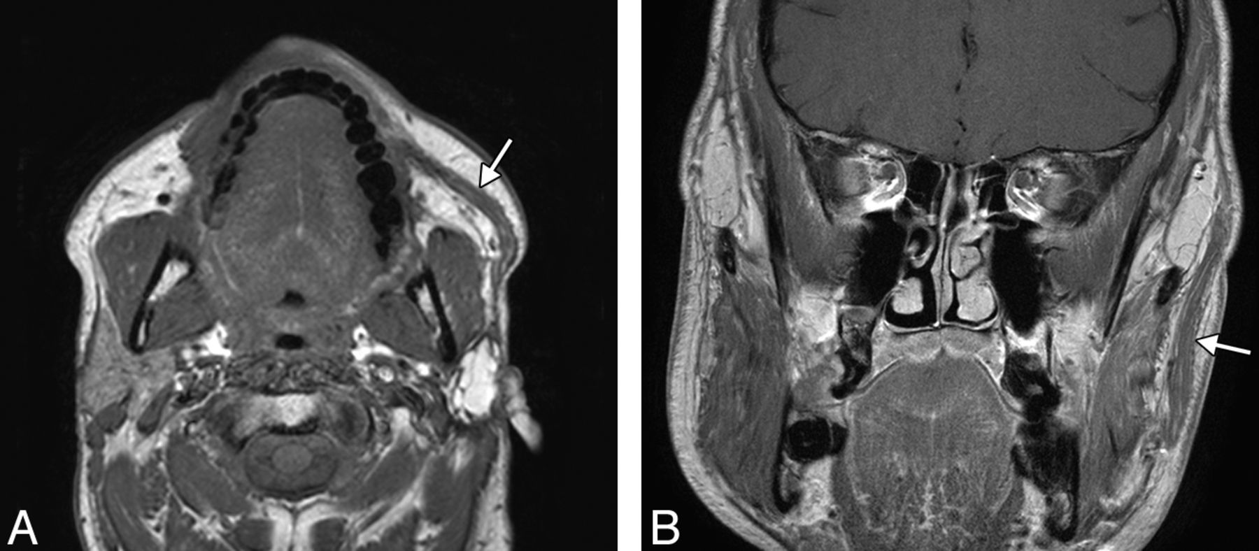
- Fig 9.
Gracilis free flap. Axial (A) and coronal (B) postcontrast T1-weighted MR images show a healthy gracilis flap (arrows), which extends from the zygomatic arch to the oral commissure following total left parotidectomy, with facial nerve sacrifice for resection of mucoepidermoid carcinoma.
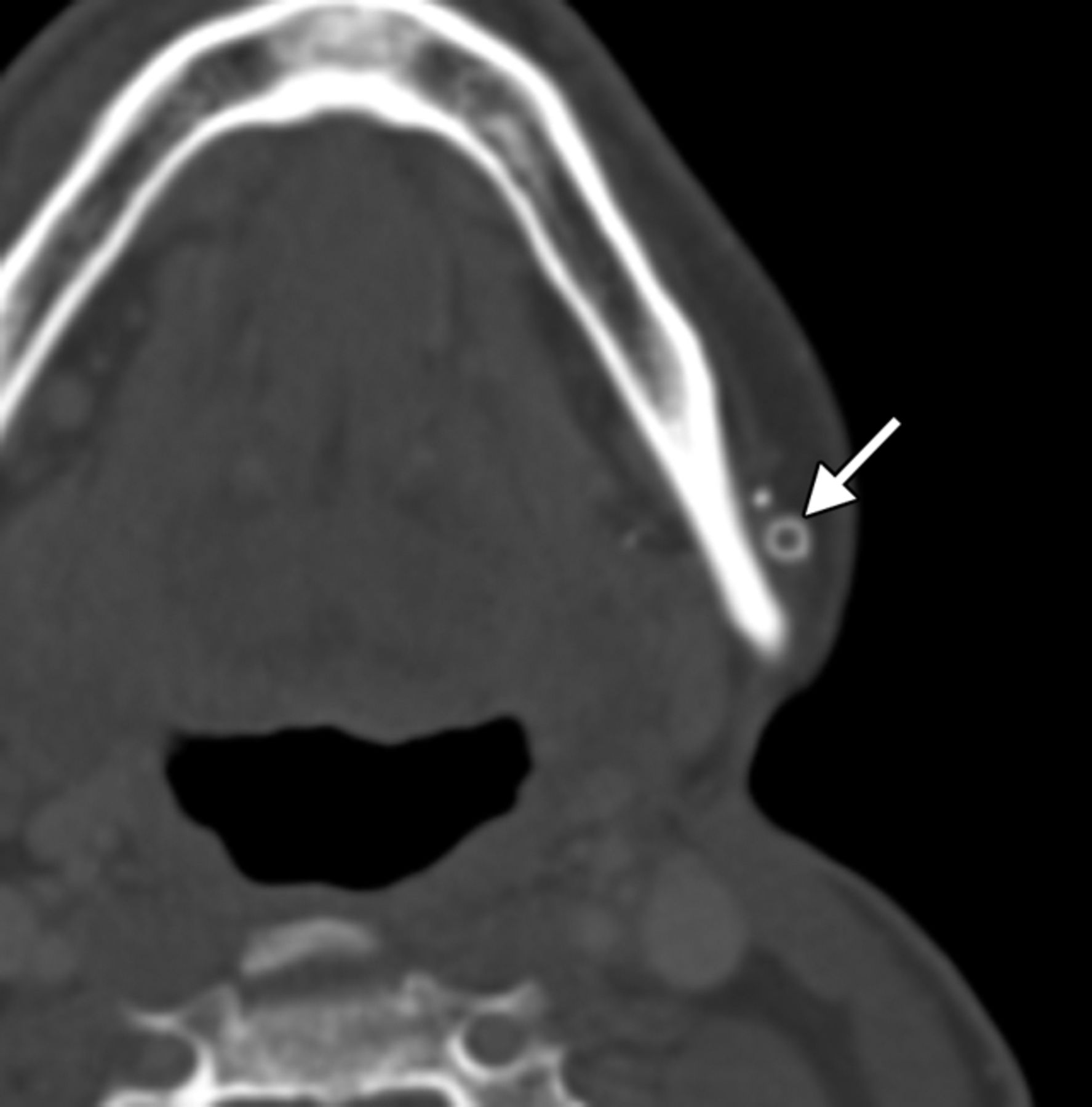
- Fig 10.
Gracilis free flap. Axial CT image shows a hyperattenuated circular ring connector (arrow) used to facilitate the vascular anastomosis.
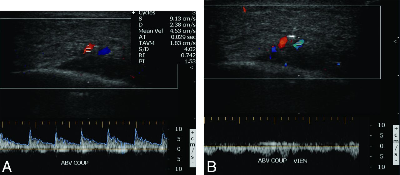
- Fig 11.
Gracilis free flap. Color Doppler sonographic images of the vascular pedicle show normal arterial (A) and venous (B) waveforms.
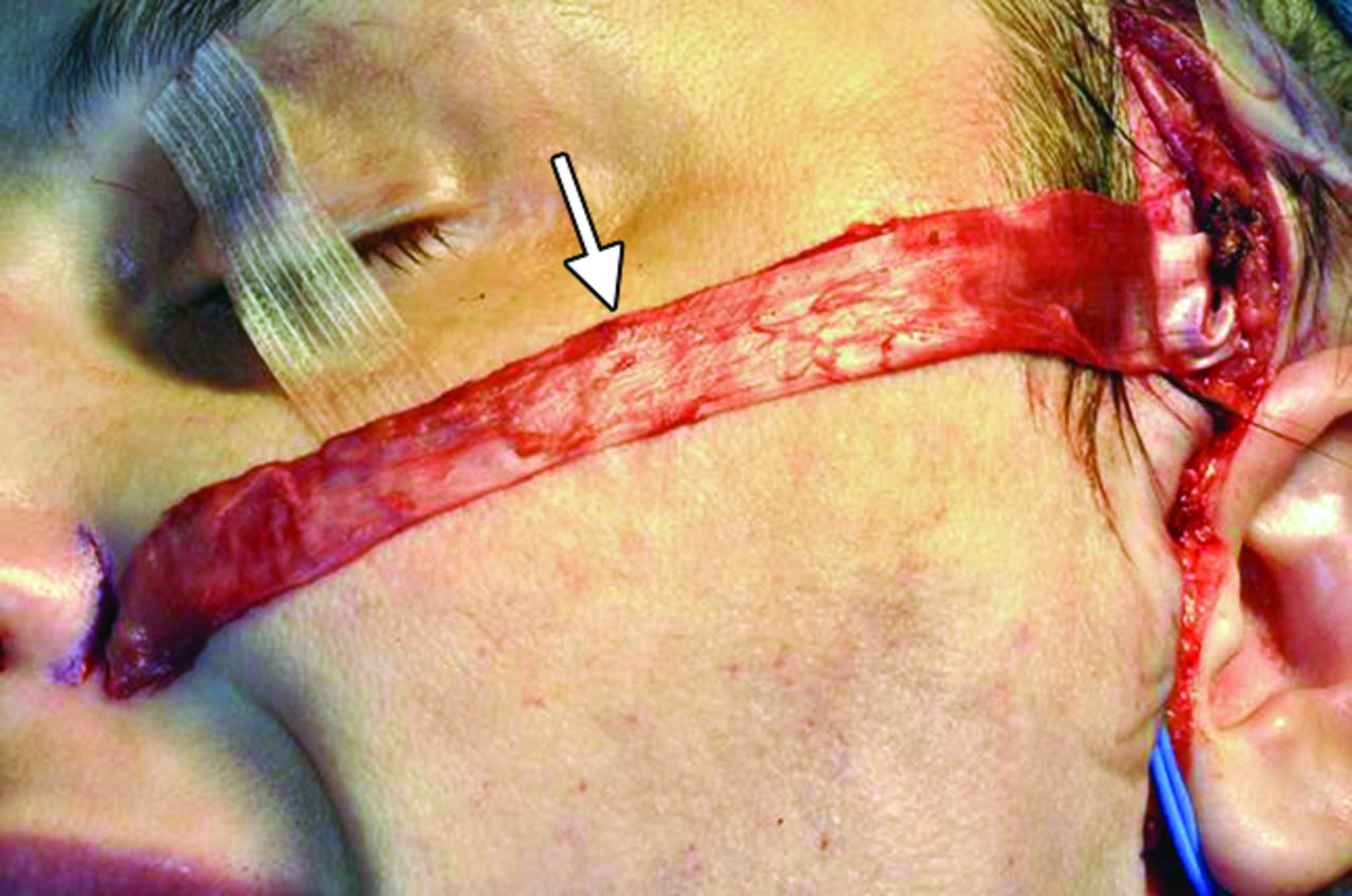
- Fig 12.
Fascia lata graft. Intraoperative photograph shows the prepared fascia lata graft (arrow) over its planned course toward the left nasal ala before implantation.
- Fig 13.
Fascia lata graft. The patient did not obtain optimal muscular function after the gracilis free flap procedure. Axial CT shows a tenuous right gracilis flap (arrowheads) and a linear band of soft tissue that extends from the gracilis flap to the right alar base, which corresponds to the fascia lata sling resuspension for external nasal valve correction (arrow).
- Fig 14.
Temporalis flap. Intraoperative photographs show that the middle portion of the temporalis muscle (arrow) has been dissected free and transposed toward the oral commissure. The flap was subsequently tunneled beneath the subcutaneous tissues.
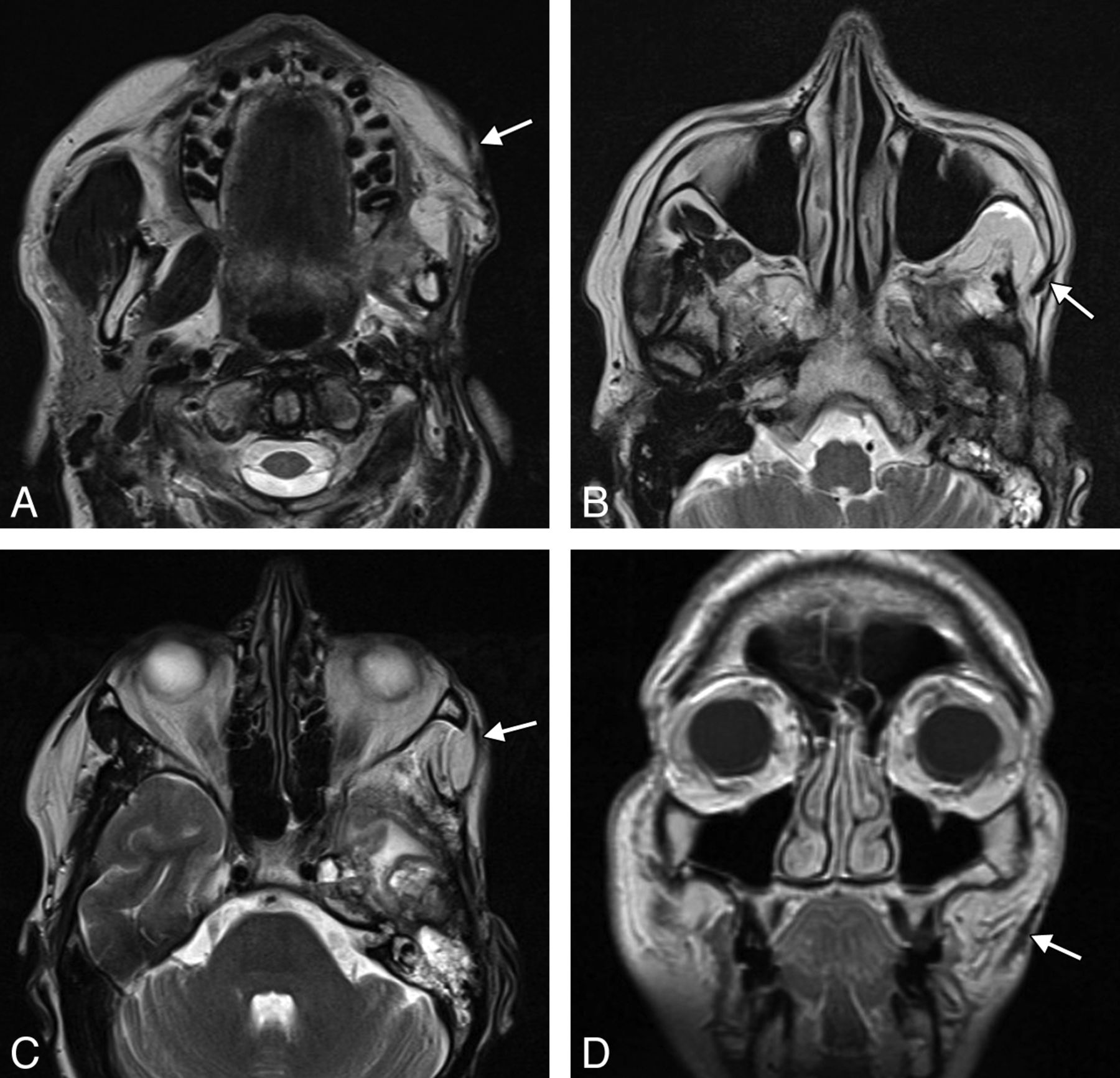
- Fig 15.
Temporalis flap. Axial T2 MR imaging (A–C) and coronal T1 MR imaging (D) show that the left temporalis muscle with the overlying fascia (arrows) is directed retrograde from the temporal fossa to the orbicularis oris.
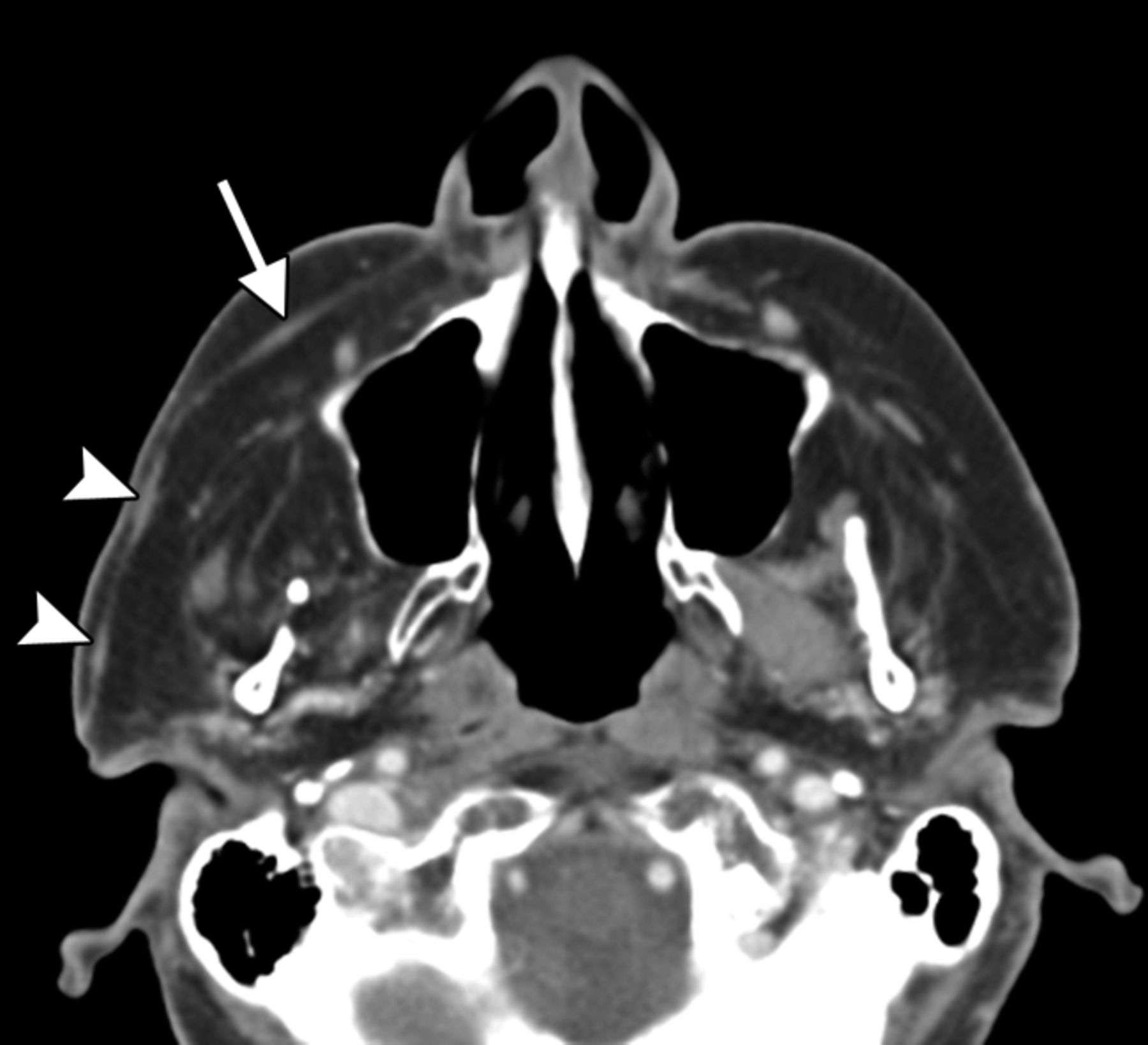
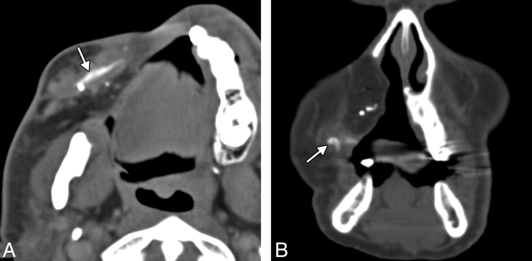
- Fig 16.
Gore-Tex sling. Axial (A) and coronal (B) CT images show the linear hyperattenuated strip of Gore-Tex that supports the right oral commissure (arrows). The patient is status post right complete maxillectomy with myocutaneous flap reconstruction.


Jump to section
Related Articles
Cited By...
- No citing articles found.