
Article Figures & Data
Figures
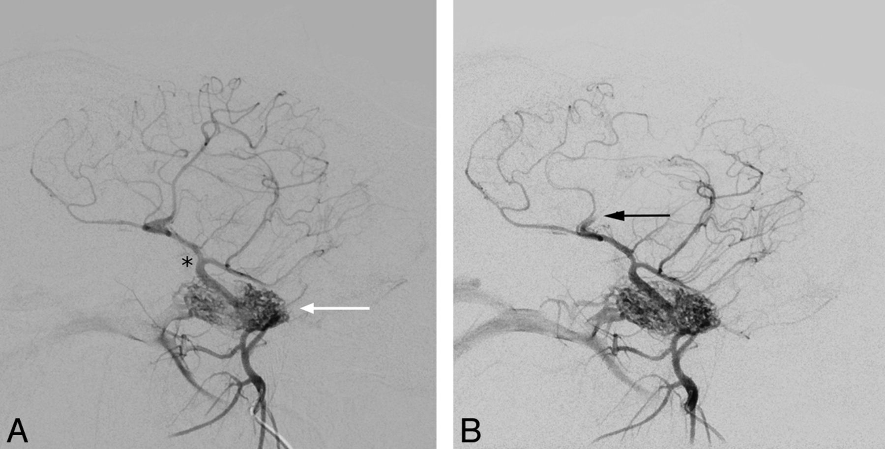
- Fig 1.
Intracranial vessels before (A) and after (B) vessel occlusion (black arrow). Contrast material was injected near the rete mirabile (white arrow in A). The MCA surfaces shortly after branching off the ICA (black asterisk in A), which also gives rise to the posterior cerebral artery. Due to early branching, the complete MCA territory was not affected.
- Fig 2.
Color maps of CBF and CBV for all protocols 3 hours after vessel occlusion on the left side. Perfusion deficits are clearly visible, but image quality is impaired by streak artifacts, especially in the fast scan. The prototype is CTP data processed with the prototype software. VPCT is CTP data processed with commercial software.
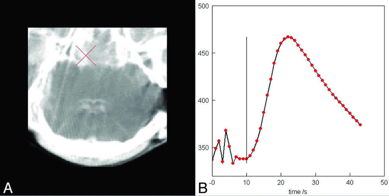
- Fig 3.
Manually chosen arterial input function in the ICA near the skull base and corresponding time-attenuation curve.


Tables
C-Arm 3 Seconds C-Arm 5 Seconds CTP Prototypea CTP VPCTb Sheep 1 Pre v v v v 0 Hours v v v v 3 Hours v v v v Sheep 2 Pre v v v v 0 Hours v v v v 3 Hours v v v v Sheep 3 Pre v v v x 0 Hours x x v v 3 Hours v v v v Sheep 4 Pre x v v v 0 Hours x x x x 3 Hours x x x x Sheep 5 x x x x x x x x x x x x Sheep 6 Pre v v v v 0 Hours v v v v 3 Hours v v v v Sheep 7 Pre v v v v 0 Hours v v v v 3 Hours v v v v Total 14 15 16 15 Note:—Pre indicates data acquired before surgery; v, sets that were acquired as the protocol demanded and that were included in the evaluation; x, excluded datasets (The reasons are explained in the text) .
↵a Datasets acquired with CTP and calculated with the prototype software.
↵b Datasets acquired with CTP and calculated with the commercial software VPCT (Siemens).
- Table 2:
Sensitivity and specificity for lesion detection shown separately for each reader and protocol
Reader 1 Reader 2 Occl. No Occl. Occl. No Occl. Fast Positive 8 0 6 0 Negative 1 5 3 5 Sensitivity 88.9% 66.7% Specificity 100% 100% Slow Positive 9 1 8 1 Negative 0 5 1 5 Sensitivity 100% 88.9% Specificity 83.3% 83.3% Prototype Positive 10 0 10 0 Negative 0 6 0 6 Sensitivity 100% 100% Specificity 100% 100% VPCT Positive 10 0 10 0 Negative 0 5 0 5 Sensitivity 100% 100% Specificity 100% 100% Note:—Occl. indicates occlusion.


Jump to section
Related Articles
Cited By...
- No citing articles found.