
Article Figures & Data
Figures
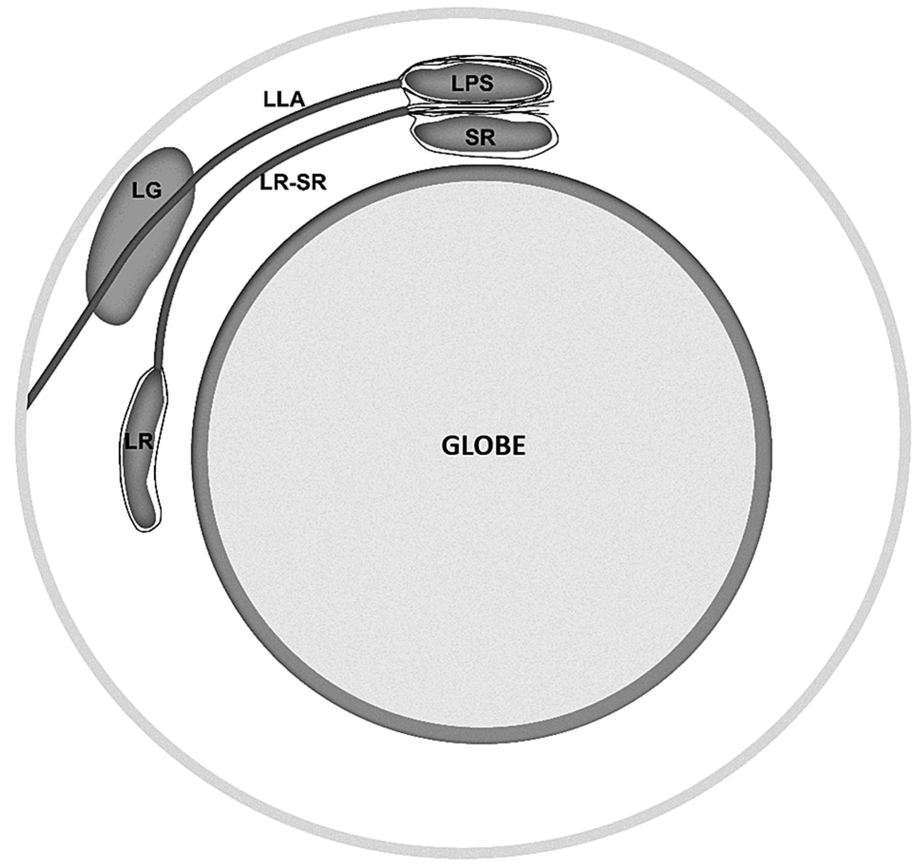
- Fig 1.
Schematic representation of orbital connective tissues within the superotemporal orbit. LG indicates lacrimal gland; LLA, lateral levator aponeurosis; LPS, levator palpebrae superioris; LR, lateral rectus muscle; SR, superior rectus muscle.
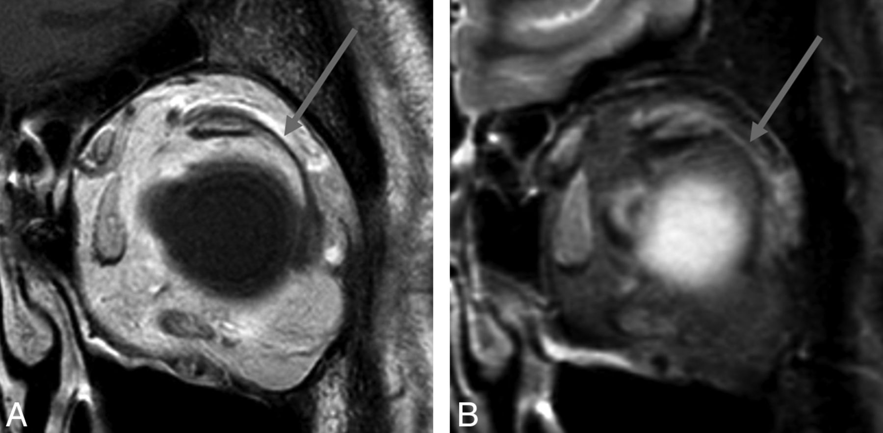
- Fig 2.
MR imaging appearance of the LR-SR band in a 44-year-old woman. Coronal T1-weighted image (A) and coronal STIR image (B) of the left orbit demonstrate the LR-SR band (arrows) as a curvilinear structure extending from the superior margin of the lateral rectus muscle to the lateral margin of the superior rectus/levator palpebrae superioris muscle complex.
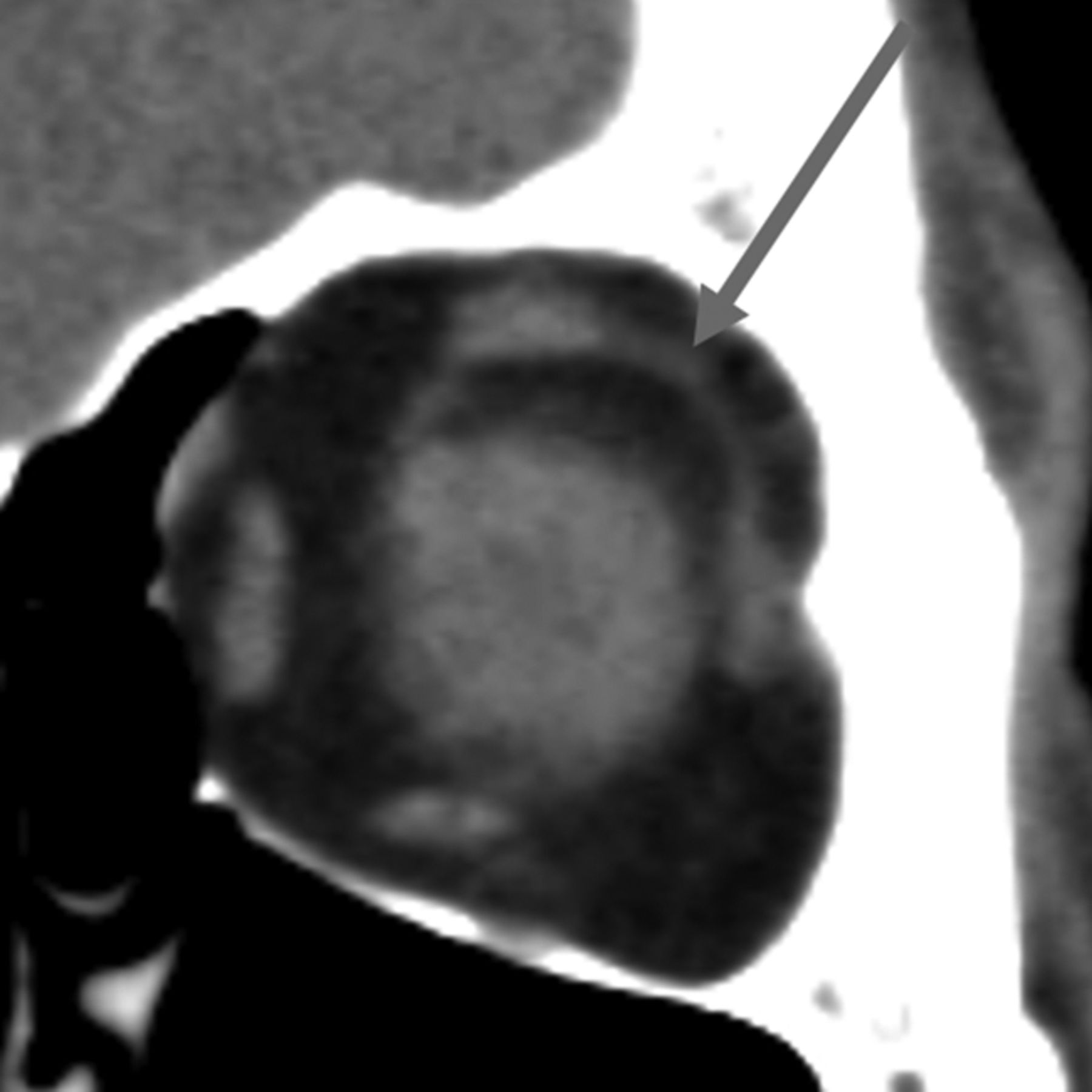
- Fig 3.
CT appearance of the LR-SR band in a 54-year-old woman. Coronal image of the left orbit demonstrates the LR-SR band (arrow).
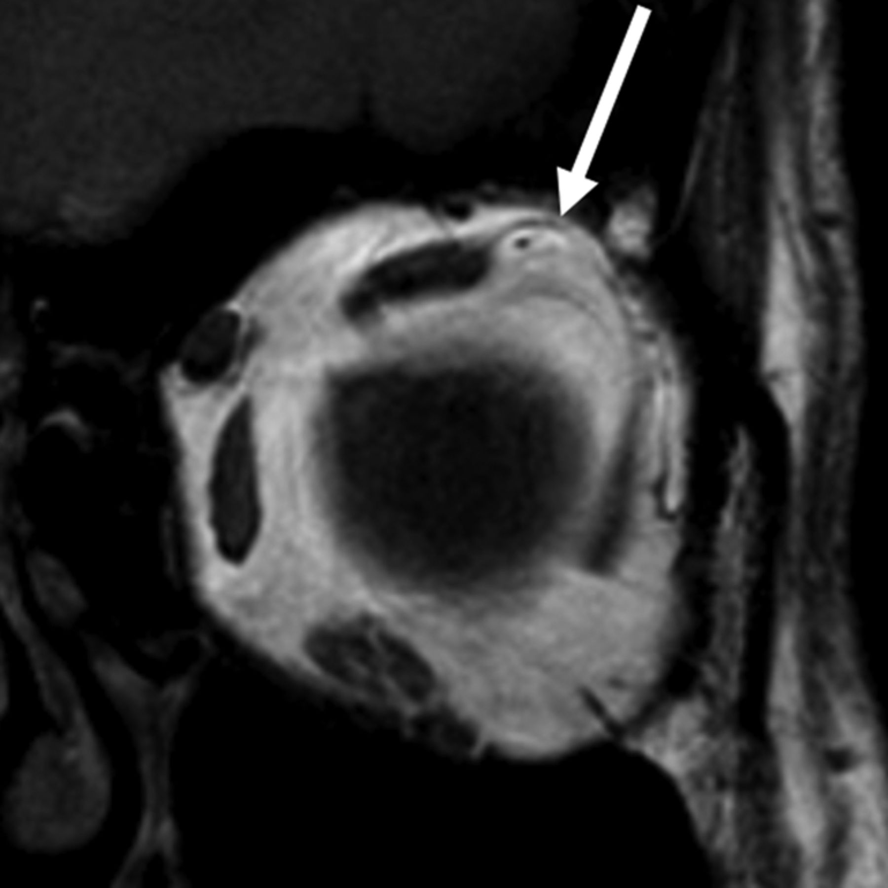
- Fig 4.
LR-SR band bowing in a 54-year-old woman. Coronal T1-weighted image of the left orbit shows superior bowing of the LR-SR band (arrow), higher than the upper margin of the superior muscle complex.
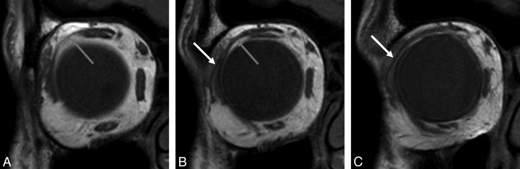
- Fig 5.
MR imaging appearance of the LR-SR band and the lateral levator aponeurosis in a 50-year-old man. Consecutive coronal T1-weighted images of the right orbit, from posterior (A) to anterior (C), show that the lateral levator aponeurosis (white arrows) lies superior and temporal to the LR-SR band (gray arrows) and traverses the lacrimal gland.
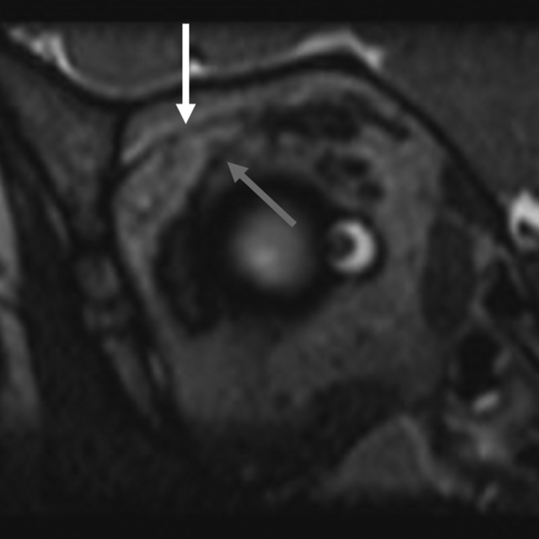
- Fig 6.
Coronal image of the right orbit from a CISS sequence in a 5-year-old boy displays the LR-SR band (gray arrow) and the lateral levator aponeurosis (white arrow).
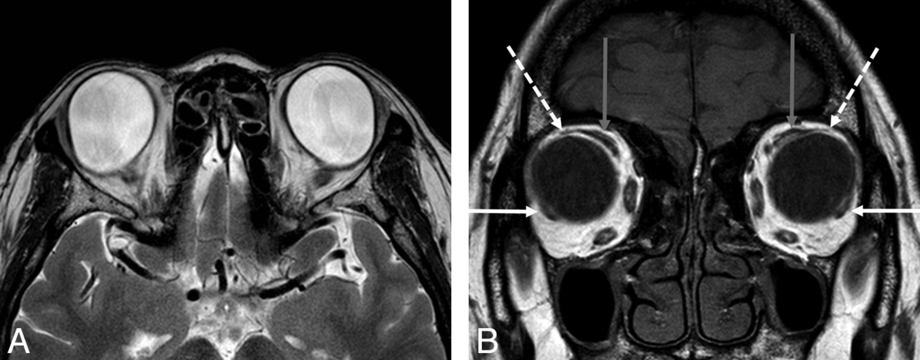
- Fig 7.
A 65-year-old woman with heavy eye syndrome who presented with high myopia and extreme esotropia (inward eye deviation), resulting in poor peripheral vision. Axial T2WI (A) shows large bilateral staphylomas and esotropia. Coronal T1WI (B) shows thinned/incomplete LR-SR bands (white dashed arrows) stretched over the enlarged globes and nasal displacement of the superior muscle complexes (gray arrows) and inferior displacement of the lateral rectus muscles (white solid arrows).
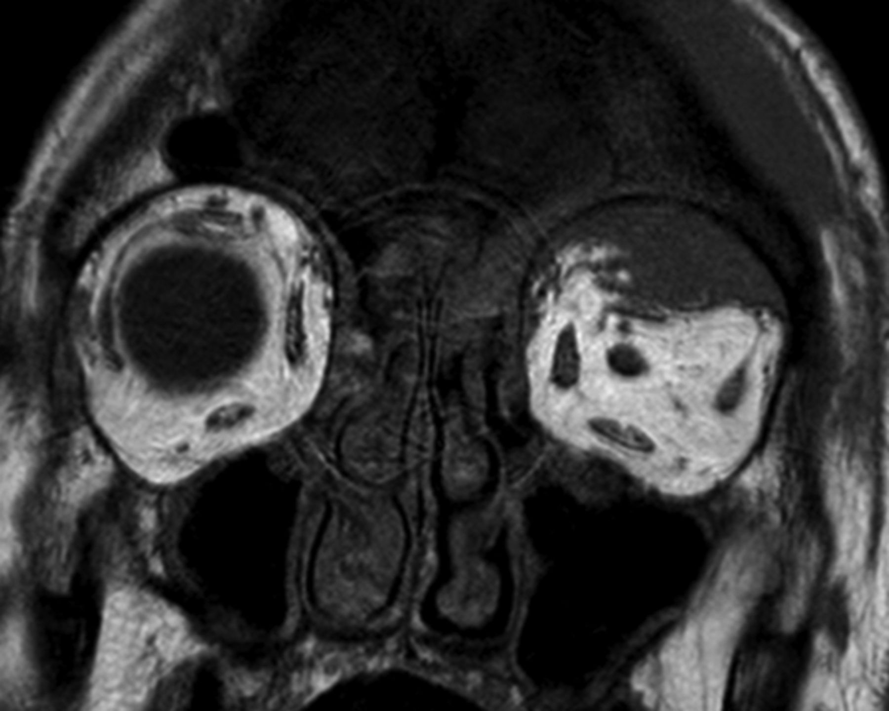
- Fig 8.
A 74-year-old man with B-cell lymphoma. Coronal precontrast T1-weighted image demonstrates lymphoma involving the superotemporal left orbit. Tumor appears to cause inferior bowing/displacement of the LR-SR band but does not definitely transgress it. Note the normal LR-SR band in the right orbit.


Tables
LR-SR Visible on T1W1a LR-SR Visible on STIRa LR-SR Visible on CTa LR-SR Continuousb LR-SR Bowingc LLA Visibled Reader 1 98% 86% 63% 97% 22% 79% Reader 2 92% 50% 76% 93% 26% 85% Note:—LLA indicates lateral levator aponeurosis.
↵a The percentage of cases in which the LR-SR band was visible on the given imaging modality/sequence.
↵b When visible, the percentage of cases in which the LR-SR band formed a continuous structure extending from the superior muscle complex to the lateral rectus muscle (ie, no gaps or discontinuities).
↵c When visible, the percentage of cases in which the LR-SR band demonstrated superotemporal bowing.
↵d The percentage of cases in which the lateral levator aponeurosis was visible, distinct from the LR-SR band.
Quartile 1 Quartile 2 Quartile 3 Quartile 4 P Value LR-SR visible on T1W1b 96% 99% 95% 90% .03 LR-SR continuousc 95% 96% 95% 91% .40 LR-SR bowingd 19% 17% 28% 34% .03 LLA visiblee 73% 77% 89% 87% .01 Note:—LLA indicates lateral levator aponeurosis.
↵a Quartile 1: 9–39 years of age; quartile 2: 40–49 years of age; quartile 3: 50–61 years of age; quartile 4: 62–81 years of age.
↵b The percentage of cases in which the LR-SR band was visible on T1WI.
↵c The percentage of cases in which the LR-SR band was visible as a continuous structure extending from the superior muscle complex to the lateral rectus muscle (ie, no gaps or discontinuities).
↵d The percentage of cases in which the LR-SR band demonstrated superotemporal bowing.
↵e The percentage of cases in which the lateral levator aponeurosis was visible, distinct from the LR-SR band.


Jump to section
Related Articles
Cited By...
- No citing articles found.