Advertisement

Main menu

  • Home
  • Content
    • Current Issue
    • Accepted Manuscripts
    • Article Preview
    • Past Issue Archive
    • AJNR Case Collection
    • Case of the Week Archive
    • Classic Case Archive
    • Case of the Month Archive
  • Special Collections
    • Spinal CSF Leak Articles (Jan 2020-June 2024)
    • 2024 AJNR Journal Awards
    • Most Impactful AJNR Articles
  • Multimedia
    • AJNR Podcast
    • AJNR Scantastics
    • Video Articles
  • For Authors
    • Submit a Manuscript
    • Author Policies
    • Fast publishing of Accepted Manuscripts
    • Graphical Abstract Preparation
    • Manuscript Submission Guidelines
    • Imaging Protocol Submission
    • Submit a Case for the Case Collection
  • About Us
    • About AJNR
    • Editorial Board
  • More
    • Become a Reviewer/Academy of Reviewers
    • Subscribers
    • Permissions
    • Alerts
    • Feedback
    • Advertisers
    • ASNR Home
  • Other Publications
    • ajnr

User menu

  • Alerts
  • Log in

Search

  • Advanced search
American Journal of Neuroradiology

American Journal of Neuroradiology

ASHNR American Society of Functional Neuroradiology ASHNR American Society of Pediatric Neuroradiology ASSR
  • Alerts
  • Log in

Advanced Search

  • Home
  • Content
    • Current Issue
    • Accepted Manuscripts
    • Article Preview
    • Past Issue Archive
    • AJNR Case Collection
    • Case of the Week Archive
    • Classic Case Archive
    • Case of the Month Archive
  • Special Collections
    • Spinal CSF Leak Articles (Jan 2020-June 2024)
    • 2024 AJNR Journal Awards
    • Most Impactful AJNR Articles
  • Multimedia
    • AJNR Podcast
    • AJNR Scantastics
    • Video Articles
  • For Authors
    • Submit a Manuscript
    • Author Policies
    • Fast publishing of Accepted Manuscripts
    • Graphical Abstract Preparation
    • Manuscript Submission Guidelines
    • Imaging Protocol Submission
    • Submit a Case for the Case Collection
  • About Us
    • About AJNR
    • Editorial Board
  • More
    • Become a Reviewer/Academy of Reviewers
    • Subscribers
    • Permissions
    • Alerts
    • Feedback
    • Advertisers
    • ASNR Home
  • Follow AJNR on Twitter
  • Visit AJNR on Facebook
  • Follow AJNR on Instagram
  • Join AJNR on LinkedIn
  • RSS Feeds

Welcome to the new AJNR, Updated Hall of Fame, and more. Read the full announcements.


AJNR is seeking candidates for the position of Associate Section Editor, AJNR Case Collection. Read the full announcement.


Research ArticleHead & Neck

High-Resolution CT Findings in Children with a Normal Pinna or Grade I Microtia and Unilateral Mild Stenosis of the External Auditory Canal

R. Jacob, S. Gupta, B. Isaacson, J.W. Kutz, P. Roland, Y. Xi and T.N. Booth
American Journal of Neuroradiology January 2015, 36 (1) 176-180; DOI: https://doi.org/10.3174/ajnr.A4067
R. Jacob
aFrom the Departments of Radiology (R.J., Y.X., T.N.B.)
  • Find this author on Google Scholar
  • Find this author on PubMed
  • Search for this author on this site
S. Gupta
bOtolaryngology (S.G., B.I., J.W.K., P.R.), Children's Medical Center of Dallas, University of Texas, Southwestern Medical Center, Dallas, Texas.
  • Find this author on Google Scholar
  • Find this author on PubMed
  • Search for this author on this site
B. Isaacson
bOtolaryngology (S.G., B.I., J.W.K., P.R.), Children's Medical Center of Dallas, University of Texas, Southwestern Medical Center, Dallas, Texas.
  • Find this author on Google Scholar
  • Find this author on PubMed
  • Search for this author on this site
J.W. Kutz Jr
bOtolaryngology (S.G., B.I., J.W.K., P.R.), Children's Medical Center of Dallas, University of Texas, Southwestern Medical Center, Dallas, Texas.
  • Find this author on Google Scholar
  • Find this author on PubMed
  • Search for this author on this site
P. Roland
bOtolaryngology (S.G., B.I., J.W.K., P.R.), Children's Medical Center of Dallas, University of Texas, Southwestern Medical Center, Dallas, Texas.
  • Find this author on Google Scholar
  • Find this author on PubMed
  • Search for this author on this site
Y. Xi
aFrom the Departments of Radiology (R.J., Y.X., T.N.B.)
  • Find this author on Google Scholar
  • Find this author on PubMed
  • Search for this author on this site
T.N. Booth
aFrom the Departments of Radiology (R.J., Y.X., T.N.B.)
  • Find this author on Google Scholar
  • Find this author on PubMed
  • Search for this author on this site
  • Article
  • Figures & Data
  • Info & Metrics
  • Responses
  • References
  • PDF
Loading

Article Figures & Data

Figures

  • Fig 1.
    • Download figure
    • Open in new tab
    • Download powerpoint
    Fig 1.

    Measurement method for the EAC diameter. Axial and coronal temporal bones at the level of the EAC. The left EAC is measured obliquely along the most medial aspect, parallel to the tympanic membrane (dotted line) in both the axial and coronal planes.

  • Fig 2.
    • Download figure
    • Open in new tab
    • Download powerpoint
    Fig 2.

    Abnormal orientation of the handle of the malleus in a 4-year-old girl with right-sided CHL (patient 6). Axial HRCT of the temporal bone, superior-to-inferior. A and B, Superior image shows a normal ossicular position. Inferiorly, abnormal orientation of the handle of the right malleus along with posterior fixation of the handle of the malleus (arrow) is present. There is asymmetric widening of the distance between the cochlear promontory (arrowhead) and the handle of the malleus. C and D, Normal ossicular anatomy is present superiorly. Inferiorly, normal orientation of the handle of the left malleus and a normal distance from the promontory to the handle (between arrows) are seen.
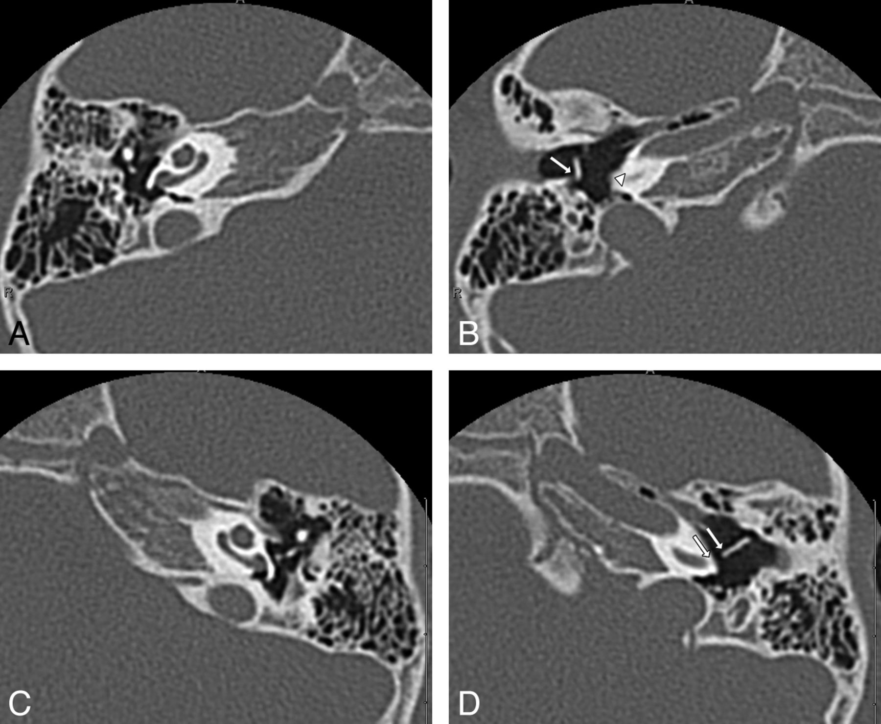
  • Fig 3.
    • Download figure
    • Open in new tab
    • Download powerpoint
    Fig 3.

    Abnormal fixation of the handle of the malleus along with increased distance of the handle of malleus to the cochlear promontory in a 4-year-old boy with right-sided CHL (patient 9). Axial HRCT of the temporal bones, superior-to-inferior. A and B, There is abnormal fusion of the right malleus and incus (long arrow) superiorly. The inferior image shows fixation of the handle of the malleus anteriorly (short arrow). Note an increased distance of the handle of the malleus to the cochlear promontory (arrowhead). C and D, Normal separation of the left ossicles is present superiorly. Inferiorly, there is a normal distance of the handle of the left malleus to the promontory in the normal ear (between arrows).
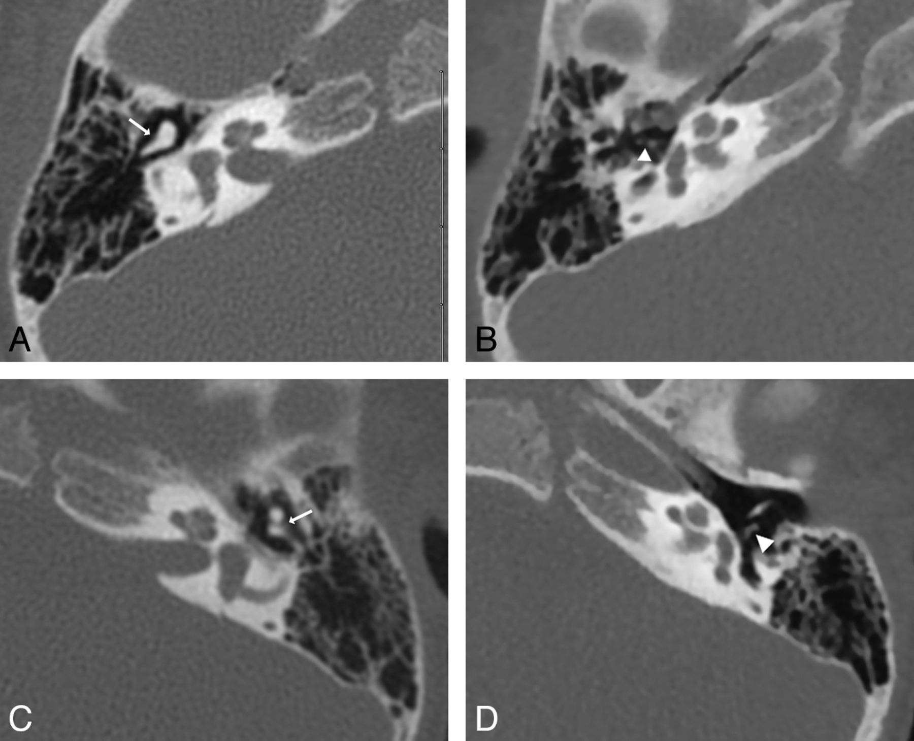
  • Fig 4.
    • Download figure
    • Open in new tab
    • Download powerpoint
    Fig 4.

    Abnormal fixation of the incus in a 5-year-old girl with left-sided CHL (patient 1). Axial HRCT of the temporal bone, superior-to-inferior. A and B, Superiorly, there is a normal incus with articulation with the stapes (arrow). Inferior image shows a normal long process of the incus with no fixation bar (arrow). C and D, Superiorly, there is fusion of the left malleus and incus (arrow). Inferior image shows a posterior fixation bar extending from the long process of the incus (long arrow) to the posterior wall of the tympanic cavity.
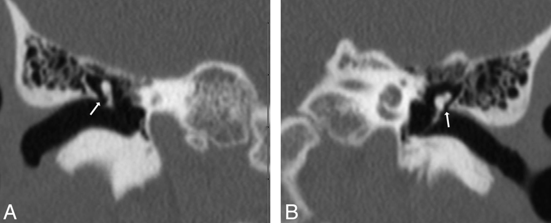
  • Fig 5.
    • Download figure
    • Open in new tab
    • Download powerpoint
    Fig 5.

    Abnormal asymmetric increased in the incudostapedial angle in a 3-year-old boy with right-sided CHL (patient 8). Coronal-reconstruction HRCT of the temporal bone at the level of the long process of the incus. A, An abnormally increased incudostapedial angle (arrow) measuring 109° is present in the right ear. B, The normal left ear has an incudostapedial angle measuring 93° (arrow).

  • Fig 6.
    • Download figure
    • Open in new tab
    • Download powerpoint
    Fig 6.

    Asymmetric narrowing of the Prussak space in a 12-year-old boy with left-sided CHL (patient 2). Coronal-reconstruction HRCT of the temporal bone at the level of the malleus. A, Normal appearance of the Prussak space in the normal right ear (arrow) is present. B, Asymmetric narrowing of the Prussak space (arrow) in the affected left ear is present with a normal volume of the tympanic cavity.
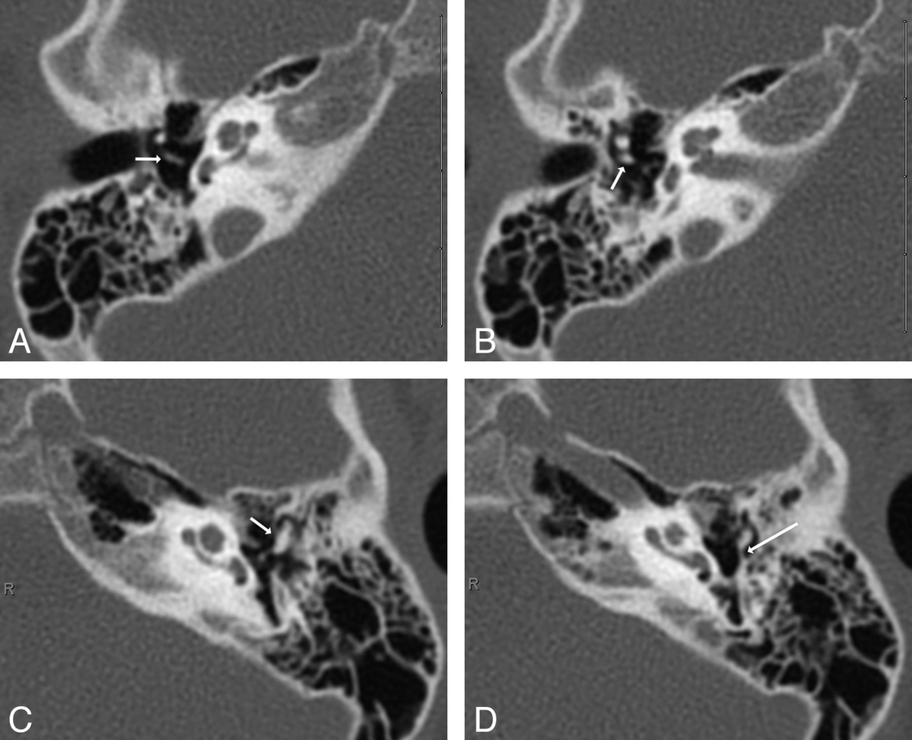
  • Fig 7.
    • Download figure
    • Open in new tab
    • Download powerpoint
    Fig 7.

    Abnormal incudomalleal and incudostapedial joints in a 3-year-old boy with right-sided CHL (patient 8). Axial HRCT of the temporal bone at the level of the ossicular articulations, superior-to-inferior. A and B, Superior image shows abnormal incudomalleal fusion (arrow). Inferiorly, abnormal incudostapedial widening (arrowhead) in the right ear is present. C and D, The normal left incudomalleal (arrow) and incudostapedial joints (arrowhead) are demonstrated.

PreviousNext
Back to top

In this issue

American Journal of Neuroradiology: 36 (1)
American Journal of Neuroradiology
Vol. 36, Issue 1
1 Jan 2015
  • Table of Contents
  • Index by author
  • Complete Issue (PDF)
Advertisement
Print
Download PDF
Email Article

Thank you for your interest in spreading the word on American Journal of Neuroradiology.

NOTE: We only request your email address so that the person you are recommending the page to knows that you wanted them to see it, and that it is not junk mail. We do not capture any email address.

Enter multiple addresses on separate lines or separate them with commas.
High-Resolution CT Findings in Children with a Normal Pinna or Grade I Microtia and Unilateral Mild Stenosis of the External Auditory Canal
(Your Name) has sent you a message from American Journal of Neuroradiology
(Your Name) thought you would like to see the American Journal of Neuroradiology web site.
CAPTCHA
This question is for testing whether or not you are a human visitor and to prevent automated spam submissions.
Cite this article
R. Jacob, S. Gupta, B. Isaacson, J.W. Kutz, P. Roland, Y. Xi, T.N. Booth
High-Resolution CT Findings in Children with a Normal Pinna or Grade I Microtia and Unilateral Mild Stenosis of the External Auditory Canal
American Journal of Neuroradiology Jan 2015, 36 (1) 176-180; DOI: 10.3174/ajnr.A4067

Citation Manager Formats

  • BibTeX
  • Bookends
  • EasyBib
  • EndNote (tagged)
  • EndNote 8 (xml)
  • Medlars
  • Mendeley
  • Papers
  • RefWorks Tagged
  • Ref Manager
  • RIS
  • Zotero
0 Responses
Respond to this article
Share
Bookmark this article
High-Resolution CT Findings in Children with a Normal Pinna or Grade I Microtia and Unilateral Mild Stenosis of the External Auditory Canal
R. Jacob, S. Gupta, B. Isaacson, J.W. Kutz, P. Roland, Y. Xi, T.N. Booth
American Journal of Neuroradiology Jan 2015, 36 (1) 176-180; DOI: 10.3174/ajnr.A4067
del.icio.us logo Twitter logo Facebook logo Mendeley logo
  • Tweet Widget
  • Facebook Like
  • Google Plus One
Purchase

Jump to section

  • Article
    • Abstract
    • ABBREVIATIONS:
    • Discussion
    • Conclusions
    • Footnotes
    • References
  • Figures & Data
  • Info & Metrics
  • Responses
  • References
  • PDF

Related Articles

  • No related articles found.
  • PubMed
  • Google Scholar

Cited By...

  • No citing articles found.
  • Crossref (9)
  • Google Scholar

This article has been cited by the following articles in journals that are participating in Crossref Cited-by Linking.

  • Anatomical measurement of the ossicles in patients with congenital aural atresia and stenosis
    Jieying Li, Keguang Chen, Chenlong Li, Dongming Yin, Tianyu Zhang, Peidong Dai
    International Journal of Pediatric Otorhinolaryngology 2017 101
  • High-resolution computed tomography evaluation of congenital aural atresia – how useful is this?
    R Gautam, J Kumar, G S Pradhan, J C Passey, R Meher, A Mehndiratta
    The Journal of Laryngology & Otology 2020 134 7
  • Measurement method for external auditory canal and clinical application in congenital aural stenosis
    Chen-long Li, Jia Li, Ying Guo, Tian-yu Zhang
    International Journal of Pediatric Otorhinolaryngology 2020 137
  • Congenital Aural Atresia: What the Radiologist Needs to Know?
    Richa Gautam, Jyoti Kumar, Gaurav S Pradhan, Ravi Meher, Shivali Arya
    Current Problems in Diagnostic Radiology 2022 51 4
  • Prevalence of middle ear malformation and outcomes of tympanoplasty and/or canalplasty in patients with and without congenital external auditory canal stenosis
    Takahiro Nakajima, Satoru Fukami, Masaomi Motegi, Hiroaki Kanaya, Hiromi Kojima, Shinichi Haruna
    Auris Nasus Larynx 2022 49 2
  • CT and MR Imaging of the Pediatric Temporal Bone: Normal Variants and Pitfalls
    Rebekah Clarke, Timothy Booth
    Current Radiology Reports 2017 5 8
  • Diagnostic Imaging: Head and Neck
    2017
  • Imaging in Otolaryngology
    2018
  • Manual of Normative Measurements in Head and Neck Imaging
    Daniel Thomas Ginat, Michael B. Gluth
    2021

More in this TOC Section

  • WHO Classification Update: Nasal&Skull Base Tumors
  • Peritumoral Signal in Vestibular Schwannomas
  • Chondrosarcoma vs Synovial Chondromatosis: Imaging
Show more HEAD & NECK

Similar Articles

Advertisement

Indexed Content

  • Current Issue
  • Accepted Manuscripts
  • Article Preview
  • Past Issues
  • Editorials
  • Editors Choice
  • Fellow Journal Club
  • Letters to the Editor

Cases

  • Case Collection
  • Archive - Case of the Week
  • Archive - Case of the Month
  • Archive - Classic Case

Special Collections

  • Special Collections

Resources

  • News and Updates
  • Turn around Times
  • Submit a Manuscript
  • Author Policies
  • Manuscript Submission Guidelines
  • Evidence-Based Medicine Level Guide
  • Publishing Checklists
  • Graphical Abstract Preparation
  • Imaging Protocol Submission
  • Submit a Case
  • Become a Reviewer/Academy of Reviewers
  • Get Peer Review Credit from Publons

Multimedia

  • AJNR Podcast
  • AJNR SCANtastic
  • Video Articles

About Us

  • About AJNR
  • Editorial Board
  • Not an AJNR Subscriber? Join Now
  • Alerts
  • Feedback
  • Advertise with us
  • Librarian Resources
  • Permissions
  • Terms and Conditions

American Society of Neuroradiology

  • Not an ASNR Member? Join Now

© 2025 by the American Society of Neuroradiology All rights, including for text and data mining, AI training, and similar technologies, are reserved.
Print ISSN: 0195-6108 Online ISSN: 1936-959X

Powered by HighWire