Advertisement

Main menu

  • Home
  • Content
    • Current Issue
    • Accepted Manuscripts
    • Article Preview
    • Past Issue Archive
    • AJNR Case Collection
    • Case of the Week Archive
    • Classic Case Archive
    • Case of the Month Archive
  • Special Collections
    • Spinal CSF Leak Articles (Jan 2020-June 2024)
    • 2024 AJNR Journal Awards
    • Most Impactful AJNR Articles
  • Multimedia
    • AJNR Podcast
    • AJNR Scantastics
    • Video Articles
  • For Authors
    • Submit a Manuscript
    • Author Policies
    • Fast publishing of Accepted Manuscripts
    • Graphical Abstract Preparation
    • Manuscript Submission Guidelines
    • Imaging Protocol Submission
    • Submit a Case for the Case Collection
  • About Us
    • About AJNR
    • Editorial Board
  • More
    • Become a Reviewer/Academy of Reviewers
    • Subscribers
    • Permissions
    • Alerts
    • Feedback
    • Advertisers
    • ASNR Home
  • Other Publications
    • ajnr

User menu

  • Alerts
  • Log in

Search

  • Advanced search
American Journal of Neuroradiology

American Journal of Neuroradiology

ASHNR American Society of Functional Neuroradiology ASHNR American Society of Pediatric Neuroradiology ASSR
  • Alerts
  • Log in

Advanced Search

  • Home
  • Content
    • Current Issue
    • Accepted Manuscripts
    • Article Preview
    • Past Issue Archive
    • AJNR Case Collection
    • Case of the Week Archive
    • Classic Case Archive
    • Case of the Month Archive
  • Special Collections
    • Spinal CSF Leak Articles (Jan 2020-June 2024)
    • 2024 AJNR Journal Awards
    • Most Impactful AJNR Articles
  • Multimedia
    • AJNR Podcast
    • AJNR Scantastics
    • Video Articles
  • For Authors
    • Submit a Manuscript
    • Author Policies
    • Fast publishing of Accepted Manuscripts
    • Graphical Abstract Preparation
    • Manuscript Submission Guidelines
    • Imaging Protocol Submission
    • Submit a Case for the Case Collection
  • About Us
    • About AJNR
    • Editorial Board
  • More
    • Become a Reviewer/Academy of Reviewers
    • Subscribers
    • Permissions
    • Alerts
    • Feedback
    • Advertisers
    • ASNR Home
  • Follow AJNR on Twitter
  • Visit AJNR on Facebook
  • Follow AJNR on Instagram
  • Join AJNR on LinkedIn
  • RSS Feeds

Welcome to the new AJNR, Updated Hall of Fame, and more. Read the full announcements.


AJNR is seeking candidates for the position of Associate Section Editor, AJNR Case Collection. Read the full announcement.


Getting new auth cookie, if you see this message a lot, tell someone!
Research ArticlePediatrics
Open Access

MRI Characteristics of Globus Pallidus Infarcts in Isolated Methylmalonic Acidemia

E.H. Baker, J.L. Sloan, N.S. Hauser, A.L. Gropman, D.R. Adams, C. Toro, I. Manoli and C.P. Venditti
American Journal of Neuroradiology January 2015, 36 (1) 194-201; DOI: https://doi.org/10.3174/ajnr.A4087
E.H. Baker
aFrom the Department of Radiology and Imaging Sciences (E.H.B.), Clinical Center
  • Find this author on Google Scholar
  • Find this author on PubMed
  • Search for this author on this site
J.L. Sloan
bGenetics and Molecular Biology Branch (J.L.S., I.M., C.P.V.)
  • Find this author on Google Scholar
  • Find this author on PubMed
  • Search for this author on this site
N.S. Hauser
eMedical Genetics and Metabolism Department (N.S.H.), Children's Hospital Central California, Madera, California
  • Find this author on Google Scholar
  • Find this author on PubMed
  • Search for this author on this site
A.L. Gropman
fDepartment of Neurology (A.L.G.), Children's National Medical Center, Washington, DC.
  • Find this author on Google Scholar
  • Find this author on PubMed
  • Search for this author on this site
D.R. Adams
cMedical Genetics Branch (D.R.A.)
  • Find this author on Google Scholar
  • Find this author on PubMed
  • Search for this author on this site
C. Toro
dUndiagnosed Diseases Program (C.T.), National Human Genome Research Institute, National Institutes of Health, Bethesda, Maryland
  • Find this author on Google Scholar
  • Find this author on PubMed
  • Search for this author on this site
I. Manoli
bGenetics and Molecular Biology Branch (J.L.S., I.M., C.P.V.)
  • Find this author on Google Scholar
  • Find this author on PubMed
  • Search for this author on this site
C.P. Venditti
bGenetics and Molecular Biology Branch (J.L.S., I.M., C.P.V.)
  • Find this author on Google Scholar
  • Find this author on PubMed
  • Search for this author on this site
  • Article
  • Figures & Data
  • Info & Metrics
  • Responses
  • References
  • PDF
Loading

Article Figures & Data

Figures

  • Tables
  • Fig 1.
    • Download figure
    • Open in new tab
    • Download powerpoint
    Fig 1.

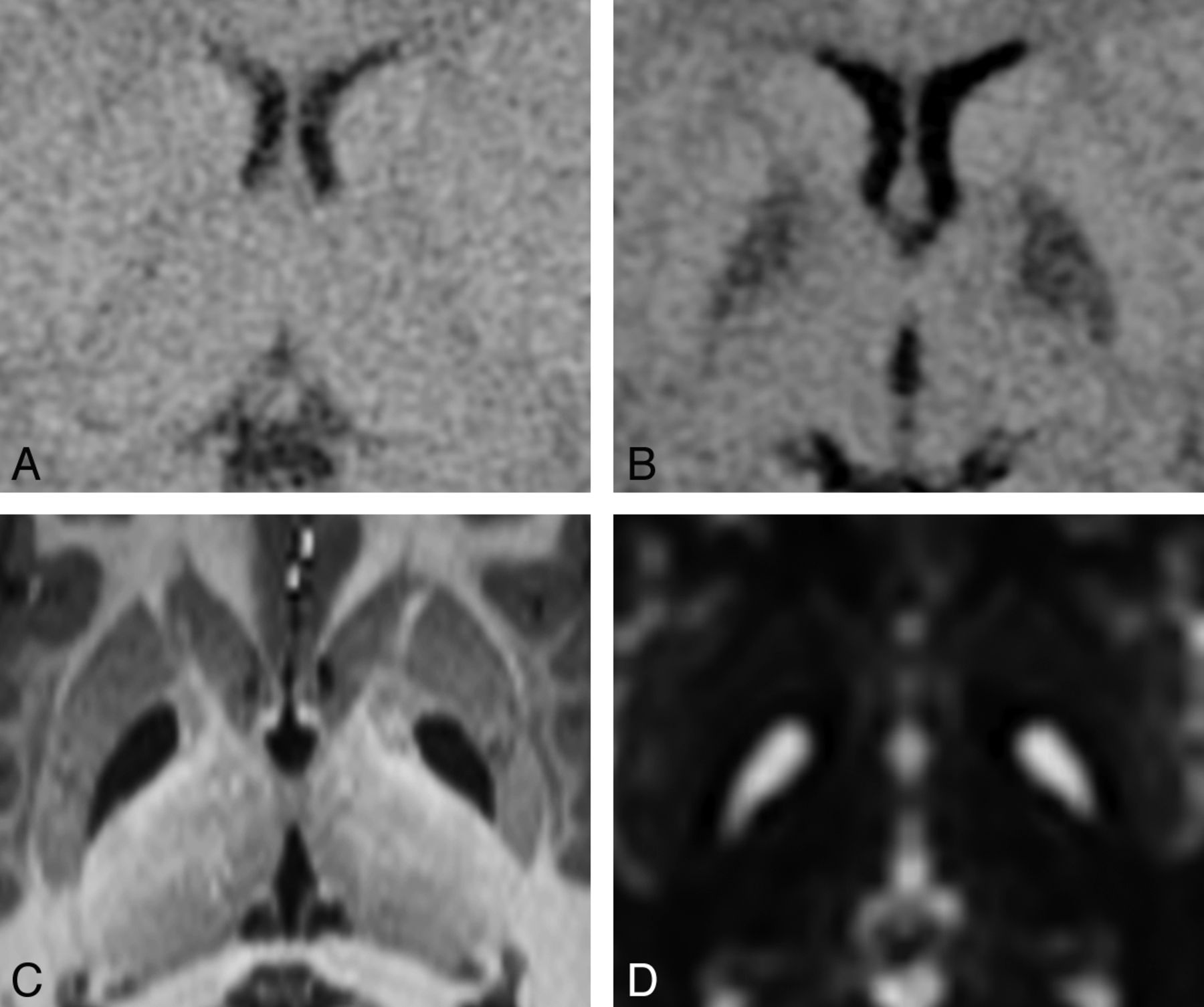
    Progression of a GP infarct. This 16-month-old patient was known to have MMA due to the cblB defect that had been diagnosed 6 months previously. Within 24 hours after onset of symptoms from gastroenteritis, she became lethargic and was taken to the emergency department. A, CT at the time of admission to the hospital appears to have normal findings. B, CT scan 35 hours later shows distinct hypoattenuating abnormalities involving the entirety of each GP. DWI performed 1 week later showed restricted diffusion in each GP (image not available). C, High-resolution MPRAGE image and D, T2-weighted image (b=0 image from DTI) acquired 7.5 years later demonstrate bilateral complete GP infarcts.

  • Fig 2.
    • Download figure
    • Open in new tab
    • Download powerpoint
    Fig 2.

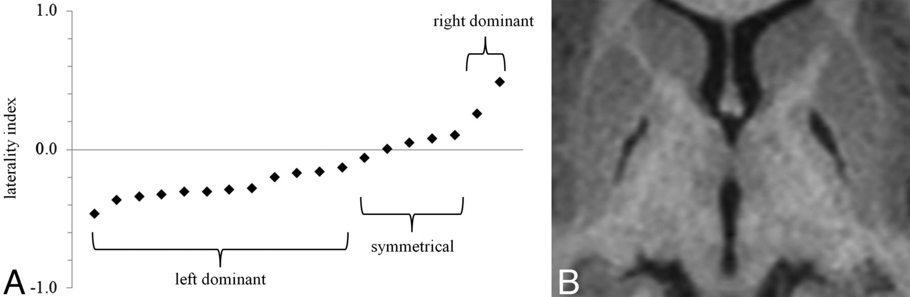
    Laterality of GP infarcts. A, Most GP infarcts were left-dominant. A few were nearly symmetrical (5 were within ±10%), and 2 were right-dominant. The average laterality index was −0.13, and the median was −0.17. B, An example of a left-dominant infarct. The laterality index in this case is −0.20.

  • Fig 3.
    • Download figure
    • Open in new tab
    • Download powerpoint
    Fig 3.

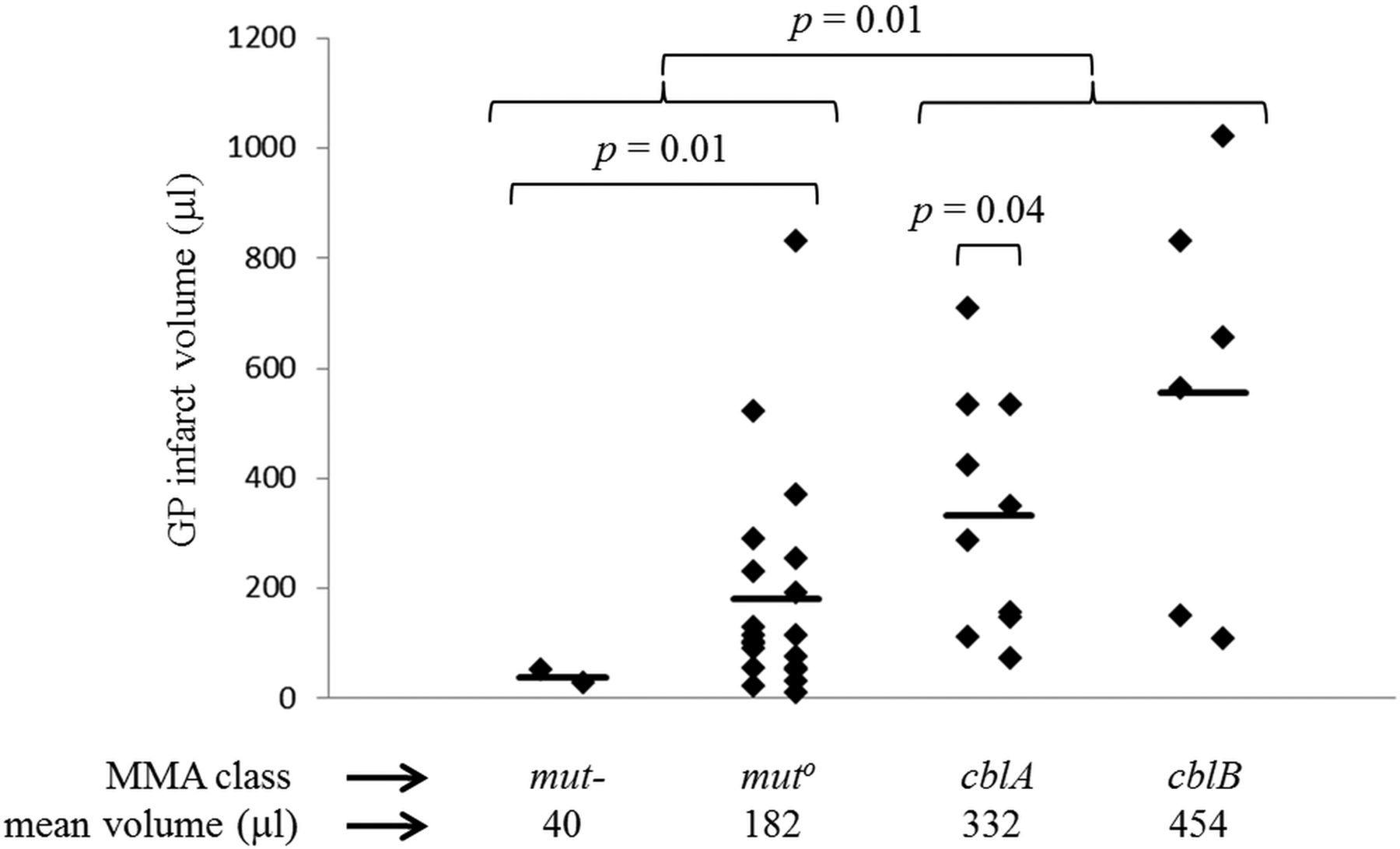
    Volume of GP infarcts. The range of GP infarct volumes is 9–1021 μL. Laterality of the infarcts is apparent when infarcts are segregated into left and right columns for each of the MMA classes. Group means (indicated by the heavy bar) differ between classes. Application of a t test suggests that there is a statistically significant difference in infarct size between the 2 mut subtypes together (mut- and muto), the 2 cbl classes together (cblA and cblB) (P = .01), and the muto subtype alone versus the 2 cbl classes together (P = .02). There was a statistically significant difference between the mut- subtype alone and each of the other categories (P = .01 versus muto, P < .01 versus cblA, and P = .02 versus cblB). There was a statistically significant difference between the left and right for the cblA class (P = .04).

  • Fig 4.
    • Download figure
    • Open in new tab
    • Download powerpoint
    Fig 4.

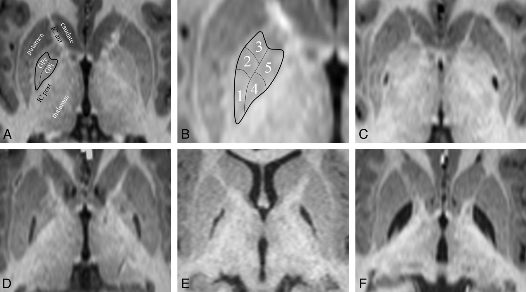
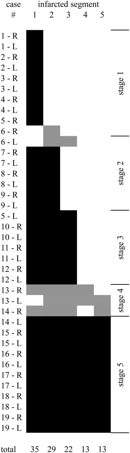
    GP infarct segments. A, Normal anatomy of the dorsal pallidum, also known as the globus pallidus. The thin line indicates the medial medullary lamina; the thick line, the boundary of the globus pallidus. Surrounding structures include the anterior limb of the internal capsule (IC ant) and the posterior limb of the internal capsule (IC post). The putamen, thalamus, and head of the caudate nucleus are also labeled. The external medullary lamina separates the putamen from the GPe. B, Infarct segments 1–5. Segment 1 seems to be the most sensitive to metabolic infarct in MMA, followed by segment 2, then segment 3; segments 4 and 5 are the least sensitive and usually infarct together. C, An example of a segment 1 infarct (posterior GPe). D, An example of an infarct of segments 1 + 2 (posterior and middle GPe). E, An example of an infarct of segments 1 + 2 + 3 (whole GPe). F, An example of an infarct of segments 1 + 2 +3 + 4 + 5 (complete GP, both externa and interna). Note that the medial medullary lamina (a white matter tract) remains intact and can be seen separating the GPe and GPi.

  • Fig 5.
    • Download figure
    • Open in new tab
    • Download powerpoint
    Fig 5.

    Graphic depiction of all 38 GP infarcts. Infarcted segments are shown in black (follows the rule) or gray (exception to the rule). The segments appear to follow a rule in which the likelihood of infarct is 1 > 2 > 3 > 4 = 5. Five infarcts (in 3 different patients) do not follow the segment-ordering rule. Infarcts can be staged by counting the number of infarcted segments, regardless of the order. In 16 of 19 patients, right and left infarcts are of the same stage, even if the volumes differ (as described in Fig 2). The location of the segments is shown in Fig 4.

  • Fig 6.
    • Download figure
    • Open in new tab
    • Download powerpoint
    Fig 6.

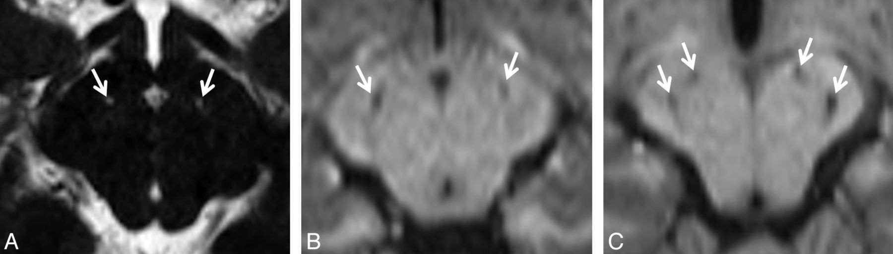
    Lacunar infarcts of the SNr. Due to the small size of the cell clusters that form the SNr (near or below the lower limit of MR imaging resolution), detection of these infarcts can be difficult. A, Tiny bilateral infarcts of the SNr are seen on a bFFE image. The resolution of this image is 0.6 × 0.6 × 1.0 mm per voxel. The infarcts measure approximately 1 mm. Both are found in the anterior location. B, Slightly larger bilateral infarcts of the SNr are seen on an MPRAGE image with a resolution of 0.9 × 0.9 × 1.0 mm per voxel. These infarcts are in the middle location. C, Bilateral infarcts at 2 sites (anterior and posterior) in the SNr are seen on an MPRAGE image. SNr infarcts were found in only 3 distinct locations, which we are designating by their relative positions; the anterior location was the most common (20 infarcts), followed by the posterior location (9 infarcts). The middle location was the least common (5 infarcts).

Tables

  • Figures
  • Demographics of infarcts in isolated MMAa

    MMA ClassNo.GP InfarctSNr InfarctGP+SNr InfarctIsolated GP InfarctIsolated SNr Infarct
    cblA75 (71%)5 (71%)411
    cblB73 (43%)3 (43%)211
    All mut subtypes2611 (42%)9 (34%)742
        mut-41 (25%)0 (0%)010
        muto2210 (45%)9 (41%)732
    All patients4019 (48%)17 (43%)1364
    • ↵a Of 40 patients scanned, 16 had both 3D-T1WI and bFFE images (most sensitive for detection of SNr infarcts), 22 had 3D-T1WI without bFFE images (less sensitive), and 2 had only 2D-T1WI (least sensitive). SNr infarcts were detected at a higher rate among patients with bFFE images (56%) than those without them (36%); therefore, SNr infarcts are probably under-reported.

PreviousNext
Back to top

In this issue

American Journal of Neuroradiology: 36 (1)
American Journal of Neuroradiology
Vol. 36, Issue 1
1 Jan 2015
  • Table of Contents
  • Index by author
  • Complete Issue (PDF)
Advertisement
Print
Download PDF
Email Article

Thank you for your interest in spreading the word on American Journal of Neuroradiology.

NOTE: We only request your email address so that the person you are recommending the page to knows that you wanted them to see it, and that it is not junk mail. We do not capture any email address.

Enter multiple addresses on separate lines or separate them with commas.
MRI Characteristics of Globus Pallidus Infarcts in Isolated Methylmalonic Acidemia
(Your Name) has sent you a message from American Journal of Neuroradiology
(Your Name) thought you would like to see the American Journal of Neuroradiology web site.
CAPTCHA
This question is for testing whether or not you are a human visitor and to prevent automated spam submissions.
Cite this article
E.H. Baker, J.L. Sloan, N.S. Hauser, A.L. Gropman, D.R. Adams, C. Toro, I. Manoli, C.P. Venditti
MRI Characteristics of Globus Pallidus Infarcts in Isolated Methylmalonic Acidemia
American Journal of Neuroradiology Jan 2015, 36 (1) 194-201; DOI: 10.3174/ajnr.A4087

Citation Manager Formats

  • BibTeX
  • Bookends
  • EasyBib
  • EndNote (tagged)
  • EndNote 8 (xml)
  • Medlars
  • Mendeley
  • Papers
  • RefWorks Tagged
  • Ref Manager
  • RIS
  • Zotero
0 Responses
Respond to this article
Share
Bookmark this article
MRI Characteristics of Globus Pallidus Infarcts in Isolated Methylmalonic Acidemia
E.H. Baker, J.L. Sloan, N.S. Hauser, A.L. Gropman, D.R. Adams, C. Toro, I. Manoli, C.P. Venditti
American Journal of Neuroradiology Jan 2015, 36 (1) 194-201; DOI: 10.3174/ajnr.A4087
del.icio.us logo Twitter logo Facebook logo Mendeley logo
  • Tweet Widget
  • Facebook Like
  • Google Plus One
Purchase

Jump to section

  • Article
    • Abstract
    • ABBREVIATIONS:
    • Materials and Methods
    • Results
    • Discussion
    • Conclusions
    • Acknowledgments
    • Footnotes
    • References
  • Figures & Data
  • Info & Metrics
  • Responses
  • References
  • PDF

Related Articles

  • No related articles found.
  • PubMed
  • Google Scholar

Cited By...

  • No citing articles found.
  • Crossref (41)
  • Google Scholar

This article has been cited by the following articles in journals that are participating in Crossref Cited-by Linking.

  • Guidelines for the diagnosis and management of methylmalonic acidaemia and propionic acidaemia: First revision
    Patrick Forny, Friederike Hörster, Diana Ballhausen, Anupam Chakrapani, Kimberly A. Chapman, Carlo Dionisi‐Vici, Marjorie Dixon, Sarah C. Grünert, Stephanie Grunewald, Goknur Haliloglu, Michel Hochuli, Tomas Honzik, Daniela Karall, Diego Martinelli, Femke Molema, Jörn Oliver Sass, Sabine Scholl‐Bürgi, Galit Tal, Monique Williams, Martina Huemer, Matthias R. Baumgartner
    Journal of Inherited Metabolic Disease 2021 44 3
  • Methylmalonic and propionic acidemias: clinical management update
    Jamie L. Fraser, Charles P. Venditti
    Current Opinion in Pediatrics 2016 28 6
  • Pathophysiology of propionic and methylmalonic acidemias. Part 1: Complications
    Hanneke A. Haijes, Judith J. M. Jans, Simone Y. Tas, Nanda M. Verhoeven‐Duif, Peter M. van Hasselt
    Journal of Inherited Metabolic Disease 2019 42 5
  • Neurological manifestations of organic acidurias
    Moacir Wajner
    Nature Reviews Neurology 2019 15 5
  • Organic acidurias in adults: late complications and management
    Ali Tunç Tuncel, Nikolas Boy, Marina A. Morath, Friederike Hörster, Ulrike Mütze, Stefan Kölker
    Journal of Inherited Metabolic Disease 2018 41 5
  • Biomarkers for drug development in propionic and methylmalonic acidemias
    Nicola Longo, Jörn Oliver Sass, Agnieszka Jurecka, Jerry Vockley
    Journal of Inherited Metabolic Disease 2022 45 2
  • Methylmalonic acidemia: Neurodevelopment and neuroimaging
    Tao Chen, Yian Gao, Shengdong Zhang, Yuanyuan Wang, Chaofan Sui, Linfeng Yang
    Frontiers in Neuroscience 2023 17
  • Neurometabolic diseases of childhood
    Zoltan Patay, Susan I. Blaser, Andrea Poretti, Thierry A. G. M. Huisman
    Pediatric Radiology 2015 45 S3
  • Mitochondrial disease, mitophagy, and cellular distress in methylmalonic acidemia
    Alessandro Luciani, Matthew C. S. Denley, Larissa P. Govers, Vincenzo Sorrentino, D. Sean Froese
    Cellular and Molecular Life Sciences 2021 78 21-22
  • Movement Disorders and Renal Diseases
    Suvorit S. Bhowmick, Anthony E. Lang
    Movement Disorders Clinical Practice 2020 7 7

More in this TOC Section

  • Comparison of Image Quality and Radiation Dose in Pediatric Temporal Bone CT Using Photon-Counting Detector CT and Energy-Integrating Detector CT
  • SyMRI & MR Fingerprinting in Brainstem Myelination
  • Pons&Vermis Localization on Fetal MRI Using U-Net
Show more Pediatrics

Similar Articles

Advertisement

Indexed Content

  • Current Issue
  • Accepted Manuscripts
  • Article Preview
  • Past Issues
  • Editorials
  • Editors Choice
  • Fellow Journal Club
  • Letters to the Editor

Cases

  • Case Collection
  • Archive - Case of the Week
  • Archive - Case of the Month
  • Archive - Classic Case

Special Collections

  • Special Collections

Resources

  • News and Updates
  • Turn around Times
  • Submit a Manuscript
  • Author Policies
  • Manuscript Submission Guidelines
  • Evidence-Based Medicine Level Guide
  • Publishing Checklists
  • Graphical Abstract Preparation
  • Imaging Protocol Submission
  • Submit a Case
  • Become a Reviewer/Academy of Reviewers
  • Get Peer Review Credit from Publons

Multimedia

  • AJNR Podcast
  • AJNR SCANtastic
  • Video Articles

About Us

  • About AJNR
  • Editorial Board
  • Not an AJNR Subscriber? Join Now
  • Alerts
  • Feedback
  • Advertise with us
  • Librarian Resources
  • Permissions
  • Terms and Conditions

American Society of Neuroradiology

  • Not an ASNR Member? Join Now

© 2025 by the American Society of Neuroradiology All rights, including for text and data mining, AI training, and similar technologies, are reserved.
Print ISSN: 0195-6108 Online ISSN: 1936-959X

Powered by HighWire