
Article Figures & Data
Figures
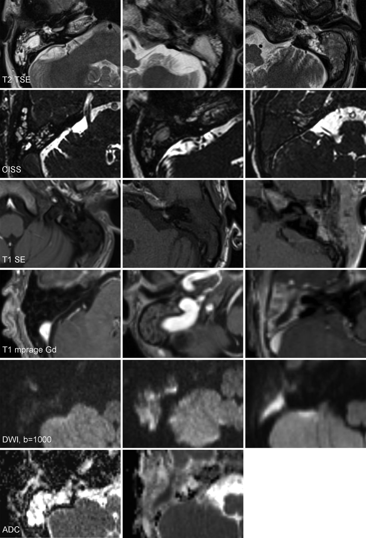
- Fig 1.
Image examples of each scoring category according to signal intensities. Categories are displayed in columns from left to right in increasing severity.
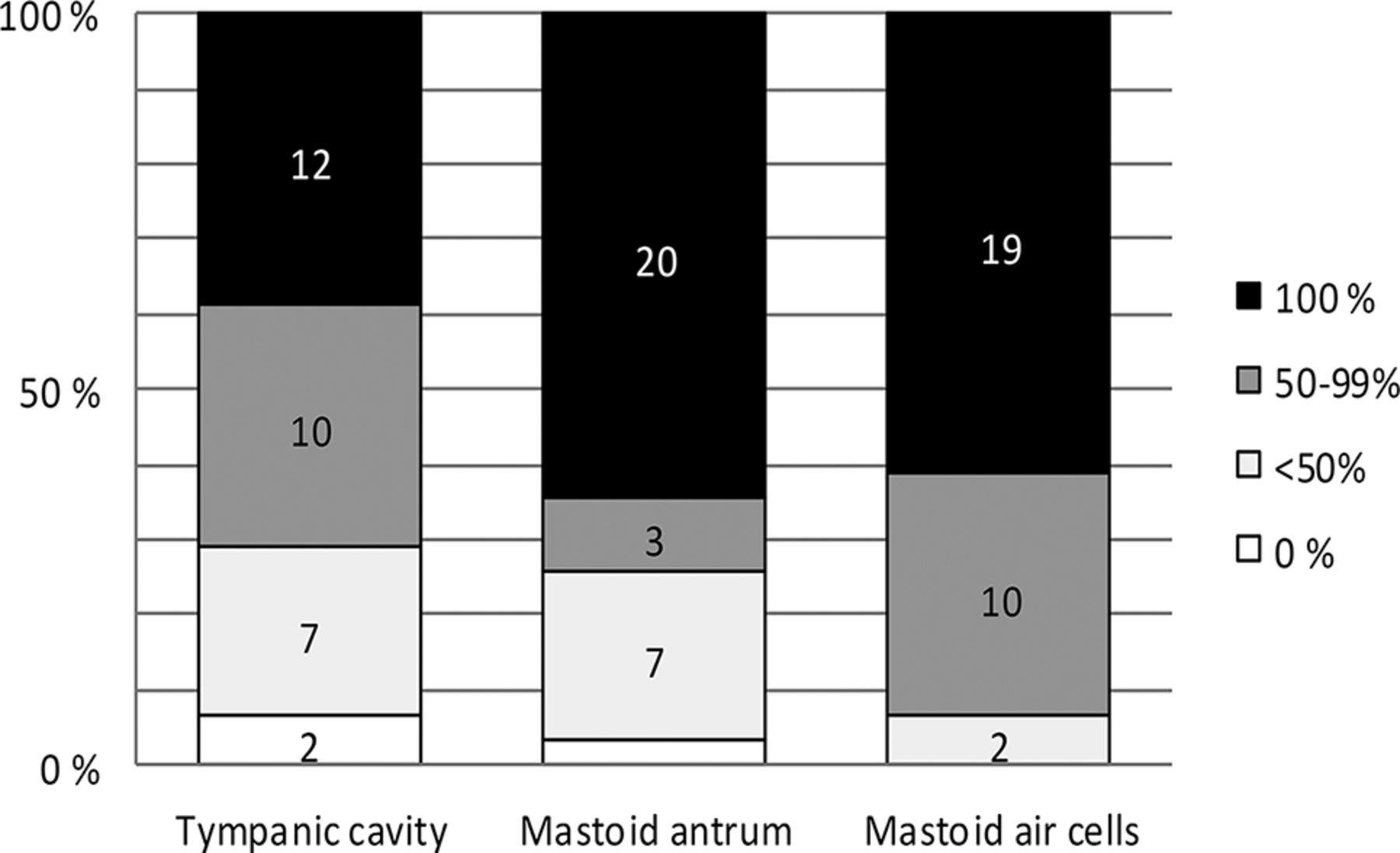
- Fig 2.
Obliteration degree in different temporal bone subregions (n = 31).
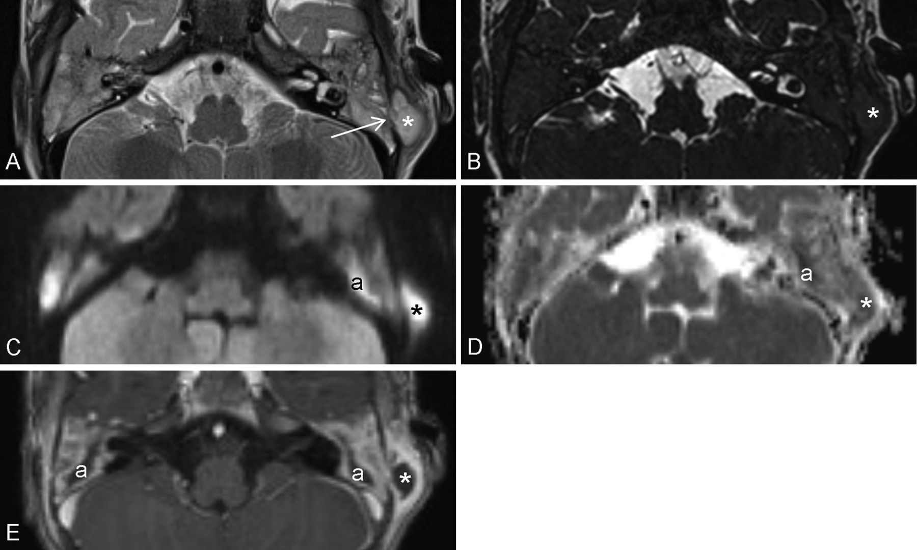
- Fig 3.
MR images of bilateral AM with duration of symptoms of 12 days on the left and fewer than 6 days (3–6 days) on the right side. T2 FSE image (A) shows total obliteration of middle ear and mastoid air spaces. SI is comparable with that of brain parenchyma. Especially on the right side, delineation of intramastoid bony septa is no longer detectable. On the left, outer cortical bone is destroyed (arrow) with subperiosteal abscess formation (asterisk). Intramastoid signal decrease, compared with CSF, becomes even more evident in CISS (B). In postgadolinium T1 MPRAGE (E), intense, thick enhancement surrounds the fluid-filled mastoid antra (a) and fills the peripheral mastoid cells. On the left, intense soft-tissue enhancement around the subperiosteal abscess and, on the right, periosteal enhancement surrounding the mastoid are visible. DWI b=1000 (C) and ADC (D) show diffusion restriction in the whole mastoid region bilaterally with foci of markedly elevated SI inside both antra (a) and the left subperiosteal abscess (asterisk).


Tables
No. No. of Patients per Category (Valid %) SI in T2 FSE and CISS Isointense to CSF Hypointense to CSF, hyperintense to WM Iso- or hypointense to WM CISS 25 1 (4) 14 (56) 10 (40) T2 FSE 31 3 (10) 24 (77) 4 (13) SI in T1 SE Isointense to CSF Hyperintense to CSF, not to WM Hyperintense to WM T1 SE 31 0 (0) 22 (71) 9 (29) SI in DWI (b=1000) Hypointense to WM Isointense to WM Hyperintense to WM 27 2 (7) 9 (33) 16 (59) SI in ADC Not lowered Lowered 26 10 (38) 16 (62) – Enhancement None Faint, thin Intense, thick 31 3 (10) 12 (39) 16 (52) Note:—No. indicates the number of patients with a specific sequence available.
Complication No. (%) All Children Adults Septal destruction 11 (35) 5 (45) 6 (30) Inner cortical bone destruction 4 (13) 2 (18) 2 (10) Outer cortical bone destruction 9 (29) 7 (64)a 2 (10)a Intratemporal abscess 7 (23) 3 (27) 4 (20) Subperiosteal abscess 6 (19) 5 (45)a 1 (5)a Labyrinth involvement 5 (16) 0 (0) 5 (25) Epidural abscess 2 (6) 2 (18) 0 (0) Soft-tissue abscess 2 (6) 1 (9) 1 (5) Generalized pachymeningitis 2 (6) 1 (9) 1 (5) Leptomeningitis 2 (6) 1 (9) 1 (5) Sinus thrombosis 1 (3) 1 (9) 0 (0) Subdural empyema 0 (0) 0 (0) 0 (0) ↵a Significant differences between adult and pediatric subgroups (P < .05).


