
Article Figures & Data
Figures
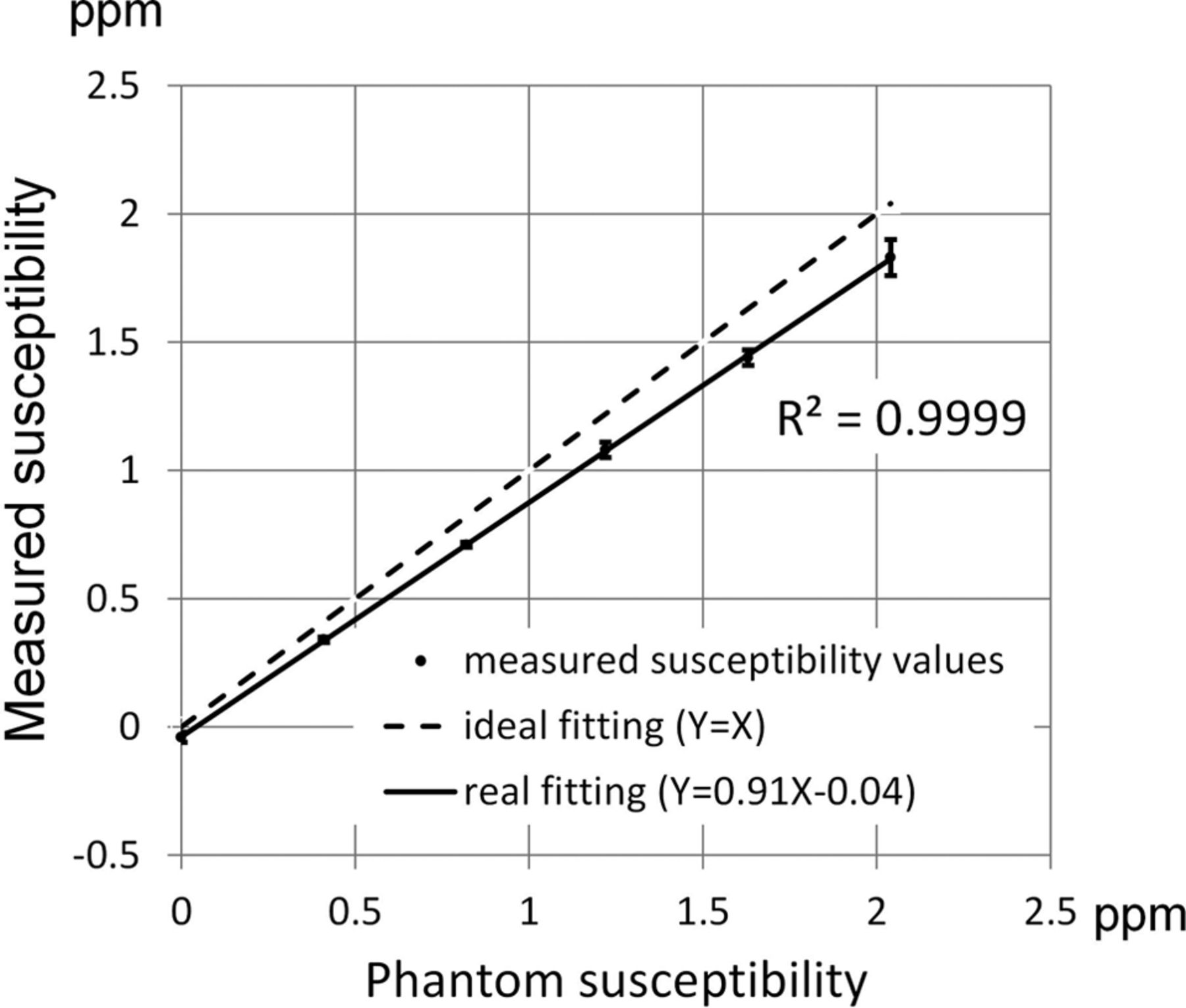
- Fig 1.
Measured susceptibility versus the phantom susceptibility values. Good linearity exists between measured susceptibility values and phantom susceptibility values in 6 test tubes of the phantom. The regression line has a slope of 0.91.
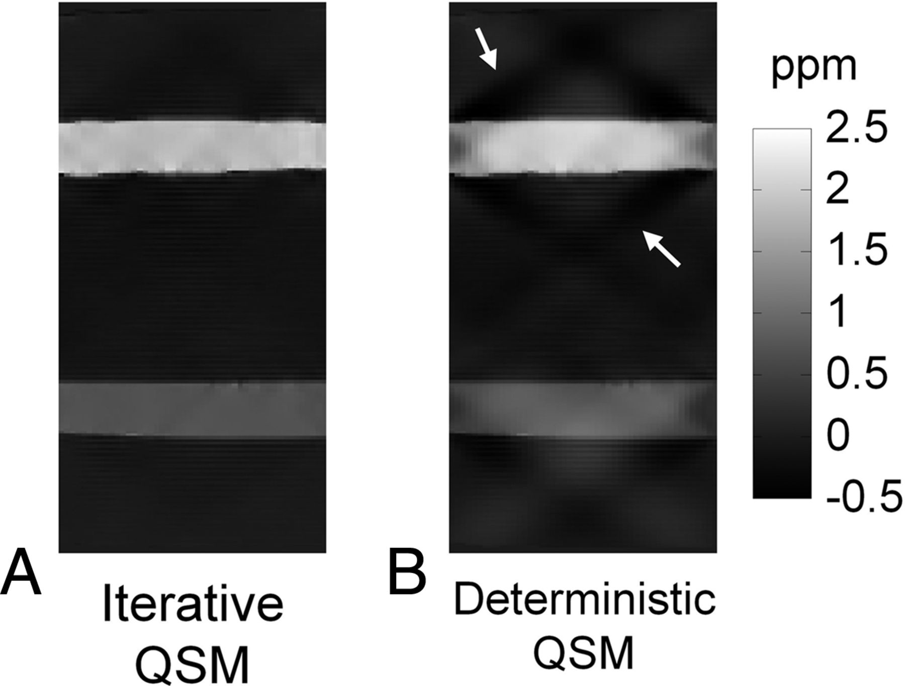
- Fig 2.
Comparison of iterative QSM and deterministic QSM in a sagittal view of the phantom. The susceptibility map from deterministic QSM presents a higher level of streaking artifacts as indicated by the white arrows.
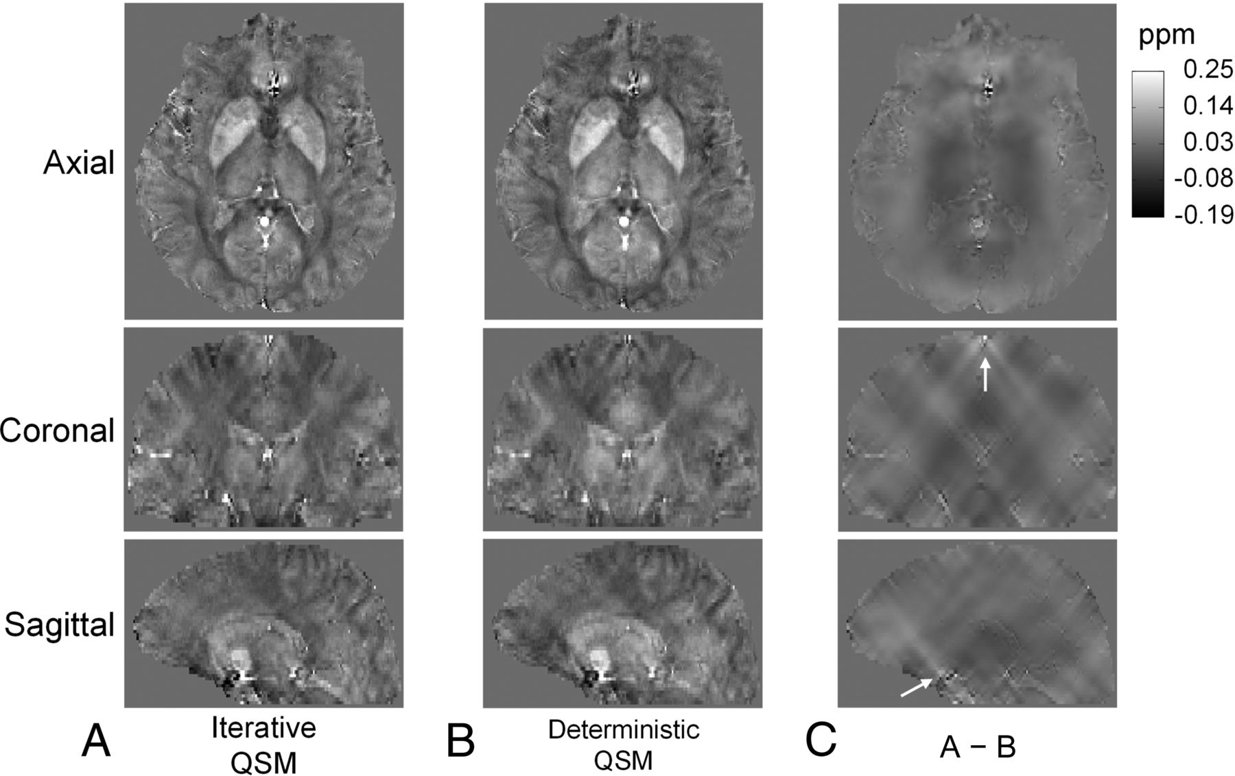
- Fig 3.
Susceptibility maps reconstructed by iterative QSM (A) and deterministic QSM (B) in 3 different views. Susceptibility maps from both methods are very similar in contrast and brain structures. C, The difference images of A and B. Note that most streaking patterns in C are centered at locations with high susceptibility values, such as the sagittal sinus and regions close to the skull base, as indicated by white arrows.
- Fig 4.
Susceptibility maps reconstructed by using iterative QSM with data from 3 scanners at sites A, B, and C. A–C, Good consistency in brain structures and susceptibility values is shown. Some difference is found at the frontal lobe as indicated by white arrows in B and C, likely due to different shimming conditions in this region.
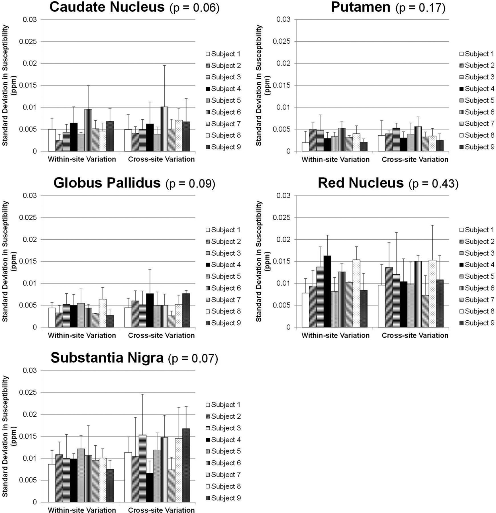
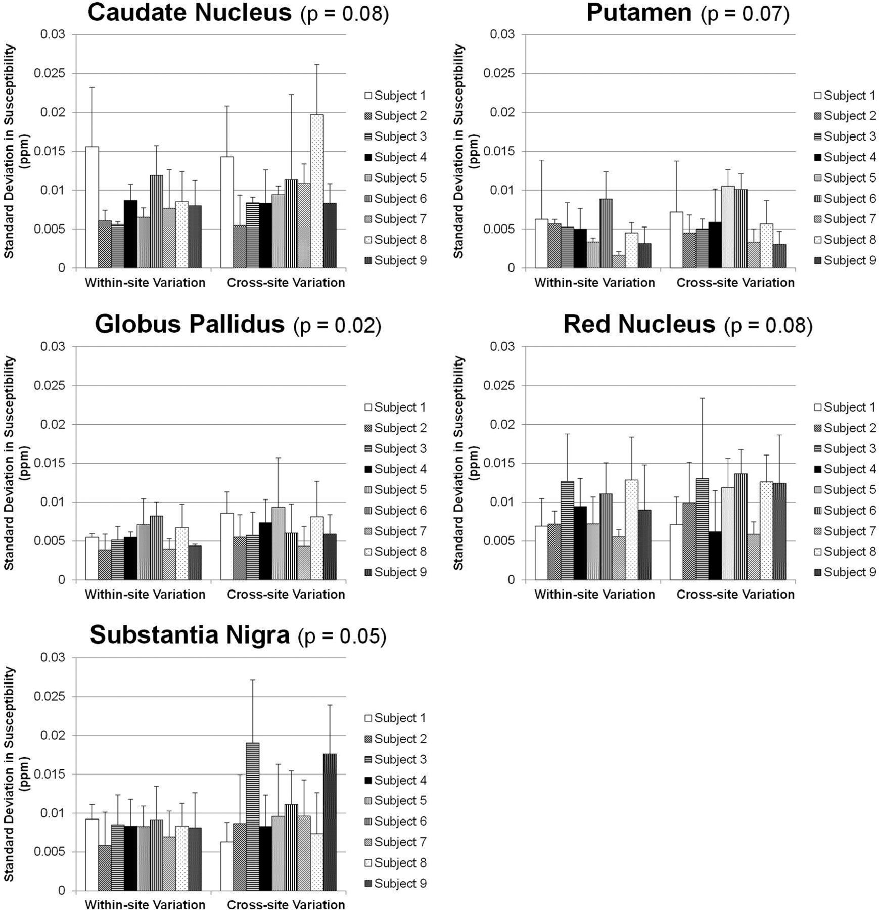
- Fig 5.
Within-site (left) and cross-site (right) imprecisions shown as SDs from multiple measurements on the 9 subjects by using the iterative QSM algorithm. The P values shown in the subplot titles stand for the difference between within-site and cross-site precisions (paired Student t test). For all 5 regions, cross-site variations are not larger than within-site variations. The putamen and globus pallidus show the highest precision, followed by the caudate nucleus, red nucleus, and substantia nigra.
- Fig 6.
Within-site (left) and cross-site (right) imprecisions shown as SDs from multiple measurements on the 9 subjects by using the deterministic QSM algorithm. The P values shown in the subplot titles stand for the difference between within-site and cross-site precisions (paired Student t test). For all 5 regions, cross-site variations are not larger than within-site variations. The putamen and globus pallidus show the highest precision, followed by the caudate nucleus, red nucleus, and substantia nigra.
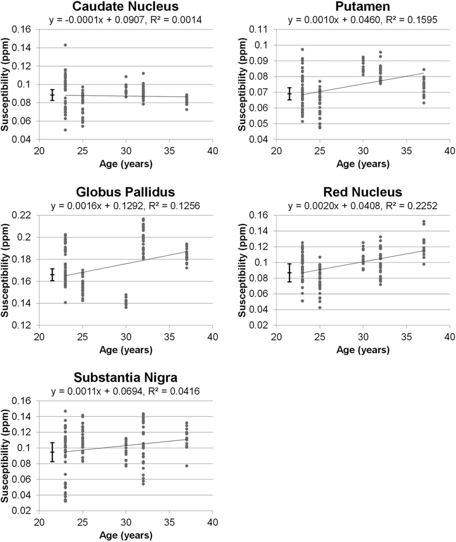
- Fig 7.
Linear regression results comparing susceptibility values versus age by using 135 datasets collected in this study. Among the 5 ROIs, the putamen, globus pallidus, and red nucleus present correlation coefficients of 0.40, 0.35, and 0.47, respectively (P < .001). The caudate nucleus shows no age association (P > .5), and the substantia nigra exhibits a marginal association that does not reach statistical significance after Bonferroni correction (P = .02). The vertical error bars shown on the left stand for the cross-site variability values of iterative QSM found for these regions.


Tables
Within-site and cross-site imprecisions averaged from 9 subjects
ROI Method Within-Site Imprecision (ppm) Cross-Site Imprecision (ppm) Caudate nucleus Iterative QSM 0.0054 (10.0%) 0.0059 (11.0%) Deterministic QSM 0.0087 (17.9%) 0.0107 (21.9%) Putamen Iterative QSM 0.0037 (9.3%) 0.0038 (9.5%) Deterministic QSM 0.0048 (8.3%) 0.0061 (10.6%) Globus pallidus Iterative QSM 0.0044 (3.4%) 0.0054 (4.1%) Deterministic QSM 0.0056 (3.4%) 0.0068 (4.1%) Red nucleus Iterative QSM 0.0113 (18.4%) 0.0115 (18.7%) Deterministic QSM 0.0091 (9.3%) 0.0103 (10.6%) Substantia nigra Iterative QSM 0.0099 (15.2%) 0.0121 (18.6%) Deterministic QSM 0.0081 (8.2%) 0.0108 (11.1%)


Jump to section
Related Articles
Cited By...
- Iron Deposition and Distribution Across the Hippocampus Is Associated with Pattern Separation and Pattern Completion in Older Adults at Risk for Alzheimer's Disease
- Validation of Data Acquisition and Phase Estimation for Quantitative Susceptibility Mapping with a Rotating-Tube Phantom
- Multi-centre, multi-vendor reproducibility of 7T QSM and R2* in the human brain: results from the UK7T study
- In Vivo MRI Mapping of Brain Iron Deposition across the Adult Lifespan