Advertisement

Main menu

  • Home
  • Content
    • Current Issue
    • Accepted Manuscripts
    • Article Preview
    • Past Issue Archive
    • AJNR Case Collection
    • Case of the Week Archive
    • Classic Case Archive
    • Case of the Month Archive
  • Special Collections
    • Spinal CSF Leak Articles (Jan 2020-June 2024)
    • 2024 AJNR Journal Awards
    • Most Impactful AJNR Articles
  • Multimedia
    • AJNR Podcast
    • AJNR Scantastics
    • Video Articles
  • For Authors
    • Submit a Manuscript
    • Author Policies
    • Fast publishing of Accepted Manuscripts
    • Graphical Abstract Preparation
    • Manuscript Submission Guidelines
    • Imaging Protocol Submission
    • Submit a Case for the Case Collection
  • About Us
    • About AJNR
    • Editorial Board
  • More
    • Become a Reviewer/Academy of Reviewers
    • Subscribers
    • Permissions
    • Alerts
    • Feedback
    • Advertisers
    • ASNR Home
  • Other Publications
    • ajnr

User menu

  • Alerts
  • Log in

Search

  • Advanced search
American Journal of Neuroradiology

American Journal of Neuroradiology

ASHNR American Society of Functional Neuroradiology ASHNR American Society of Pediatric Neuroradiology ASSR
  • Alerts
  • Log in

Advanced Search

  • Home
  • Content
    • Current Issue
    • Accepted Manuscripts
    • Article Preview
    • Past Issue Archive
    • AJNR Case Collection
    • Case of the Week Archive
    • Classic Case Archive
    • Case of the Month Archive
  • Special Collections
    • Spinal CSF Leak Articles (Jan 2020-June 2024)
    • 2024 AJNR Journal Awards
    • Most Impactful AJNR Articles
  • Multimedia
    • AJNR Podcast
    • AJNR Scantastics
    • Video Articles
  • For Authors
    • Submit a Manuscript
    • Author Policies
    • Fast publishing of Accepted Manuscripts
    • Graphical Abstract Preparation
    • Manuscript Submission Guidelines
    • Imaging Protocol Submission
    • Submit a Case for the Case Collection
  • About Us
    • About AJNR
    • Editorial Board
  • More
    • Become a Reviewer/Academy of Reviewers
    • Subscribers
    • Permissions
    • Alerts
    • Feedback
    • Advertisers
    • ASNR Home
  • Follow AJNR on Twitter
  • Visit AJNR on Facebook
  • Follow AJNR on Instagram
  • Join AJNR on LinkedIn
  • RSS Feeds

Welcome to the new AJNR, Updated Hall of Fame, and more. Read the full announcements.


AJNR is seeking candidates for the position of Associate Section Editor, AJNR Case Collection. Read the full announcement.


Getting new auth cookie, if you see this message a lot, tell someone!
Research ArticleEXTRACRANIAL VASCULAR

Appropriate Minimal Dose of Gadobutrol for 3D Time-Resolved MRA of the Supra-Aortic Arteries: Comparison with Conventional Single-Phase High-Resolution 3D Contrast-Enhanced MRA

S.H. Bak, H.G. Roh, W.-J. Moon, J.W. Choi and H.S. An
American Journal of Neuroradiology July 2017, 38 (7) 1383-1390; DOI: https://doi.org/10.3174/ajnr.A5176
S.H. Bak
aFrom the Department of Radiology (S.H.B., H.G.R., W.-J.M., J.W.C.), Konkuk University Medical Center, Konkuk University School of Medicine, Seoul, Korea
bDepartment of Radiology (S.H.B.), Kangwon National University Hospital, Chuncheon, Korea
  • Find this author on Google Scholar
  • Find this author on PubMed
  • Search for this author on this site
  • ORCID record for S.H. Bak
H.G. Roh
aFrom the Department of Radiology (S.H.B., H.G.R., W.-J.M., J.W.C.), Konkuk University Medical Center, Konkuk University School of Medicine, Seoul, Korea
  • Find this author on Google Scholar
  • Find this author on PubMed
  • Search for this author on this site
  • ORCID record for H.G. Roh
W.-J. Moon
aFrom the Department of Radiology (S.H.B., H.G.R., W.-J.M., J.W.C.), Konkuk University Medical Center, Konkuk University School of Medicine, Seoul, Korea
  • Find this author on Google Scholar
  • Find this author on PubMed
  • Search for this author on this site
  • ORCID record for W.-J. Moon
J.W. Choi
aFrom the Department of Radiology (S.H.B., H.G.R., W.-J.M., J.W.C.), Konkuk University Medical Center, Konkuk University School of Medicine, Seoul, Korea
  • Find this author on Google Scholar
  • Find this author on PubMed
  • Search for this author on this site
  • ORCID record for J.W. Choi
H.S. An
cDepartment of Radiology (H.S.A.), Armed Forces Yangju Hospital, Yangju, Korea.
  • Find this author on Google Scholar
  • Find this author on PubMed
  • Search for this author on this site
  • ORCID record for H.S. An
  • Article
  • Figures & Data
  • Info & Metrics
  • Responses
  • References
  • PDF
Loading

Article Figures & Data

Figures

  • Tables
  • Fig 1.
    • Download figure
    • Open in new tab
    • Download powerpoint
    Fig 1.

    Flow chart of the study population.
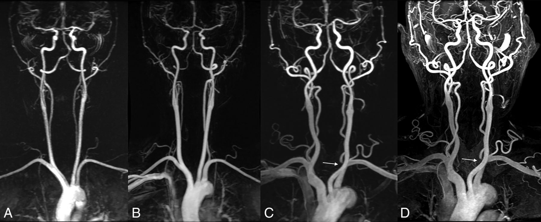
  • Fig 2.
    • Download figure
    • Open in new tab
    • Download powerpoint
    Fig 2.

    A, TR-MRA with 1 mL of gadobutrol with subtracted coronal MIP images shows good segmental visualization with minimal blurring or undulation of both CCAs, the extracranial ICA, and the vertebral artery. TR-MRA with 2 (B) and 3 mL (C) of gadobutrol with subtraction coronal MIP images shows clear arterial visualization. D, CE-MRA shows mild venous contamination with no influence on the diagnostic interpretation at the intracranial area. TR-MRA with 3 mL of gadobutrol (C) and CE-MRA (D) show severe stenosis of the left vertebral artery origin (arrow) in a 75-year-old man with suspected stroke.
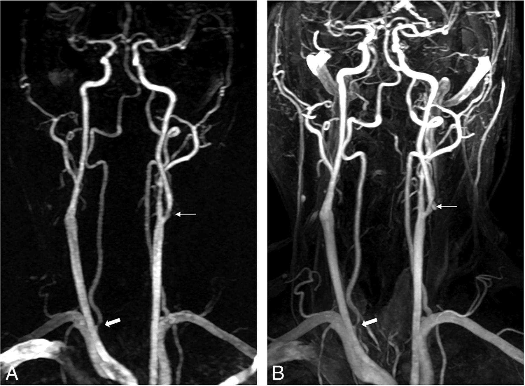
  • Fig 3.
    • Download figure
    • Open in new tab
    • Download powerpoint
    Fig 3.

    A 52-year-old man with suspected transient ischemic attack. TR-MRA with 1 mL of gadobutrol (A) and CE-MRA (B) show moderate stenosis at the origin of the right vertebral artery (thick arrow) and mild stenosis of the left proximal extracranial ICA (thin arrow).

  • Fig 4.
    • Download figure
    • Open in new tab
    • Download powerpoint
    Fig 4.

    A 74-year-old man with suspected stroke. TR-MRA with 2 mL of gadobutrol (A) and CE-MRA (B) show moderate stenosis in the proximal extracranial ICA (thick arrow).

Tables

  • Figures
    • View popup
    Table 1:

    Demographic characteristicsa

    Group A (1 mL) (n = 139)Group B (2 mL) (n = 169)Group C (3 mL) (n = 154)P Value
    Age (yr)60.3 ± 13.662.2 ± 13.463.2 ± 14.5.184
    Male/female80:5994:7583:71.821
    Body weight (kg)63.4 ± 11.663.9 ± 10.362.4 ± 10.1.498
    Contrast dose of CE-MRA (mL)6.3 ± 1.26.4 ± 1.06.2 ± 1.0.498
    Time between CE-MRA and TR-MRA (sec)224 ± 21251 ± 42233 ± 36.532
    • ↵a Data are means.

    • View popup
    Table 2:

    Comparison of image quality between 2 MRA techniques in the 3 groups

    Arterial SegmentGroup AGroup BGroup C
    CE-MRATR-MRAPCE-MRATR-MRAPCE-MRATR-MRAP
    MeanRangeMeanRangeMeanRangeMeanRangeMeanRangeMeanRange
    Right brachiocephalic trunk3.963–43.582–4.0003.973–43.913–4.0013.973–43.933–4.034
    Right subclavian artery3.932–43.581–4.0003.921–43.921–4.6553.932–43.943–4.414
    Left subclavian artery3.771–43.271–4.0003.902–43.852–4.0503.943–43.903–4.058
    Right CCA3.912–43.531–4.0003.953–43.913–4.0523.933–43.923–4.655
    Right extracranial ICA3.993–43.543–4.0003.993–43.963–4.0593.993–44.004–4.317
    Right intracranial ICA3.391–43.672–4.0003.452–43.883–4.0003.562–43.912–4.000
    Left CCA3.832–43.221–4.0003.923–43.793–4.0003.943–43.923–4.180
    Left extracranial ICA3.993–43.613–4.0003.983–43.963–4.1803.993–44.004–4.157
    Left intracranial ICA3.381–43.632–4.0003.542–43.893–4.0003.622–43.903–4.000
    Right vertebral artery orifice3.791–43.331–4.0003.742–43.752–4.3173.773–43.783–4.705
    Right vertebral artery3.511–43.221–4.0003.622–43.812–4.0003.622–43.903–4.000
    Left vertebral artery orifice3.723–43.191–4.0003.763–43.752–4.4053.773–43.773–4.808
    Left vertebral artery3.551–43.202–4.0003.682–43.792–4.0063.722–43.902–4.000
    Basilar artery3.851–43.692–4.0003.882–43.882–41.0003.911–43.941–4.157
    • View popup
    Table 3:

    Distribution of venous contamination at CE-MRA and TR-MRA in the 3 groupsa

    Arterial SegmentGroup AGroup BGroup C
    Grade 0Grade 1Grade 2Grade 3Grade 0Grade 1Grade 2Grade 3Grade 0Grade 1Grade 2Grade 3
    Right brachiocephalic trunk138/1371/10/10/0164/1654/41/00/0154/1540/00/00/0
    Right subclavian artery61/11877/191/20/064/123104/401/60/059/11593/352/40/0
    Left subclavian artery128/13611/00/30/0164/1685/10/00/0142/14710/62/10/0
    Right CCA110/13429/40/10/0146/15623/120/10/0112/13941/141/10/0
    Right extracranial ICA25/101114/380/00/021/119148/500/00/05/111148/431/00/0
    Right intracranial ICA24/108109/316/00/024/120138/496/01/030/114120/394/10/0
    Left CCA125/13314/50/10/0158/16711/20/00/0140/14813/51/10/0
    Left extracranial ICA28/103111/360/00/024/111145/580/00/015/109137/452/00/0
    Left intracranial ICA25/97102/4012/20/019/115142/538/10/032/112115/427/00/0
    Right vertebral artery orifice122/13717/10/10/0151/16017/61/30/0132/14522/70/20/0
    Right vertebral artery54/11284/271/00/068/139101/290/10/045/126104/275/10/0
    Left vertebral artery orifice132/1397/00/00/0166/1673/10/10/0143/15110/30/01/0
    Left vertebral artery51/11586/242/00/063/145105/241/00/051/131101/222/10/0
    Basilar artery29/119103/166/41/051/142105/2513/20/036/134111/207/00/0
    • ↵a Data are number of segments for CE-MRA/TR-MRA.

    • View popup
    Table 4:

    Evaluation of stenosis at CE-MRA and TR-MRA in the 3 groups

    CE-MRAGroup A TR-MRAGroup B TR-MRAGroup C TR-MRA
    0123Total0123Total0123Total
    0176623217732149100215019471001948
    1657406709820100110210104
    2146917533760820475079
    3000313100034341002425
    Total17736376341946215210278342366194910776242156
    • View popup
    Table 5:

    Comparison of SNR between 2 MRA techniques in the 3 groupsa

    Arterial SegmentGroup AGroup BGroup C
    CE-MRATR-MRAPCE-MRATR-MRAPCE-MRATR-MRAP
    Right brachiocephalic trunk211.2 ± 50.6144.0 ± 47.6.000203.0 ± 45.3211.2 ± 61.1.019204.0 ± 116.0230.8 ± 65.9.011
    Right subclavian artery150.8 ± 41.097.8 ± 32.0.000144.2 ± 36.6154.0 ± 81.9.099135.2 ± 37.0167.4 ± 110.7.000
    Left subclavian artery163.3 ± 35.3100.9 ± 29.3.000157.0 ± 34.0154.0 ± 40.4.276152.3 ± 36.6174.9 ± 45.7.000
    Right CCA172.1 ± 45.5103.9 ± 32.6.000164.8 ± 42.1156.4 ± 48.7.017157.1 ± 44.9173.8 ± 47.3.000
    Right extracranial ICA190.4 ± 63.9107.0 ± 38.9.000177.4 ± 53.5166.4 ± 57.4.003176.5 ± 54.5193.1 ± 61.3.001
    Right intracranial ICA280.1 ± 77.4136.3 ± 46.6.000289.9 ± 172.2226.3 ± 74.7.000274.5 ± 71.3280.1 ± 77.2.364
    Left CCA200.6 ± 48.8112.9 ± 33.3.000189.7 ± 45.9172.2 ± 43.2.000183.6 ± 50.1200.2 ± 52.0.000
    Left extracranial ICA220.8 ± 66.0120.1 ± 38.5.000204.2 ± 59.3184.9 ± 62.0.000199.8 ± 61.5224.1 ± 70.8.000
    Left intracranial ICA300.5 ± 73.2144.9 ± 47.1.000295.3 ± 63.5240.4 ± 66.1.000291.1 ± 70.8301.7 ± 82.5.137
    Right vertebral artery165.3 ± 52.971.8 ± 27.7.000158.4 ± 57.3115.8 ± 57.9.000158.0 ± 55.2136.2 ± 56.4.000
    Left vertebral artery169.6 ± 60.773.7 ± 25.6.000167.4 ± 53.8122.1 ± 49.8.000170.6 ± 55.3149.1 ± 52.2.000
    Basilar artery223.2 ± 59.580.7 ± 29.5.000229.9 ± 55.4136.1 ± 62.8.000230.5 ± 46.9165.5 ± 54.9.000
    • ↵a Data are means.

PreviousNext
Back to top

In this issue

American Journal of Neuroradiology: 38 (7)
American Journal of Neuroradiology
Vol. 38, Issue 7
1 Jul 2017
  • Table of Contents
  • Index by author
  • Complete Issue (PDF)
Advertisement
Print
Download PDF
Email Article

Thank you for your interest in spreading the word on American Journal of Neuroradiology.

NOTE: We only request your email address so that the person you are recommending the page to knows that you wanted them to see it, and that it is not junk mail. We do not capture any email address.

Enter multiple addresses on separate lines or separate them with commas.
Appropriate Minimal Dose of Gadobutrol for 3D Time-Resolved MRA of the Supra-Aortic Arteries: Comparison with Conventional Single-Phase High-Resolution 3D Contrast-Enhanced MRA
(Your Name) has sent you a message from American Journal of Neuroradiology
(Your Name) thought you would like to see the American Journal of Neuroradiology web site.
CAPTCHA
This question is for testing whether or not you are a human visitor and to prevent automated spam submissions.
Cite this article
S.H. Bak, H.G. Roh, W.-J. Moon, J.W. Choi, H.S. An
Appropriate Minimal Dose of Gadobutrol for 3D Time-Resolved MRA of the Supra-Aortic Arteries: Comparison with Conventional Single-Phase High-Resolution 3D Contrast-Enhanced MRA
American Journal of Neuroradiology Jul 2017, 38 (7) 1383-1390; DOI: 10.3174/ajnr.A5176

Citation Manager Formats

  • BibTeX
  • Bookends
  • EasyBib
  • EndNote (tagged)
  • EndNote 8 (xml)
  • Medlars
  • Mendeley
  • Papers
  • RefWorks Tagged
  • Ref Manager
  • RIS
  • Zotero
0 Responses
Respond to this article
Share
Bookmark this article
Appropriate Minimal Dose of Gadobutrol for 3D Time-Resolved MRA of the Supra-Aortic Arteries: Comparison with Conventional Single-Phase High-Resolution 3D Contrast-Enhanced MRA
S.H. Bak, H.G. Roh, W.-J. Moon, J.W. Choi, H.S. An
American Journal of Neuroradiology Jul 2017, 38 (7) 1383-1390; DOI: 10.3174/ajnr.A5176
del.icio.us logo Twitter logo Facebook logo Mendeley logo
  • Tweet Widget
  • Facebook Like
  • Google Plus One
Purchase

Jump to section

  • Article
    • Abstract
    • ABBREVIATIONS:
    • Materials and Methods
    • Results
    • Discussion
    • Conclusions
    • References
  • Figures & Data
  • Info & Metrics
  • Responses
  • References
  • PDF

Related Articles

  • No related articles found.
  • PubMed
  • Google Scholar

Cited By...

  • A Novel Collateral Imaging Method Derived from Time-Resolved Dynamic Contrast-Enhanced MR Angiography in Acute Ischemic Stroke: A Pilot Study
  • Crossref (8)
  • Google Scholar

This article has been cited by the following articles in journals that are participating in Crossref Cited-by Linking.

  • Reducing Contrast Agent Dose in Cardiovascular MR Angiography with Deep Learning
    Javier Montalt‐Tordera, Michael Quail, Jennifer A Steeden, Vivek Muthurangu
    Journal of Magnetic Resonance Imaging 2021 54 3
  • A Novel Collateral Imaging Method Derived from Time-Resolved Dynamic Contrast-Enhanced MR Angiography in Acute Ischemic Stroke: A Pilot Study
    H.G. Roh, E.Y. Kim, I.S. Kim, H.J. Lee, J.J. Park, S.B. Lee, J.W. Choi, Y.S. Jeon, M. Park, S.U. Kim, H.J. Kim
    American Journal of Neuroradiology 2019 40 6
  • Collateral and permeability imaging derived from dynamic contrast material-enhanced MR angiography in prediction of PH 2 hemorrhagic transformation after acute ischemic stroke: a pilot study
    Taek-Jun Lee, Hong Gee Roh, Joo Hyun Kim, Sang Bong Lee, Jeong Jin Park, Hyung Jin Lee, Yoo Sung Jeon, Jin Woo Choi, Young Il Chun, Yu Jin Jung, Seon Young Ryu, Ji Sung Lee, Hyun Jeong Kim
    Neuroradiology 2021 63 9
  • Fast MRI in Acute Ischemic Stroke: Applications of MRI Acceleration Techniques for MR-Based Comprehensive Stroke Imaging
    Sung-Hye You, Byungjun Kim, Bo Kyu Kim, Sang Eun Park
    Investigative Magnetic Resonance Imaging 2021 25 2
  • Ferumoxytol-enhanced ultrashort TE MRA and quantitative morphometry of the human kidney vasculature
    Liam Timms, Tianyi Zhou, Yue Lyu, Ju Qiao, Vishala Mishra, Rita Maria Lahoud, Gayatri Veeramani Jayaraman, Andrew S. Allegretti, David Drew, Ravi T. Seethamraju, Mukesh Harisinghani, Srinivas Sridhar
    Abdominal Radiology 2021 46 7
  • Technique and protocols for cardiothoracic time-resolved contrast-enhanced magnetic resonance angiography sequences: a systematic review
    M. Zanardo, F. Sardanelli, L. Rainford, C.B. Monti, J.G. Murray, F. Secchi, A. Cradock
    Clinical Radiology 2021 76 2
  • Clinical feasibility of deep learning-driven magnetic resonance angiography collateral map in acute anterior circulation ischemic stroke
    Ye Jin Jeon, Hong Gee Roh, Sumin Jung, Hyun Yang, Hee Jong Ki, Jeong Jin Park, Taek-Jun Lee, Na Il Shin, Ji Sung Lee, Jin Tae Kwak, Hyun Jeong Kim
    Scientific Reports 2025 15 1
  • Preoperative assessment of peripheral vascular invasion of pancreatic ductal adenocarcinoma based on high-resolution MRI
    Xiaoqi Zhou, Danyang Xu, Meng Wang, Ruixia Ma, Chenyu Song, Zhi Dong, Yanji Luo, Jifei Wang, Shi-Ting Feng
    BMC Cancer 2023 23 1

More in this TOC Section

  • Proximal Vertebral Artery Variants and Embryology
  • High-Risk Plaque Features in Carotid MRI
  • Nonstenotic Carotid Plaques and Stroke Review
Show more Extracranial Vascular

Similar Articles

Advertisement

Indexed Content

  • Current Issue
  • Accepted Manuscripts
  • Article Preview
  • Past Issues
  • Editorials
  • Editors Choice
  • Fellow Journal Club
  • Letters to the Editor

Cases

  • Case Collection
  • Archive - Case of the Week
  • Archive - Case of the Month
  • Archive - Classic Case

Special Collections

  • Special Collections

Resources

  • News and Updates
  • Turn around Times
  • Submit a Manuscript
  • Author Policies
  • Manuscript Submission Guidelines
  • Evidence-Based Medicine Level Guide
  • Publishing Checklists
  • Graphical Abstract Preparation
  • Imaging Protocol Submission
  • Submit a Case
  • Become a Reviewer/Academy of Reviewers
  • Get Peer Review Credit from Publons

Multimedia

  • AJNR Podcast
  • AJNR SCANtastic
  • Video Articles

About Us

  • About AJNR
  • Editorial Board
  • Not an AJNR Subscriber? Join Now
  • Alerts
  • Feedback
  • Advertise with us
  • Librarian Resources
  • Permissions
  • Terms and Conditions

American Society of Neuroradiology

  • Not an ASNR Member? Join Now

© 2025 by the American Society of Neuroradiology All rights, including for text and data mining, AI training, and similar technologies, are reserved.
Print ISSN: 0195-6108 Online ISSN: 1936-959X

Powered by HighWire