
Article Figures & Data
Figures
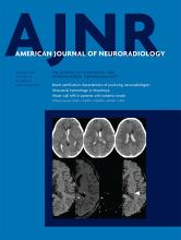
- Fig 1.
Diagram used for the classification of the site of bleeding. The periventricular area was defined as subependymal tissue and the white matter area located within 10 mm from the wall of the lateral ventricles except for the thalamus, basal ganglia, and corpus callosum. Th indicates thalamus; BgIC, basal ganglia and internal capsule; Cc, corpus callosum.
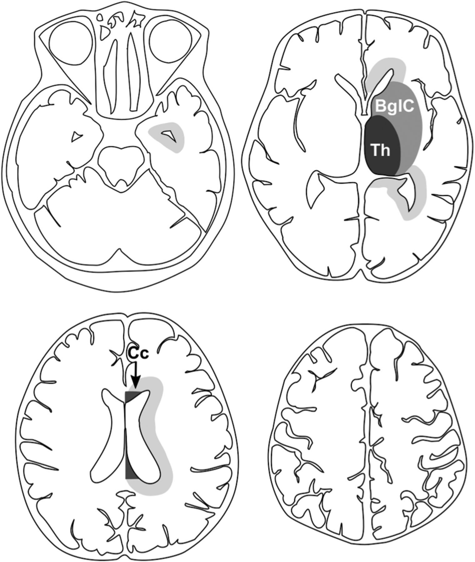
- Fig 2.
Diagrams show the distribution map of bleeding points and responsible vessels. Squares represent bleeding points derived from lenticulostriate arteries, black circles represent those from thalamic perforators, and white circles represent those from choroidal arteries. A, All bleeding points are drawn in a diagram. B–D, Diagrams show bleeding points derived from lenticulostriate arteries, the thalamic perforator, and choroidal artery, respectively. Bleeding points are on the left hemisphere.
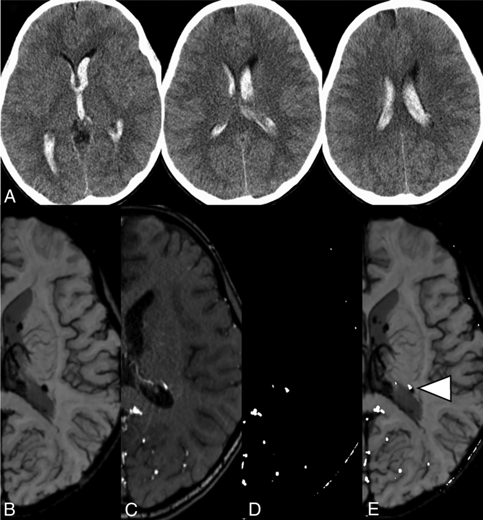
- Fig 3.
A, CT images from a 7-year-old boy with acute intraventricular hemorrhage. Images of SWI (B) and TOF-MRA (C), contrast-adjusted TOF-MRA (D), and a fusion image of contrast-adjusted TOF-MRA and SWI (E) for intraventricular hemorrhage in the same patient (chronic phase). The arrowhead shows the bleeding point at which the signal from the abnormally extended artery on TOF-MRA overlaps the hypointense spot on SWI. This point was defined as the bleeding point for the present case.
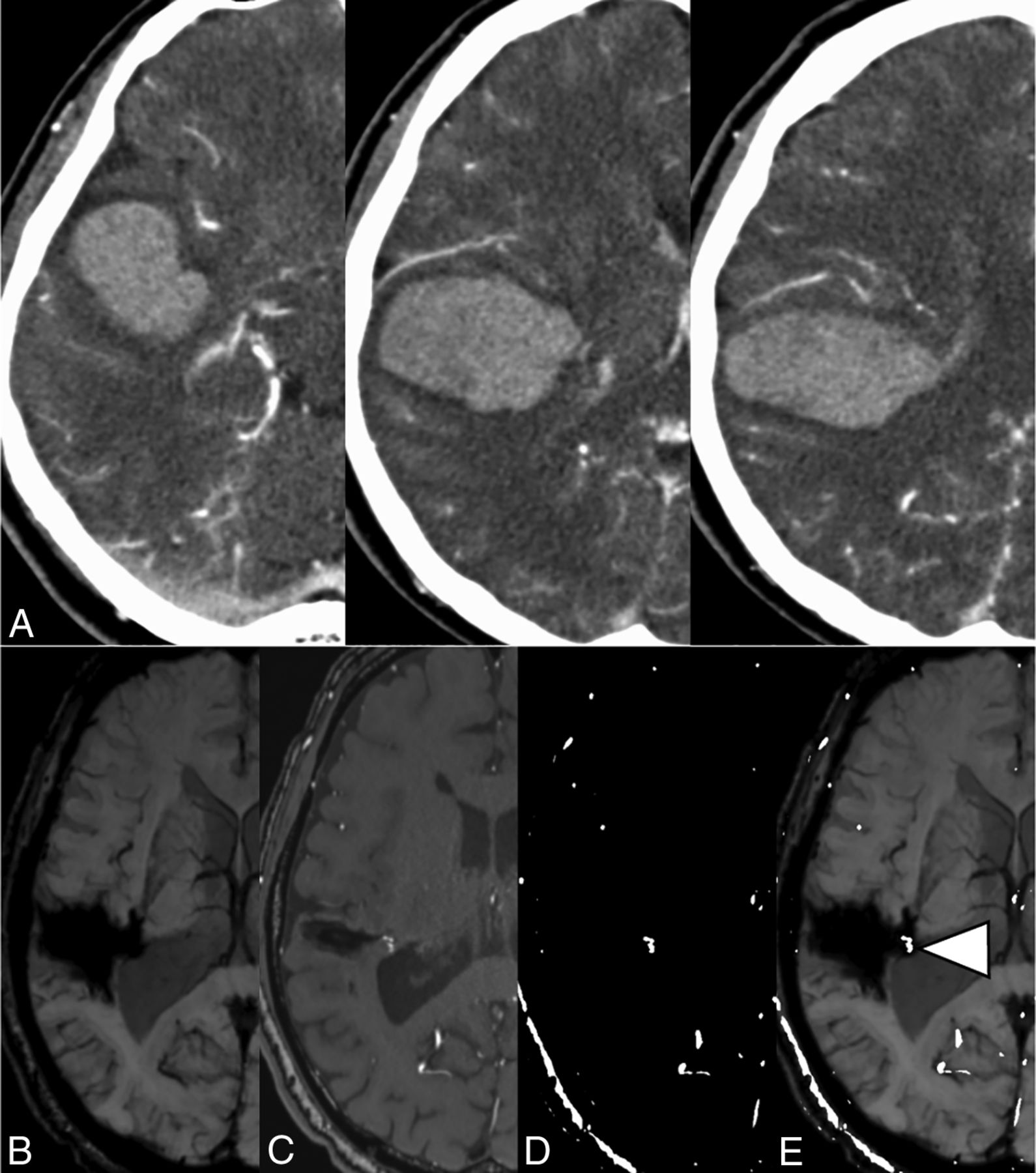
- Fig 4.
A, Contrast-enhanced CT of a 54-year-old man with acute lobar hemorrhage. Images of SWI (B) and TOF-MRA (C), contrast-adjusted TOF-MRA (D), and a fusion image of contrast-adjusted TOF-MRA and SWI (E) in the same patient (chronic phase). The arrowhead shows the signal of an abnormally dilated choroidal artery.
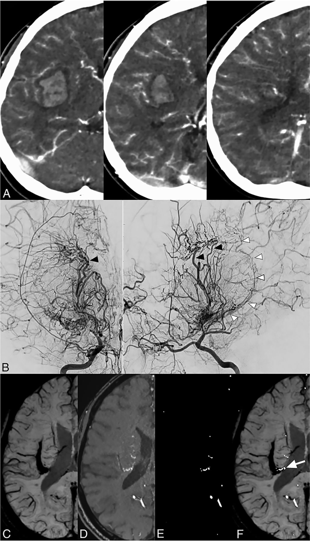
- Fig 5.
A, Contrast-enhanced CT of a 29-year-old woman with acute putaminal hemorrhage. B, Right internal carotid angiogram of the same patient (acute phase) shows abnormal dilation and extension of the lenticulostriate arteries (black arrowheads) and choroidal artery (white arrowheads). Images of SWI (C) and TOF-MRA (D), contrast-adjusted TOF-MRA (E), and fusion image of contrast-adjusted TOF-MRA and SWI (F) in the same patient (chronic phase). The arrow shows the dilated choroidal artery, which might be disrupted in the periventricular area in this case.


Tables
Reliability of detection of the bleeding point and vessel-related hemorrhagea
Rater 1 (n = 48) Rater 2 (n = 48) IE (95% CI) IA (95% CI) Presence of responsible vessel 38 (79.2%) 35 (72.9%) 0.83 (0.65–1) 0.86 (0.68–1) Location of bleeding point (n = 38) (n = 35) 0.92 (0.82–1) 0.85 (0.72–0.99) Thalamus 3 (7.9%) 4 (11.4%) Basal ganglia and internal capsule 9 (23.7%) 7 (20%) Periventricular area 25 (65.8%) 23 (65.7%) Corpus callosum 1 (2.6%) 1 (2.9%) Other 0 0 Origin of responsible vessels (n = 38) (n = 35) 0.84 (0.69–1) 0.90 (0.78–1) Lenticulostriate artery 9 (23.7%) 10 (28.6%) Thalamic perforator 7 (18.4%) 4 (11.4%) Choroidal artery 22 (57.9%) 21 (55.3%) Other 0 0 Note:—IE indicates interrater; IA, intrarater.
↵a Data are κ-agreements and correlation coefficients.


