
Article Figures & Data
Figures
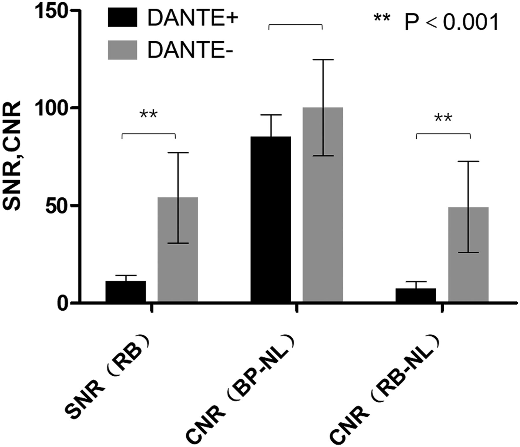
- Fig 1.
SNR of residual blood (RB), CNR between brain parenchyma (BP) and normal lumen (NL), and CNR between RB and NL in the healthy volunteer group. SNR of RB and RB-to-NL CNR were significantly reduced on BTI DANTE+ images compared with those on BTI DANTE- images. Double asterisks denote P < .001.
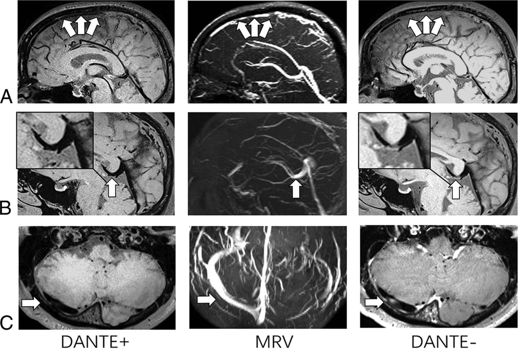
- Fig 2.
BTI (with/without DANTE preparation) images and MRV images of 3 patients with CVT. On BTI DANTE− images, isointense signals appear in the superior sagittal sinus (arrows, A), vein of Galen (arrow, B), and right transvers sinus (arrow, C). However, they are not shown on the BTI DANTE+ images (arrows). MRVs for these 3 patients demonstrate no filling defects on corresponding segments (arrows).
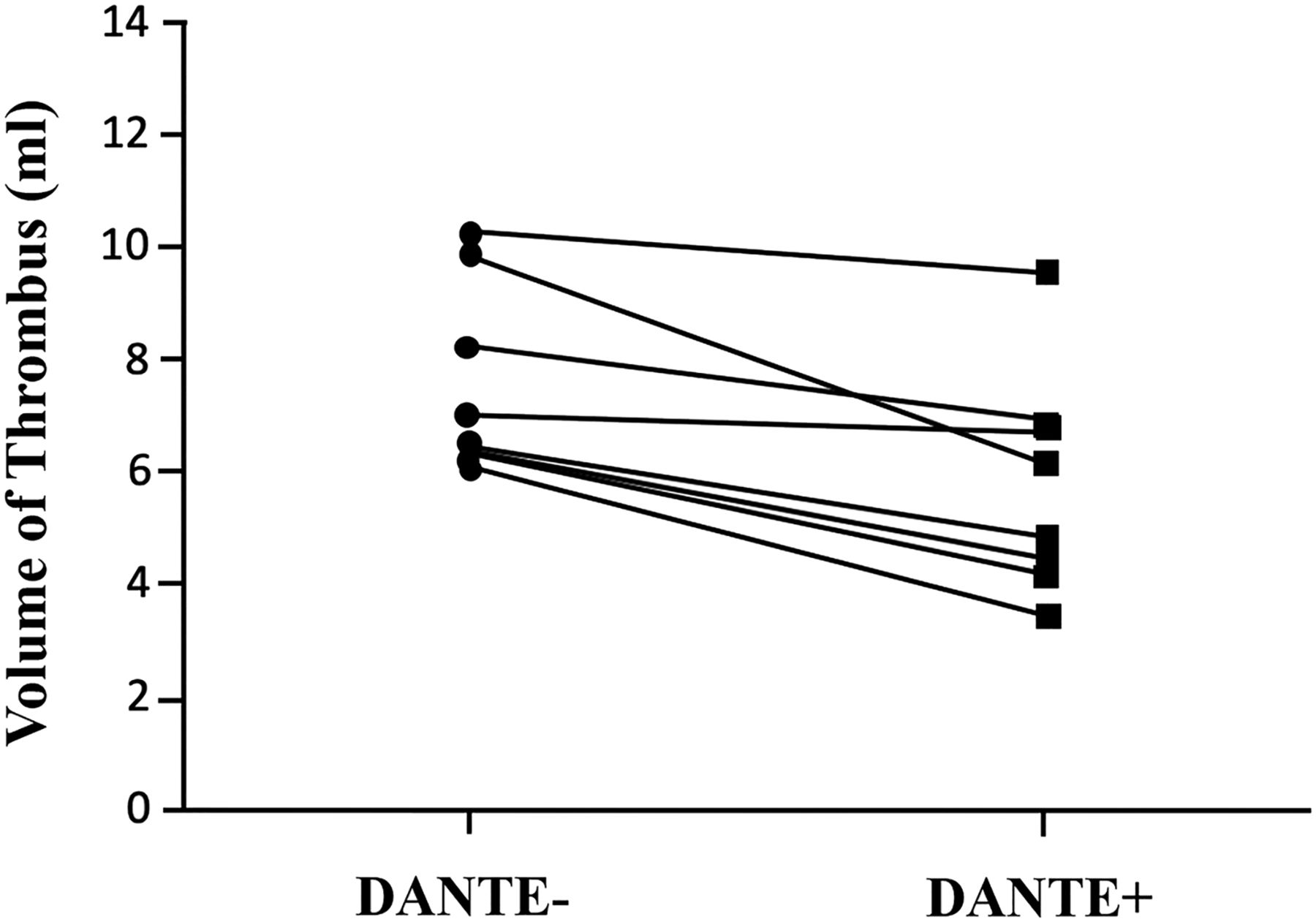
- Fig 3.
Thrombus volumes on BTI DANTE+ and BTI DANTE− images. In the 8 patients who were diagnosed with apparent thrombi in the superior sagittal sinus, the measured thrombus volume was significantly lowered (4.814 ± 2.278 mL versus 6.341 ± 2.302 mL, P =.008) when using the DANTE preparation.
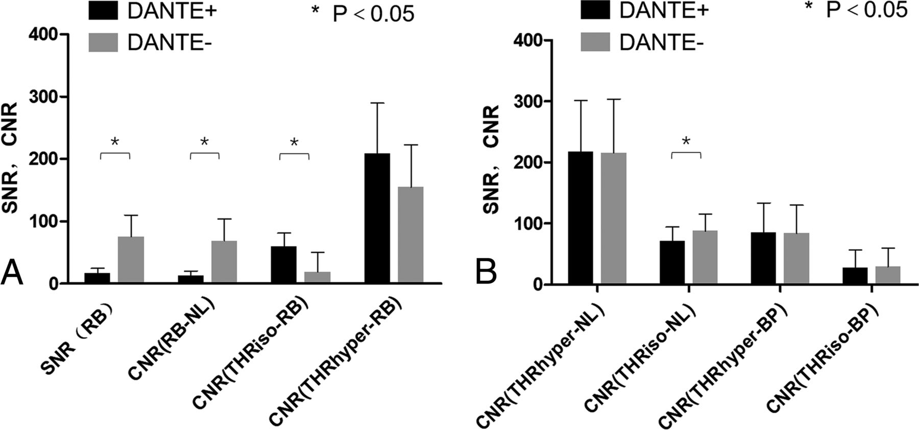
- Fig 4.
A, SNR of residual blood (RB), CNR between RB and normal lumen (NL), and CNR between RB and thrombus in the patient group. SNR of RB and RB-to-NL CNR was significantly reduced on BTI DANTE+ images compared with BTI DANTE− images. RB-to-thrombus CNR was significantly improved for the isointense thrombus type. B, CNR between thrombus and NL and CNR bet-ween thrombus and brain parenchyma (BP). When one used the DANTE preparation, the CNR between thrombus and NL or BP was not significantly sacrificed except for the CNR between isointense thrombus and NL. THRiso indicates isointense thrombus; THRhyper, hyperintense thrombus; asterisk, P < .05.


Tables
Patient Sex Age (yr) Symptom Duration Conventional Imaging Methods 1 F 47 Headache 2 days TSE, SWI, CE-MRV, TOF-MRV, CT 2 M 19 Headache 10 days TSE, TOF-MRV, CT 3 M 54 Focal neurologic deficit 13 days TSE, SWI, CE-MRV, TOF-MRV, CT 4 M 30 Headache, focal neurological deficit 20 days TSE, CE-MRV, TOF-MRV, CT 5 M 44 Seizures 25 days TSE, TOF-MRV 6 F 53 Headache 27 days TSE, SWI, CE-MRV, TOF-MRV, CT 7 M 42 Headache 1 mo TSE, CE-MRV, TOF-MRV 8 F 32 Headache 1 mo + 22 days TSE, SWI, CE-MRV, TOF-MRV, CT 9 M 19 Headache 2 mo TSE, TOF-MRV, CT 10 F 28 Headache 4 mo TSE, TOF-MRV, CT 11 F 45 Headache, papilledema 4 mo SWI, TOF-MRV, CT 12 F 36 Headache 1 year TSE, CE-MRV, TOF-MRV, CT 13 F 19 Headache 2 days TSE, TOF-MRV, CE-MRV - Table 2:
Locations of thrombi and residual flow artifacts identified on BTI with and without DANTE preparationa
Pt. No. Segments SSS ISS VG SS CS RTS LTS RSS LSS ICV BVR VL RC LC RJV LJV 1 = = = = = 2 = = 3 +/= = + = = + + 4 = = = +/= = = + = = = = 5 6 = = = = = = = = 7 = = = = +/= = = 8 +/= + +/= +/= +/= = = = = + +/= +/= 9 = = = = = = = = = 10 +/= +/= +/= +/= +/= = 11 12 = = = = = = = 13 = = = Note:—SSS indicates superior sagittal sinus; ISS, inferior sagittal sinus; VG, vein of Galen; SS, straight sinus; CS, confluence of sinus; RTS, right transverse sinus; LTS, left transverse sinus; RSS, right sigmoid sinus; LSS, left sigmoid sinus; ICV, internal cerebral vein; BVR, basal vein of Rosenthal; VL, vein of Labbé; RC, right cortical vein; LC, left cortical vein; RJV, right jugular vein; LJV, left jugular vein; =, iso-intense thrombus; +, hyperintense thrombus; +/=, hybrid thrombus.
↵a Shadow indicates that residual flow artifacts are present in the segment on BTI without DANTE preparation but not on BTI with DANTE preparation.
- Table 3:
Diagnostic performance of BTI with/without DANTE for the detection of CVT on the per-segment level
Dante+ Dante− Sensitivity 100% 100% Specificity 96.3% 83.8% PPV 93.5% 76.6% NPV 100% 100% FP 3.7% 16.2% Note:—PPV indicates positive predictive value; NPV, negative predictive value; FP, false-positive.


Jump to section
Related Articles
Cited By...
- No citing articles found.