
Article Figures & Data
Figures
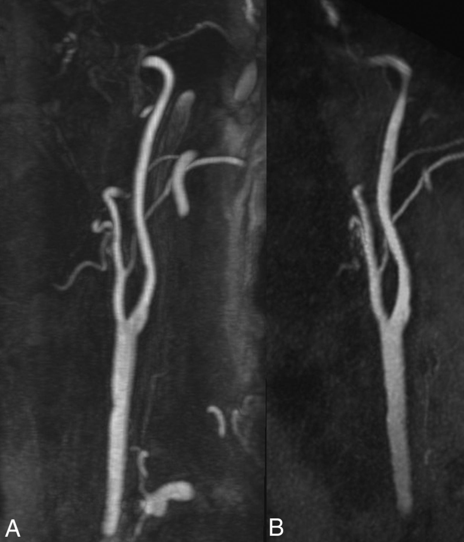
- Fig 1.
Example of an excellent imaging quality (grade 3) without any venous contamination (grade 0). Maximum intensity projection (MIP) with angulation to the left carotid bifurcation of the CE-MRA (A, slice thickness: 14.5 mm) and the ungated QISS-MRA (B, slice thickness: 14.1 mm) of a 76-year-old patient with clinically suspected infarction of the right hemisphere and suspected stenosis of the right cervical internal carotid artery by sonography (same patient as in Fig 5).
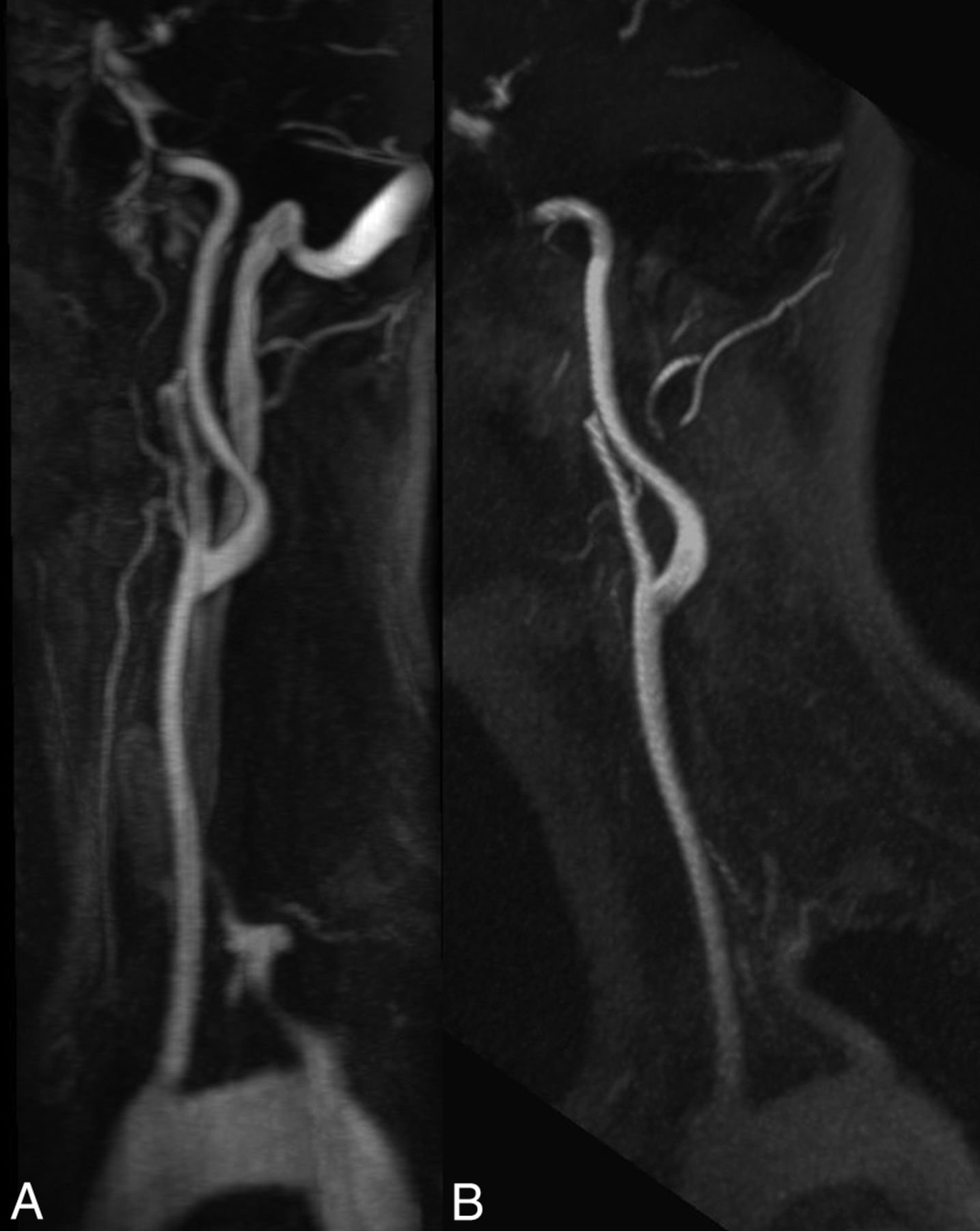
- Fig 2.
The effect of venous contamination on the image quality. MIP with angulation to the left carotid bifurcation of the CE-MRA (A, slice thickness: 13.9 mm) and the QISS-MRA (B, slice thickness: 13.5 mm) of a 33-year-old patient with suspected cerebral infarction. In the CE-MRA, the bolus is slightly missed, resulting in a severe venous contamination, whereas the QISS-MRA shows no venous signal.
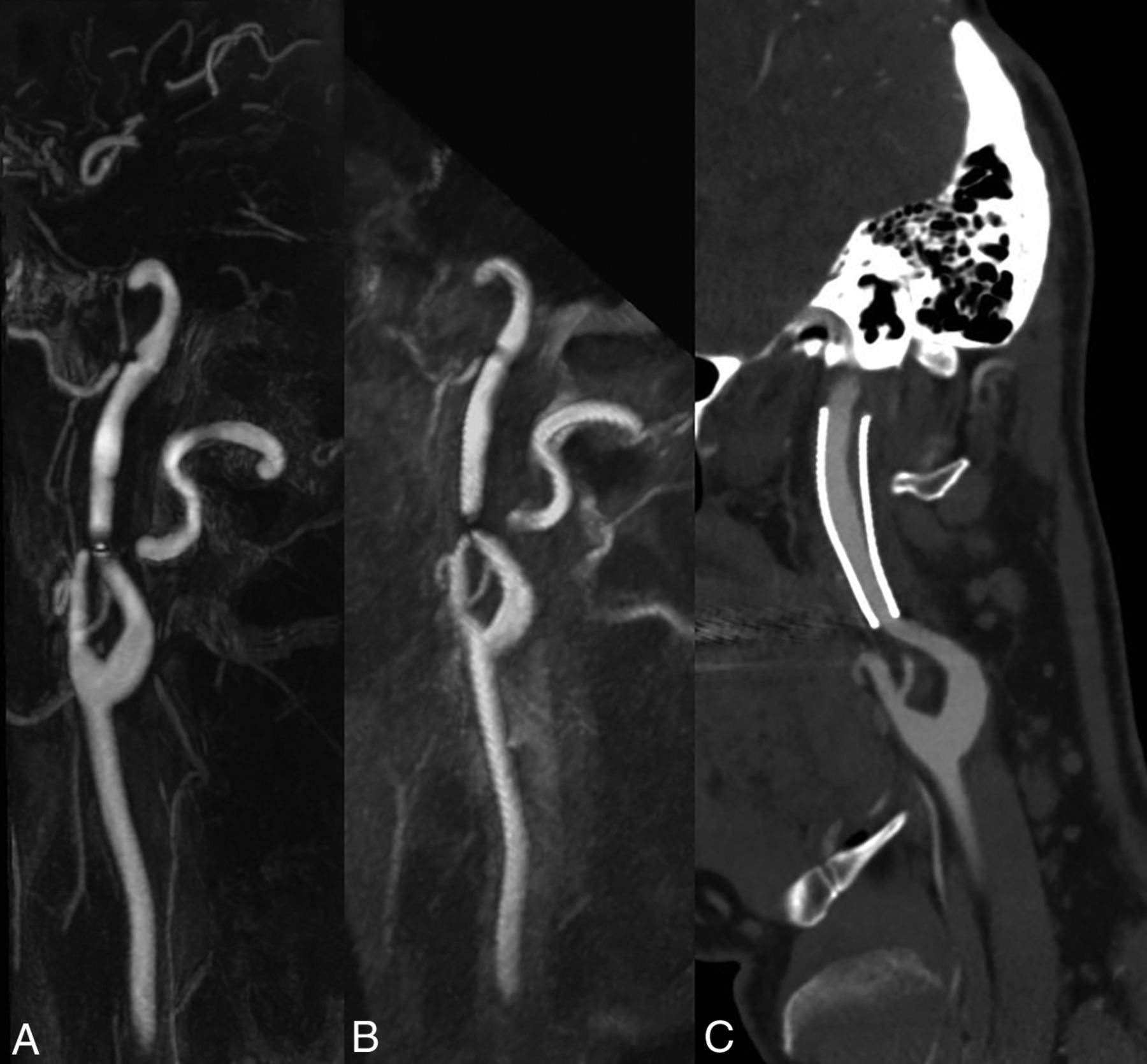
- Fig 3.
Influence of an implanted stent on the image quality. MIP of the CE-MRA (A, slice thickness: 13.0 mm) and the ungated QISS-MRA (B, slice thickness: 13.0 mm) with angulation to the left internal carotid artery of a 50-year-old patient who was stented 5 years ago due to a carotid artery dissection. The corresponding MIP of a CE-CTA (C, slice thickness: 1.4 mm) was obtained 2 years, and DSA, 1 year after stent placement. In both MRA techniques, there are just slight artifacts at the ends of the stent, and the lumen is well visualized. This patient was not included in this study.
- Fig 4.
Visualization of internal carotid artery stenosis using CE-MRA and ungated QISS-MRA compared with CE-CTA. MIP with angulation to the left carotid bifurcation of the CE-MRA (A, slice thickness: 13.1 mm), QISS-MRA (B, slice thickness: 13.0 mm), and CE-CTA (C, slice thickness: 13.0 mm) of a 55-year-old patient with confirmed infarction of the left hemisphere and suspected stenosis of the left internal carotid artery using sonography. All 3 techniques verified the diagnosis of carotid stenosis (white arrows).
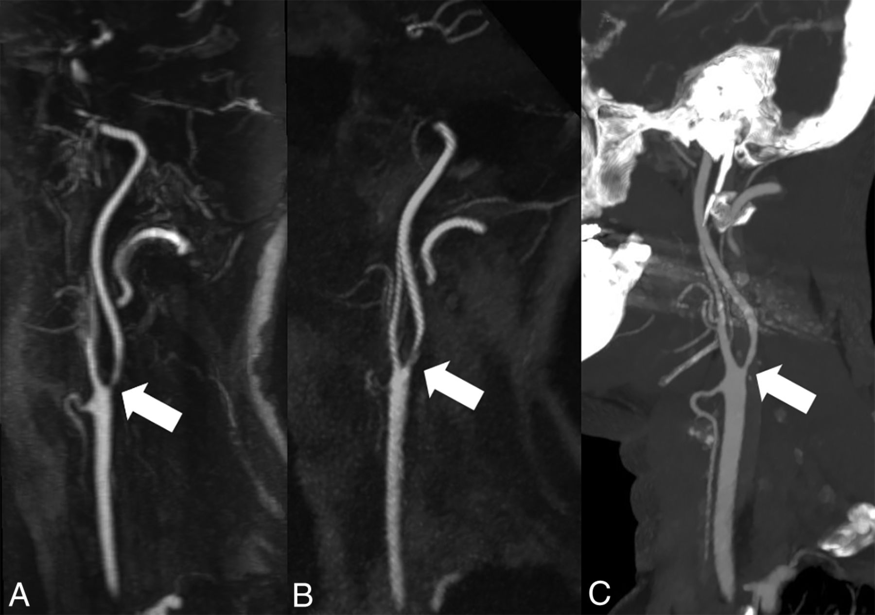
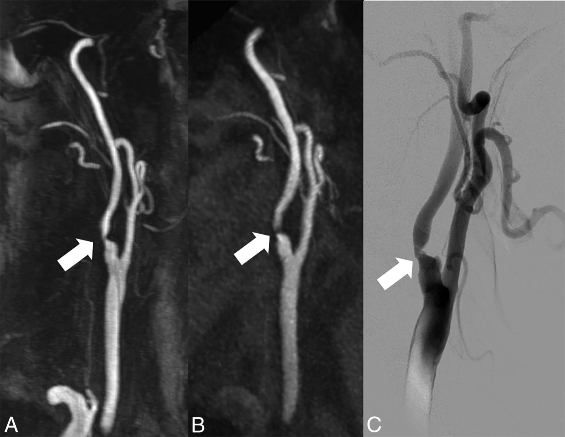
- Fig 5.
Visualization of internal carotid artery stenosis using CE-MRA and ungated QISS-MRA compared with invasive DSA. MIP with angulation to the right carotid bifurcation of the CE-MRA (A, slice thickness: 14.0 mm) and the QISS-MRA (B, slice thickness: 13.5 mm) of a 76-year-old patient with clinically suspected infarction of the right hemisphere and suspected stenosis of the cervical internal carotid artery on the right by sonography (same patient as in Fig 1). The corresponding DSA of the right carotid bifurcation (C) before stent angioplasty confirmed the stenosis (white arrows).


Tables
Parameter Ungated QISS-MRA CE-MRA Imaging mode 2D 3D FLASH TR/TE (ms) 15.0/4.7 3.09/1.2 QISS sequence TR (ms) 1100.8 – Acquisition matrix (Px) 384 × 384 512 × 512 Acquisition pixel (mm2) 0.5 × 0.5 0.6 × 0.6 In-plane interpolation On On Slice thickness (mm) 2.0 1.0 No. of slices 128 80 Slice distance factor (%) −33 20 No. of averages 1 1 Receiver bandwidth (Hz/Px) 303 540 Flip angle 30° 30° Slice orientation Tilted transversal to coronal (45° tilt) Coronal K-space trajectory Radial Cartesian No. of radial projections 204 – No. of shots per slice 3 – Phase oversampling (%) 0 40 Filter Distortion correction (2D); prescan normalizer Distortion correction (3D); prescan normalizer B0 shim mode Heart Tune-up Asymmetric echo Off On RF pulse type Normal Normal Gradient mode Fast Fast RF spoiler On On iPAT modus (acceleration factor/No. of reference lines) – 2/24 Partial Fourier (phase and slice) – 6th/8th Venous saturation slab thickness (mm) 100 – Distance between venous saturation and imaging slab (mm) 10 – TI (ms) 530 – Acquisition time (min:sec) 7:03 0:20 Note:—iPAT indicates integrated parallel imaging technique; TI, time from in-plane and venous saturation to the acquisition of central k-space (ky = 0); Px, pixel; –, sequence parameter is not available; TR, repetition time; TE, echo time; RF, radiofrequency; Hz, Hertz.
- Table 2:
Evaluation of ungated QISS-MRA versus CE-MRA based on the introduced 3-, 4-, and 5-point scale scoring systems in the section “Image Analysis” using the Wilcoxon signed rank test
Variablea QISS-MRAb CE-MRAb P Value (QISS-MRA vs CE-MRA) Image quality 2 (1–3) 2 (1–3) .46 Venous contamination 0 (0–2) 1 (0–3) <.0001 Global quality of arterial visualization 2 (1–4) 3 (1–4) <.0001 Stenosis grading Right 1 (1–5) 1 (1–5) .64 Left 1 (1–5) 1 (1–5) .73 Segmental quality of arterial visualization Right side Origin of brachiocephalic artery (1) 3 (1–4) 3 (1–4) <.0001 Origin of CCA (2) 3 (1–4) 3 (1–4) <.0001 CCA (3) 3 (1–4) 4 (1–4) .03 Bifurcation of CCA (4) 3 (1–4) 4 (1–4) .002 ICA-C1 (cervical) (5) 3 (1–4) 4 (1–4) .011 ECA (superior thyroid artery) (6) 1 (1–3) 2 (1–4) .007 ECA (lingual artery) (7) 1 (1–3) 2 (1–4) .0002 ECA (facial artery) (8) 2 (1–3) 2 (1–4) .0003 ECA (occipital artery) (9) 2 (1–3) 2 (1–4) .043 ECA (posterior auricular artery) (10) 1 (1–3) 1 (1–4) .16 ECA (suprafacial temporal artery) (11) 2 (1–3) 2 (1–4) .002 ECA (maxillary artery) (12) 2 (1–3) 2 (1–4) .001 ECA (ascending pharyngeal artery) (13) 1 (1–3) 1 (1–4) .39 Origin of subclavian artery (14) 2 (1–4) 3 (1–4) <.0001 Origin of vertebral artery (V0) (15) 2 (1–4) 2 (1–4) .19 V1 (preforaminal) (16) 3 (1–4) 3 (1–4) .064 V2 (foraminal) (17) 3 (1–4) 3 (1–4) .51 V3 (atlantic, extradural, or extraspinal) (18) 3 (1–4) 3 (1–4) .097 Left side Origin of CCA (1) 2 (1–4) 3 (1–4) .0003 CCA (2) 3 (2–4) 4 (1–4) .01 Bifurcation of CCA (3) 3 (2–4) 4 (1–4) .008 ICA-C1 (cervical) (4) 3 (1–4) 4 (1–4) .02 ECA (superior thyroid artery) (5) 2 (1–3) 2 (1–3) .02 ECA (lingual artery) (6) 1 (1–3) 2 (1–3) .002 ECA (facial artery) (7) 2 (1–3) 2 (1–4) <.0001 ECA (occipital artery) (8) 2 (1–4) 2 (1–3) .34 ECA (posterior auricular artery) (9) 1 (1–3) 1 (1–3) .98 ECA (suprafacial temporal artery) (10) 2 (1–3) 2 (1–4) .0003 ECA (maxillary artery) (11) 2 (1–3) 2 (1–4) .0008 ECA (ascending pharyngeal artery) (12) 1 (1–3) 1 (1–3) .34 Origin of subclavian artery (13) 2 (1–4) 3 (1–4) <.0001 Origin of vertebral artery (V0) (14) 2 (1–4) 2 (1–4) .001 V1 (preforaminal) (15) 3 (1–4) 3 (1–4) .002 V2 (foraminal) (16) 3 (1–4) 3 (1–4) .88 V3 (atlantic, extradural, or extraspinal) (17) 3 (1–4) 3 (1–4) .11 - Table 3:
Interobserver agreement for the evaluation of QISS-MRA and CE-MRA based on the introduced 3-, 4-, and 5-point scale scoring systems in the “Image Analysis” sectiona
Variable Interobserver Agreement Image quality 0.54 (0.46–0.62) Venous contamination 0.86 (0.80–0.91) Quality of global arterial visualization Right side 0.72 (0.70–0.74) Left side 0.71 (0.69–0.72) ICA stenosis Right side 0.94 (0.89–0.97) Left side 0.95 (0.90–0.98) ↵a Data are agreement (95% CI).
- Table 4:
Comparison of ungated QISS-MRA and CE-MRA for assessment of the stenosis grade of the extracranial carotid arteriesa
Right Sideb Left Sideb 1 2 3 1 2 3 Sensitivity (%) 66.7 (9.4–99.2) 100.0 (15.8–100.0) 50.0 (1.3–98.7) 100 (39.8–100.0) 83.3 (35.9–99.6) 100.0 (39.8–100.0) All readers 71.4 (29.0–96.3) 92.9 (66.1–99.8) Both sides 85.7 (63.7–97.0) Specificity (%) 89.3 (71.8–97.7) 86.2 (68.3–96.1) 89.7 (72.7–97.8) 92.6 (75.7–99.1) 88.0 (68.8–97.5) 96.3 (81.0–99.9) All readers 87.7 (78.5–93.3) 92.4 (84.2–97.2) Both sides 90.0 (84.3–94.2)


Jump to section
Related Articles
Cited By...
- No citing articles found.