
Article Figures & Data
Figures
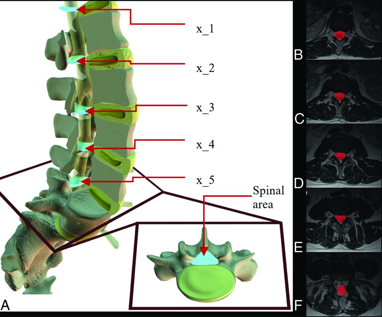
- Fig 1.
Variation of spinal canal area with level. A, This 3D model represents a generic lumbar spine where light blue objects represent an area of the central canal at each lumbar level at the midsection of a disc. The square frame (red) zooms in on the intervertebral disc (yellow) below L5 to give an axial view of where the central canal area (light blue) is located. In a randomly selected T2-MR imaging, each picture in this series B–F depicts 1 section of spinal cord segmentation (red) from each level. Tissues within the canal but outside the thecal sac are not segmented.
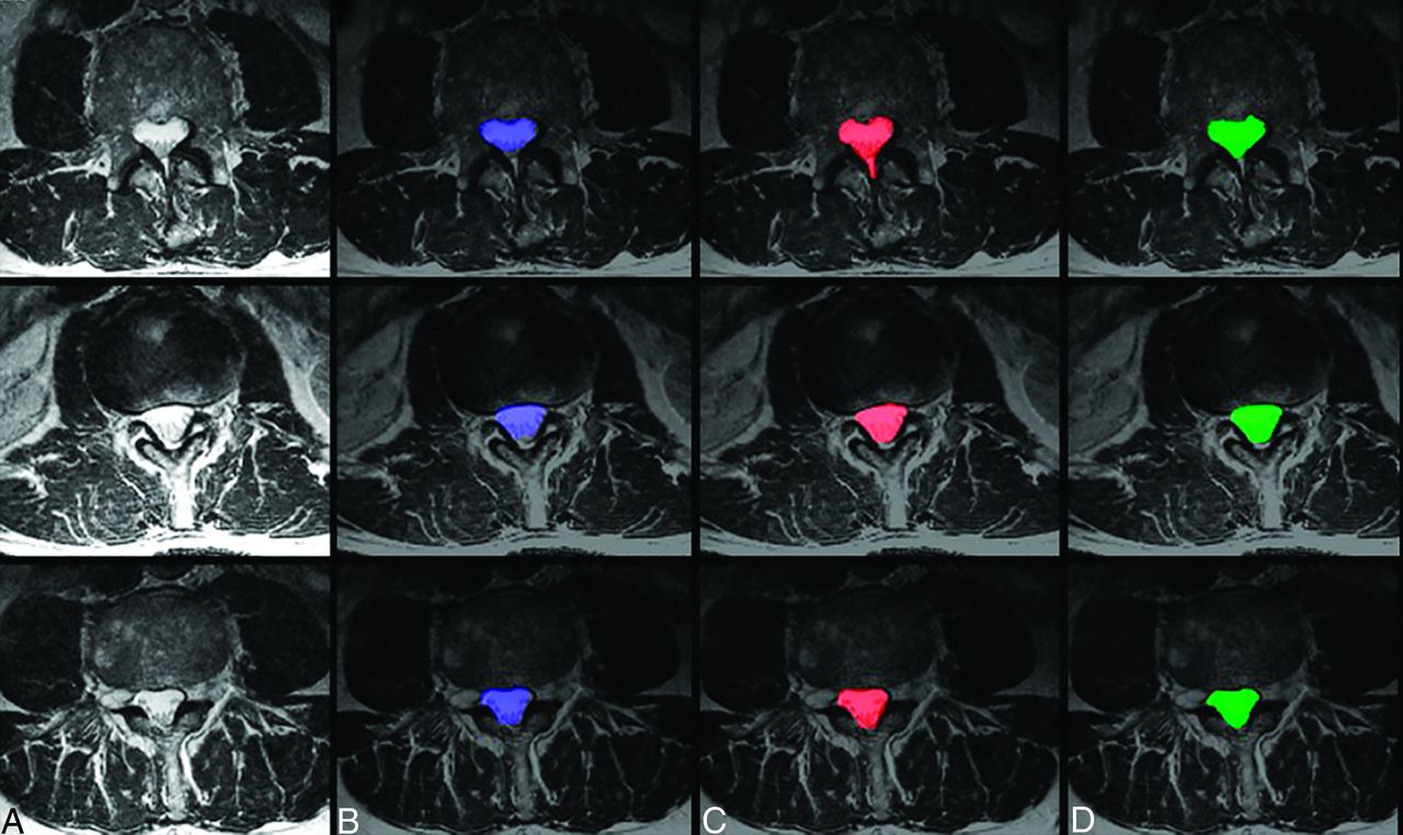
- Fig 2.
Sample case images of central canal segmentations. Three case images of axial T2 MR imaging (A) randomly selected from the dataset are shown alongside their resulting segmentations (blue) of the spinal canal using the proposed ensemble technique (B), segmentation (red) by manual rater 1 (C), and segmentation (green) by manual rater 2 (D).
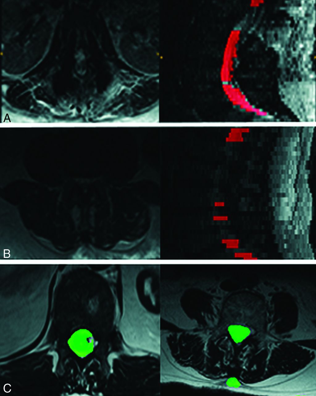
- Fig 3.
Modes of segmentation failure of the proposed algorithm compared with U-Net results. Two scans using SVM + ERT failed (Dice score <0.7). The mode of failure was complete lack of segmentation as seen in rows A and B, thereby making it easy to eliminate such cases automatically. In contrast U-Net failures are more subtle and can involve under and oversegmentation as shown in row C.


Tables
- Table 1:
Comparison of automated spinal canal segmentations in a validation dataset of 109 axial MRIsa
Centrality Auto vs Rater 1 Auto vs Rater 2 Rater 1 vs Rater 2 Dice ratio Mean 0.84 ± 0.08 0.83 ± 0.08 0.9 ± 0.05 Median 0.87 0.85 0.92 Hausdorff distance (mm) Mean 7.89 ± 9.42 9.41 ± 11.2 7.90 ± 9.62 Median 4.59 5.64 4.66 Average surface distance (mm) Mean 0.84 ± 0.08 0.83 ± 0.08 0.9 ± 0.05 Median 0.10 0.14 0.07 ↵a Data are means and medians.
- Table 2:
Age, gender, and height were analyzed using a mixed effects model Canal Area ~ age + gender + height + gender*height + (1 | subject). This table represents the Random Effects fita
Random Effects Groups Name Variance SD Group (Intercept) 2868 53.55 Residual 2365 48.63 No. of obs 8775 No. of subjects 1755 Note:—obs indicates observers.
↵a The model fit is summarized. Height is the only variable that is statistically significantly related to canal areas.
- Table 3:
Age, gender, and height were analyzed using a mixed effects model Canal Area ~ age + gender + height + gender*height + (1 | subject). This table represents the Intercept fit - equivalent to traditional regressiona
Fixed Effects Estimate Standard Error T Value Pr (>|t|) (Intercept) −278.18495 43.20594 −6.439 0.00 Age 0.05158 0.08429 0.612 0.54 Sex −0.63539 62.56533 −0.01 0.10 Height 8.00079 0.66184 12.089 0.00 Sex:height −0.46311 0.93188 −0.497 0.62 Note:—Pr (>|t|) indicates p-value.
↵a The model fit is summarized. Height is the only variable that is statistically significantly related to canal areas.
- Table 4:
Age, gender, and height were analyzed using a mixed effects model Canal Area ~ age + gender + height + gender*height + (1 | subject). This table represents the correlation of fixed effectsa
Correlation of Fixed Effects (Intercept) Age Sex Height Age −0.203 Sex −0.662 0.001 Height −0.995 0.11 0.672 Sex:height 0.692 −0.009 −0.998 −0.703 ↵a The model fit is summarized. Height is the only variable that is statistically significantly related to canal areas.


Jump to section
Related Articles
Cited By...
- No citing articles found.